. 2016 Apr 12;113(15):4045-50.
doi: 10.1073/pnas.1524404113. Epub 2016 Mar 28.

Host-parasite oscillation dynamics and evolution in a compartmentalized RNA replication system

Affiliations

Host-parasite oscillation dynamics and evolution in a compartmentalized RNA replication system

Yohsuke Bansho et al. Proc Natl Acad Sci U S A. .

Abstract

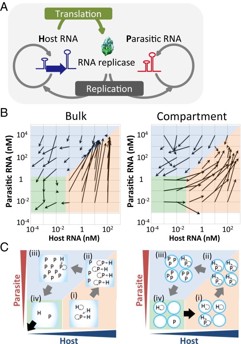
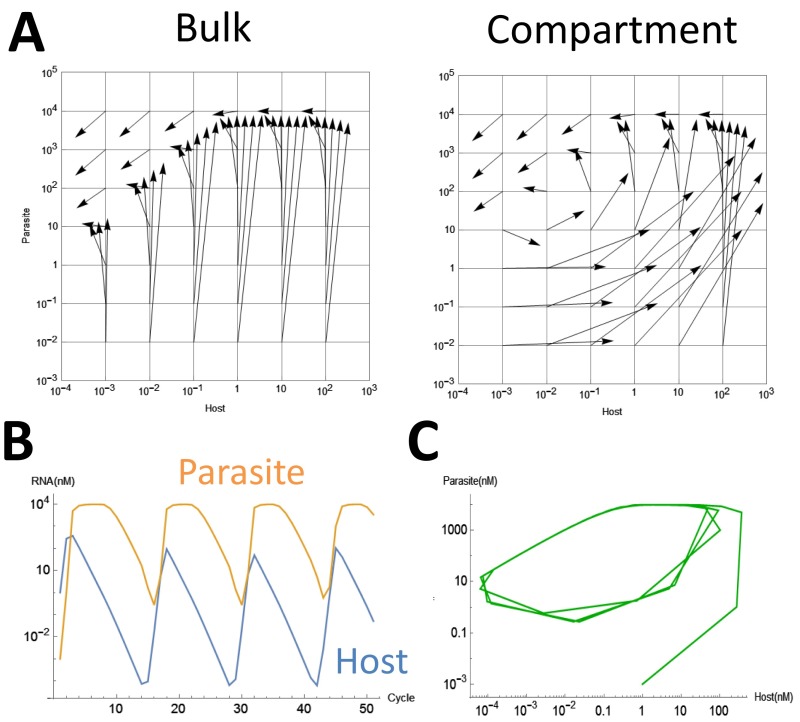
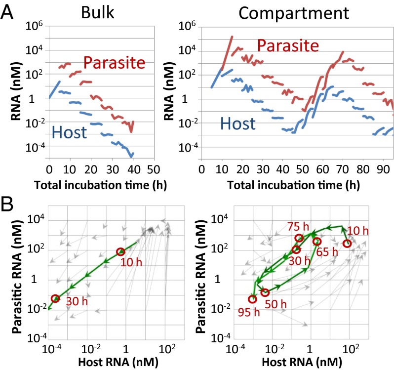
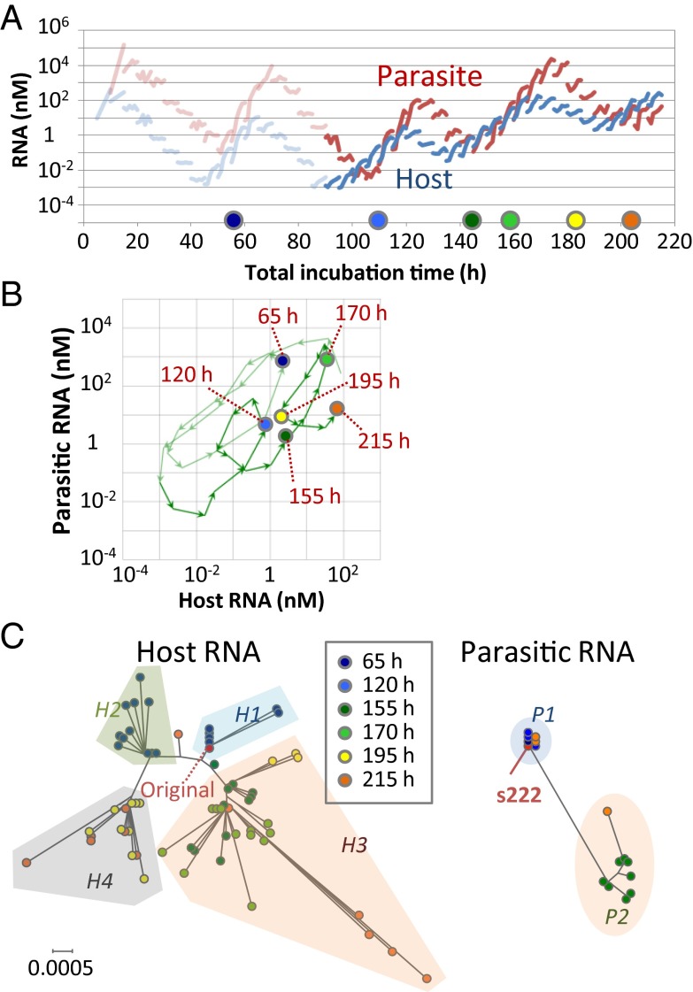
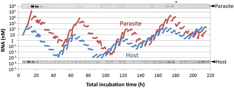
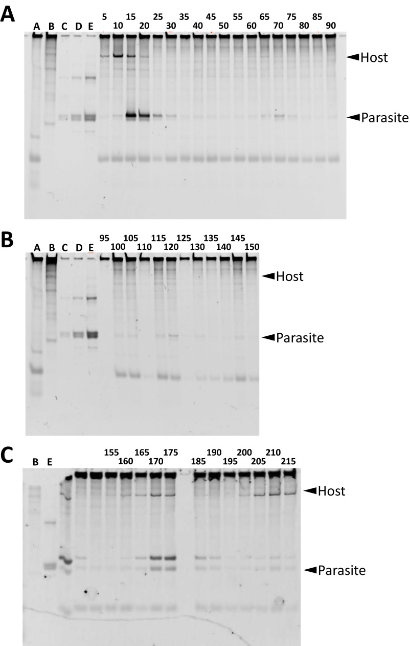
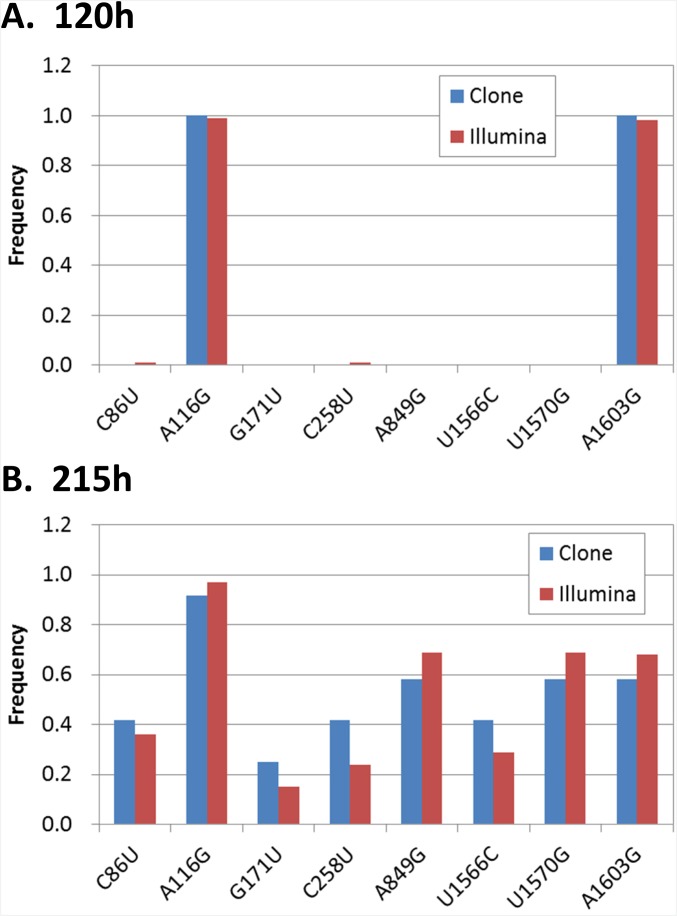
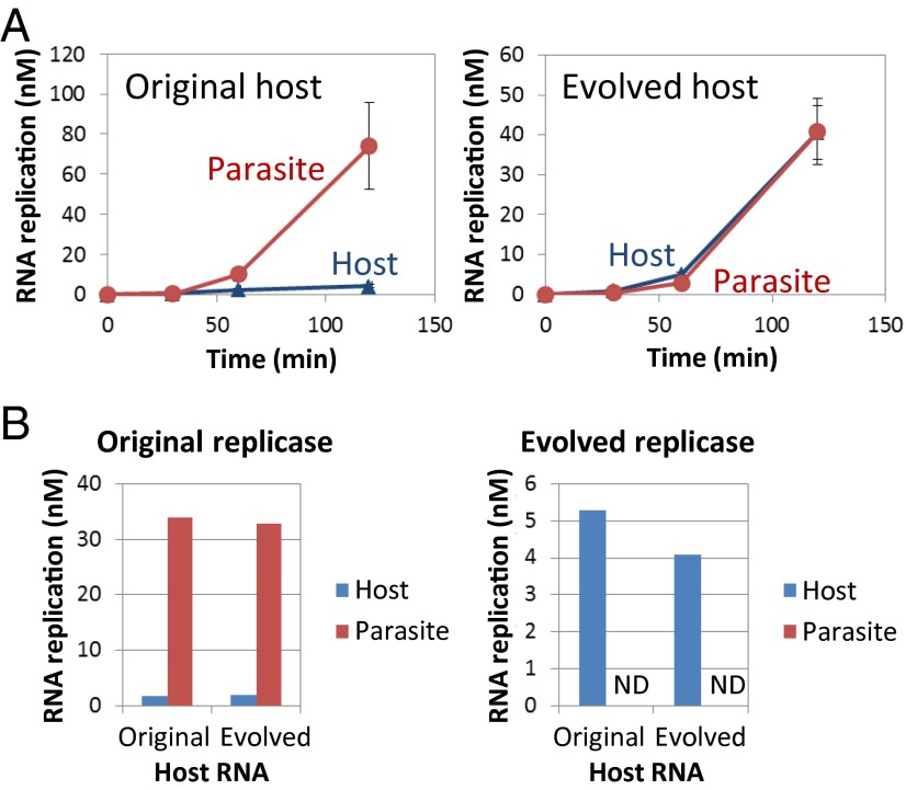
To date, various cellular functions have been reconstituted in vitro such as self-replication systems using DNA, RNA, and proteins. The next important challenges include the reconstitution of the interactive networks of self-replicating species and investigating how such interactions generate complex ecological behaviors observed in nature. Here, we synthesized a simple replication system composed of two self-replicating host and parasitic RNA species. We found that the parasitic RNA eradicates the host RNA under bulk conditions; however, when the system is compartmentalized, a continuous oscillation pattern in the population dynamics of the two RNAs emerges. The oscillation pattern changed as replication proceeded mainly owing to the evolution of the host RNA. These results demonstrate that a cell-like compartment plays an important role in host-parasite ecological dynamics and suggest that the origin of the host-parasite coevolution might date back to the very early stages of the evolution of life.

Keywords: RNA replication; compartment; evolution; oscillation; parasite.


Conflict of interest statement

The authors declare no conflict of interest.

Figures

Fig. 1.
Fig. 1.
RNA replication system and single-round replication assay. (A) Replication scheme of the host and parasitic RNAs. (B) Vector plots of the host and parasitic RNA replications in a single round of the transfer experiment. The initial and averaged final concentrations are indicated by roots and heads of arrows (n = 2–4). (C) Schematic drawing of the host and parasitic RNA replications in each region.
Fig. S1.
Fig. S1.
Size distribution of the water droplets used in this study. The water droplets were prepared by the same method as used for the single-round assay as shown in Fig. 1B and described in Materials and Methods. The sizes of 100 water droplets in water-in-oil emulsion were measured using an optical microscope. The error bars indicate SDs (n = 3).
Fig. S2.
Fig. S2.
Simulations of the replication of the host and parasitic RNAs. (A) We performed simulations the replication of the host and parasitic RNAs at various starting concentrations under bulk or compartmentalized conditions according to the method in SI Materials and Methods. The initial and final concentrations after concentrations after dilution are indicated by roots and heads of arrows, respectively. (B) Simulation of an oscillation in the host and parasitic RNA concentrations. We performed the simulation of replication of the host and parasitic RNA according to the method in SI Materials and Methods. We started the simulation at host = 1 nM and parasite = 10−3 nM. (C) Replot of the host and parasitic RNA concentrations on the host–parasite plane. We also started the simulation from 36 combinations of initial concentrations from 10−3 to 102 nM and found that the trajectories converged to the same.
Fig. 2.
Fig. 2.
Transfer experiments. (A) The host and parasitic RNA concentrations during incubation in the transfer experiments. (B) Trajectory of the host and parasitic RNA concentrations on a host–parasite plane.
Fig. 3.
Fig. 3.
Changes of the oscillation dynamics under the compartmentalized condition. (A) The host and parasitic RNA concentrations in the continued transfer experiments. At the time indicated by the colored circles, the host and parasitic RNAs were cloned and sequenced. (B) Trajectory of the host and parasitic RNA populations on a host–parasite plane. (C) Phylogenetic trees of the host and parasitic RNA clones; the trees were drawn on the same scale. For parasitic RNA, the frequently amplified s222 RNA is also indicated.
Fig. S3.
Fig. S3.
Comparison between the quantification results of the host and parasitic RNAs using RNA staining after polyacrylamide gel electrophoresis. The water droplets during the transfer experiment were collected and applied to polyacrylamide gel electrophoresis and RNAs were stained with SYBR green II. The parts of the gel corresponding to the parasite and the host were aligned with the quantitation data of Fig. 3A. *No sample was applied here because, unfortunately, the sample was lost. The original gel data are shown in Fig. S4.
Fig. S4.
Fig. S4.
The native polyacylamide gel electrophoresis of the reaction mixture during the transfer experiment. The number on the top of the gels indicates the incubation time during the transfer [(A) 5–90 h, (B) 95–150 h, and (C) 155–215 h]. Lane A is the reaction mixture without any host or parasite (i.e., only the reconstituted translation system). Lane B is pure original host RNA. Lanes C, D, and E are three different amounts of the original parasitic RNA. Both the host and parasitic RNAs exhibit multiple bands caused by structural heterogeneity. The major bands indicated in Fig. S3 have been marked in this figure with arrowheads.
Fig. S5.
Fig. S5.
Comparison of frequency of mutations between different sequencing methods. The frequency of each fixed mutation in the host RNA at (A) 120 h and (B) 215 h were compared between the two different methods: sequencing by Sanger method after cloning (Clone, the same data as Table S1) and a large-scale sequencing method using Illumina Miseq without cloning (Illumina). For the Illumina analysis, more than 1400 reads were obtained at all of the mutation sites, and no other fixed mutations that existed in more than 50% of the reads were found.
Fig. 4.
Fig. 4.
Competition experiments between the host and parasitic RNAs. (A) Translation-coupled replication competition experiments. The error bars indicate the SD (n = 3). (B) Competition experiments with purified replicases. When using the evolved replicase, the parasitic RNA was not detected (ND) with either the original or evolved host RNAs.
Fig. S6.
Fig. S6.
Translation of the evolved host RNA. To estimate the kinetic parameter of translation of the replicase, we performed translation with the original or evolved host RNAs in the translation system containing [35S]methionine. In this experiment, we omitted UTP to prevent replication of the host RNA. After incubation for the indicated periods, aliquots were subjected to SDS polyacrylamide gel electrophoresis, and the synthesized β-subunit of the replicase was measured after autoradiography as described previously (20). The estimated translation rates were shown in Table 1.
Fig. S7.
Fig. S7.
Estimation of kinetic parameters of replication. We performed the replication reaction with several combinations of the purified replicases and the host and parasitic RNAs to estimate the kinetic parameters of replication, kcat and Km, shown in Table 1. We mixed the original or evolved purified replicase with the original or evolved hosts or the parasitic RNA in the translation system containing [32P]UTP to trace the amplification of both the RNAs. In this experiment, we omitted tyrosine and cysteine to prevent the translation of the replicase. After incubation for the indicated periods, aliquots were subjected to polyacrylamide gel electrophoresis, and the synthesized RNA was measured by autoradiography as described in SI Materials and Methods. The initial RNA synthesis rates were plotted and fitted by Michaelis–Menten curve (gray lines) to estimate kcat and Km values.

Similar articles

Cited by

References

    1. Szostak JW, Bartel DP, Luisi PL. Synthesizing life. Nature. 2001;409(6818):387–390. - PubMed
    1. Forster AC, Church GM. Towards synthesis of a minimal cell. Mol Syst Biol. 2006;2:45. - PMC - PubMed
    1. Lincoln TA, Joyce GF. Self-sustained replication of an RNA enzyme. Science. 2009;323(5918):1229–1232. - PMC - PubMed
    1. Cheng LK, Unrau PJ. Closing the circle: Replicating RNA with RNA. Cold Spring Harb Perspect Biol. 2010;2(10):a002204. - PMC - PubMed
    1. Vaidya N, et al. Spontaneous network formation among cooperative RNA replicators. Nature. 2012;491(7422):72–77. - PubMed

Publication types

LinkOut - more resources