. 2016 Apr 19;113(16):E2267-76.
doi: 10.1073/pnas.1525465113. Epub 2016 Mar 28.

Functional architecture of the Reb1-Ter complex of Schizosaccharomyces pombe

Affiliations

Functional architecture of the Reb1-Ter complex of Schizosaccharomyces pombe

Rahul Jaiswal et al. Proc Natl Acad Sci U S A. .

Abstract

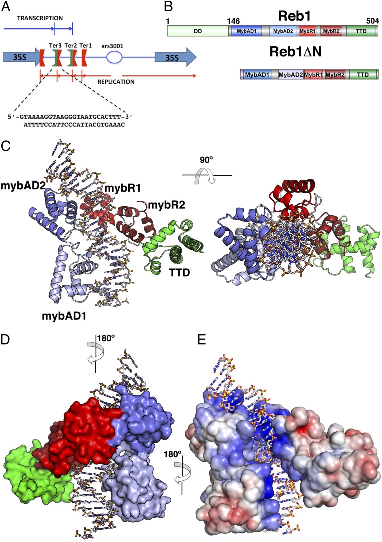
Reb1 ofSchizosaccharomyces pomberepresents a family of multifunctional proteins that bind to specific terminator sites (Ter) and cause polar termination of transcription catalyzed by RNA polymerase I (pol I) and arrest of replication forks approaching the Ter sites from the opposite direction. However, it remains to be investigated whether the same mechanism causes arrest of both DNA transactions. Here, we present the structure of Reb1 as a complex with a Ter site at a resolution of 2.7 Å. Structure-guided molecular genetic analyses revealed that it has distinct and well-defined DNA binding and transcription termination (TTD) domains. The region of the protein involved in replication termination is distinct from the TTD. Mechanistically, the data support the conclusion that transcription termination is not caused by just high affinity Reb1-Ter protein-DNA interactions. Rather, protein-protein interactions between the TTD with the Rpa12 subunit of RNA pol I seem to be an integral part of the mechanism. This conclusion is further supported by the observation that double mutations in TTD that abolished its interaction with Rpa12 also greatly reduced transcription termination thereby revealing a conduit for functional communications between RNA pol I and the terminator protein.

Keywords: RNA polymerase I; crystal structure; protein–DNA interaction; replication termination; transcription termination.


Conflict of interest statement

The authors declare no conflict of interest.

Figures

Fig. 1.
Fig. 1.
Structure of the Reb1-DNA complex. (A) Diagram showing the S. pombe rRNA nontranscribed spacer region containing three Ter sites and the fork-pausing site RFP4. Reb1 binds to Ter2 and Ter3 sites arresting replication approaching from one direction (red arrows). Transcription is terminated in the opposite direction (blue arrows). Also shown is the sequence of the Ter3 DNA used for crystallization of the complex. (B) Modular domain organization of Reb1FL and Reb1ΔN proteins. DD, dimerization domain (light green); MybAD1, Myb-associated domain 1 (blue); MybAD2, Myb-associated domain2 (light blue); MybR1, Myb repeat 1 (red); MybR2, Myb repeat 2 (dark red); TTD, transcription termination domain (green). Limits of domains are based on the crystal structure. (C) Crystal structure of Reb1ΔN-Ter3 DNA complex in two orientations (rotation of 90° along the indicated axis). Domains are colored according to diagram shown in B. Orientation of DNA in the complex shows that transcription will be approaching from left and replication from right. (D) Surface representation of Reb1ΔN-Ter3 complex rotated by 180° with relative to the view shown in C. (E) Electrostatic potential surface of Reb1ΔN-Ter3 complex. The electropositive DNA binding surface is blue, neutral is white and negatively charged shown in red. DNA is shown as a stick representation.
Fig. S1.
Fig. S1.
Superposition of the two complexes in the asymmetric unit. Superposition of the two complexes was carried out using the program LSQMAN. Reb1 molecules superimposed with an RMSD of 1.09 Å, whereas the two Ter3 DNA molecules superimpose with an RMSD of 0.95 Å. Complexes A and B are colored blue and red, respectively.
Fig. 2.
Fig. 2.
Structure of the quadripartite Reb1 DNA-binding domain. (A) Domain structure of quadripartite Reb1 DBD consisting in MybAD1 (teal), MybAD2 (blue), MybR1 (red), and MyR2 (dark red). The four domains form a U-shaped structure. (Left) Ribbon representation. (Right) Surface rendering. (B) (Left) Superposition of MybAD domains. Although sequence homology is only 15%, the two domains superimposed with an RMSD of 1.36 Å. (Right) Superposition of Reb1 MybAD2 (blue) and MATα2 (magenta) reveals that MybAD αB helix is an addition to the classical HTH motif. (C) Sequence alignment of Reb1 MybADs with MATα2. Sequence coloring highlights regions that are highly conserved (red) and moderately conserved (orange); positions of the α-helices are shown above the sequences as blue bars. (D) Structural alignment of Reb1 Myb repeats (red) and c-Myb repeats (green) represented as ribbon diagrams. MybAD2 loop 2 (l2) is shown as a dotted connection. (E) Sequence alignment of Reb1 Myb repeats with c-Myb repeats and TRF2. Residues in red indicate highly conserved; orange, moderate conserved.
Fig. 3.
Fig. 3.
Reb1-Ter3 interactions. (A) Contacts are colored according to the different domains: MybAD1, teal; MybAD2, blue; MybR1, red; MybR2, orange. Bases directly contacted are colored yellow. Dotted lines depict hydrophobic interactions. (B) Close-up view of MybAD1-Ter3 interaction. R216 is shown making a hydrophobic interaction with the methyl group of thymine18. R212 is shown in two conformations, one is making interaction a phosphate group and the second makes hydrogen bonds with Guanine8’. (C) Details of MybAD2-Ter3 interface showing the residues involved in DNA recognition. K297 makes a bidentate interaction with Guanine8, whereas H301 interacts with thymine18’. Also shown is Tyr-300 that interacts with the phosphate backbone. (D) MybR1 sequence-specific interaction of Ter3 DNA. R350 makes bidentate hydrogen bonds with Guanine12, whereas R354 interacts in a similar way with Guanine13. Paired cytosines at positions 16’ and 15’ from the opposite strand make interactions with N347 and D351, respectively. (E) A close-up view of MybR2 recognition helix interacting with the Ter3 site. R407 makes bidentate hydrogen bonds with Guanine14. L408 interacts via hydrophobic interactions with the methyl group of both Thymine15 and Thymine12’.
Fig. 4.
Fig. 4.
Role of Reb1 domains in DNA binding. (A) Binding isotherms of Reb1ΔN (black circles), Reb1ΔNΔTTD (green squares), Reb1MybAds (red triangles), and Myb repeats (blue triangles). Fluorescein-labeled Ter3 DNA site was titrated with different protein concentrations. Fluorescence anisotropy was read, and the DNA fraction bound was plotted as a function of Reb1 concentration. For the experiments of MybADs and Myb repeats, titration was done up to a concentration of 3 μM to achieve saturation. (B) Superposition of Ter3 DNA from the crystal structure (white sugar backbone) onto an ideal B-DNA Ter3 (blue sugar backbone). Dark blue line represents the calculated DNA axis.
Fig. 5.
Fig. 5.
Reb1 has an extended conformation in absence of DNA. (A) apo-Reb1ΔN ab initio molecular envelope calculated with Dammin with the docked model calculated with the program CORAL. (B) Structure of Reb1ΔN in the bound conformation. (C) Fit of the theoretical scattering profile of the CORAL rigid model shown in A (red), with the experimental scattering profile (circles).
Fig. 6.
Fig. 6.
TTD is a transcription termination domain. (A) Ribbon diagrams showing two views of the TTD domain (residues 425–504). (B) Schematic representation of the template and the probes used to measure the extent of transcriptional read through past the Ter site. (C) Transcription run on assay showing that in the absence of the C-terminal region of Reb1 caused significant loss of termination at the Ter site. (D) Bar chart of results shown in C.
Fig. S2.
Fig. S2.
Structure-based sequence alignment of the eukaryotic transcription and/or replication terminator family. Secondary structure elements are shown above the sequence and color-coded according to Fig. 1 with MybAD1(αA–αD), MybAD2(αA′–αD′), MybR1(α1–α3), MybR2(α4–α6), and TTD(αE–αI). Sequence coloring highlights regions that are highly conserved (red) and moderately conserved (orange). Reb1_Sp, S. pombe Reb1; RTF1_Sp, S. pombe replication termination factor 1; Nsi1_Sc, S. cerevisiae NTS1 silencing protein 1 also known as Ydr026c; Reb1p_Sc, S. cerevisiae Reb1 protein; TTFI_Hs, Homo sapiens transcription termination factor I; TTFI_Mm, Mus musculus transcription termination factor I; Rib2_Xt, X. tropicalis.
Fig. 7.
Fig. 7.
Brewer–Fangman 2D gels. 2D gels of the (A) WT, (B) truncated Reb1 (1–418) lacking the TTD, and (C) the reb1Δ control showing that deletion of the C-terminal region (419–504) that contains the TTD did not detectably alter the fork arrests at Ter2 and Ter3 that are caused by Reb1 binding.
Fig. 8.
Fig. 8.
Disruption of protein–protein interaction between the TTD and Rpa12 by double point mutations in TTD significantly reduced termination of transcription at Ter. (A) Ribbon diagram of the TTD showing with the location of the L485 (Left) and Trp465 (Right) point mutants that disrupted its interaction with Rpa12. (B) ELISA data showing that TTD and to a lesser extent the N-terminal domain (1–145) of Reb1 interacted with Rpa12. (C) ELISA data showing that Reb1 double mutant failed to interact with Rpa12. (D and E) Representative TRO analysis showing that in the WT Reb1, transcription termination occurred at the Ter site, whereas in the double mutant, a significant amount of transcripts passed through the Ter site.
Fig. S3.
Fig. S3.
Rpa12 interaction with WT and mutant form of 419–504 Reb1. Y2H data (β-galactosidase activities) showing that single mutants, W460R and L485P of 419–504 reb1, mostly retain their ability to interact with Rpa12 (>80%); in contrast, double mutant W460R-L485P has greatly reduced (just above background level) interaction with Rpa12.
Fig. S4.
Fig. S4.
DNA terminator sequence motif across species. Alignment of the different terminator DNA sequences recognize by Terminator proteins. Shown is from top to bottom: Ter3 from S. pombe recognize by Reb1; Ter from S. cerevisiae recognized by Reb1p; REP4 from S. pombe recognize by RTF1; T0 and T1 from M. musculus recognized by murine TTF-I and T1 from H. sapiens recognized by TTF-I. Binding sites can be divided in three modules that interact with MybAD2 (box 1), MybRepeats (box 2), and MybAD1 (box 3).

Similar articles

Cited by

References

    1. Sollner-Webb B, Tower J. Transcription of cloned eukaryotic ribosomal RNA genes. Annu Rev Biochem. 1986;55:801–830. - PubMed
    1. Clos J, Normann A, Ohrlein A, Grummt I. The core promoter of mouse rDNA consists of two functionally distinct domains. Nucleic Acids Res. 1986;14(19):7581–7595. - PMC - PubMed
    1. Reeder RH. Regulation of RNA polymerase I transcription in yeast and vertebrates. Prog Nucleic Acid Res Mol Biol. 1999;62:293–327. - PubMed
    1. Richard P, Manley JL. Transcription termination by nuclear RNA polymerases. Genes Dev. 2009;23(11):1247–1269. - PMC - PubMed
    1. Németh A, et al. RNA polymerase I termination: Where is the end? Biochim Biophys Acta. 2013;1829(3-4):306–317. - PubMed

Publication types

MeSH terms

Associated data

LinkOut - more resources