. 2016 Apr 12;113(15):E2124-33.
doi: 10.1073/pnas.1603388113. Epub 2016 Mar 28.

Genome-wide kinetics of DNA excision repair in relation to chromatin state and mutagenesis

Affiliations

Genome-wide kinetics of DNA excision repair in relation to chromatin state and mutagenesis

Sheera Adar et al. Proc Natl Acad Sci U S A. .

Abstract

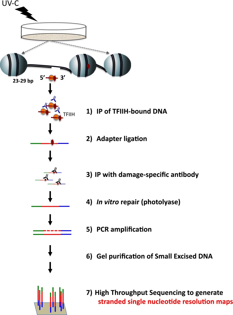
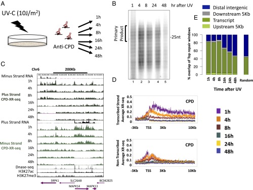
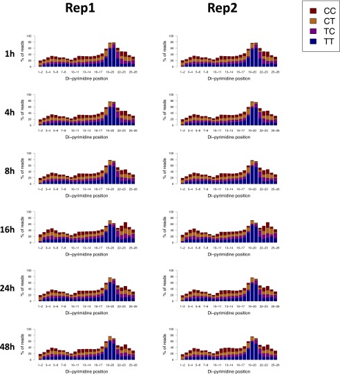
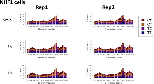
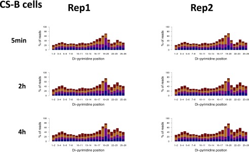
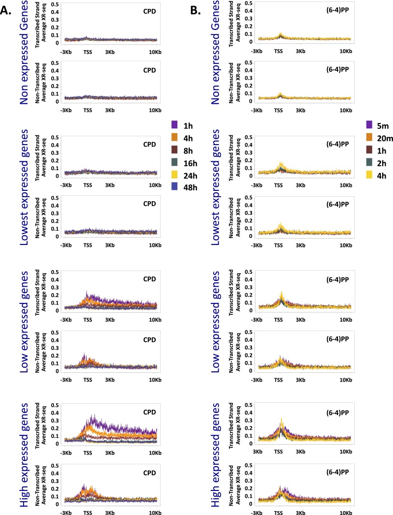
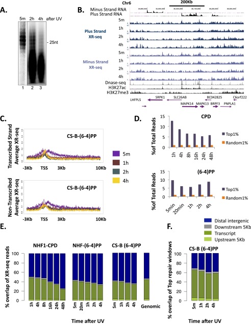
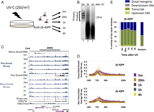
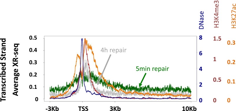
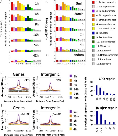
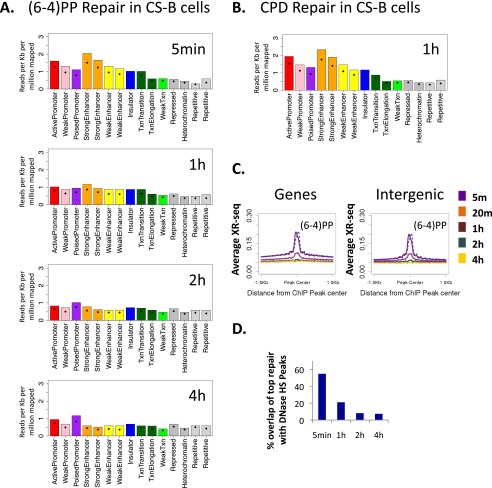
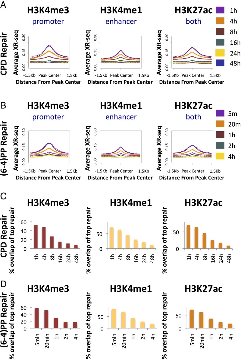
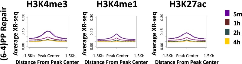
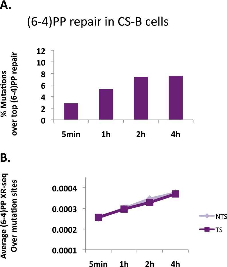
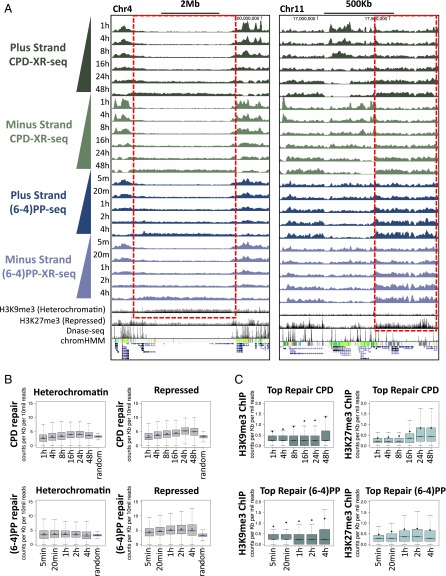
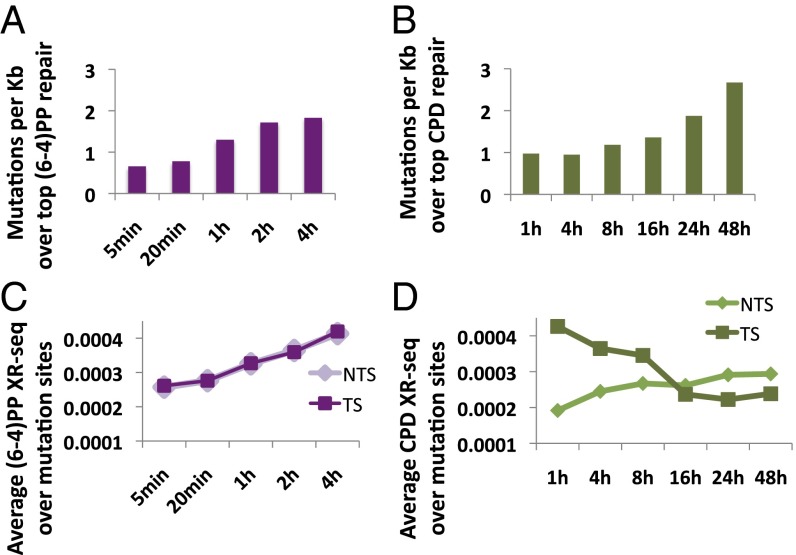
We recently developed a high-resolution genome-wide assay for mapping DNA excision repair named eXcision Repair-sequencing (XR-seq) and have now used XR-seq to determine which regions of the genome are subject to repair very soon after UV exposure and which regions are repaired later. Over a time course, we measured repair of the UV-induced damage of cyclobutane pyrimidine dimers (CPDs) (at 1, 4, 8, 16, 24, and 48 h) and (6-4)pyrimidine-pyrimidone photoproducts [(6-4)PPs] (at 5 and 20 min and 1, 2, and 4 h) in normal human skin fibroblasts. Each type of damage has distinct repair kinetics. The (6-4)PPs are detected as early as 5 min after UV treatment, with the bulk of repair completed by 4 h. Repair of CPDs, which we previously showed is intimately coupled to transcription, is slower and in certain regions persists even 2 d after UV irradiation. We compared our results to the Encyclopedia of DNA Elements data regarding histone modifications, chromatin state, and transcription. For both damage types, and for both transcription-coupled and general excision repair, the earliest repair occurred preferentially in active and open chromatin states. Conversely, repair in regions classified as "heterochromatic" and "repressed" was relatively low at early time points, with repair persisting into the late time points. Damage that remains during DNA replication increases the risk for mutagenesis. Indeed, late-repaired regions are associated with a higher level of cancer-linked mutations. In summary, we show that XR-seq is a powerful approach for studying relationships among chromatin state, DNA repair, genome stability, mutagenesis, and carcinogenesis.

Keywords: DNA damage; DNA repair; chromatin; mutation; transcription.


Conflict of interest statement

The authors declare no conflict of interest.

Figures

Fig. S1.
Fig. S1.
Schematic of the XR-seq assay. For details, see ref. .
Fig. 1.
Fig. 1.
Kinetics of the genome-wide repair of CPD in normal human fibroblasts. (A) Schematic of time course of excision repair of the UV-induced CPD. (B) After irradiation with 10 J/m2 UV-C, normal NHF1 fibroblasts were incubated for the indicated time, and the excised oligonucleotides were immunoprecipitated with anti-CPD antibody, radiolabeled at the 3′ end with 32P-cordycepin and analyzed on sequencing gels. (C) Distribution of the XR-seq signal at each time point, separated by strand, for CPD repair along a 400-Kb region of chr6. ENCODE total stranded RNA-seq, DNase-seq, and ChIP-seq signal for H3K27ac and H3K27me3 from NHDF cells are plotted in black. Arrows in the bottom indicate position and directions of annotated genes. Bold arrows indicate expressed genes. (D) Average CPD XR-seq repair signal 3 Kb upstream and 10 Kb downstream of the annotated TSS of genes with the highest quartile of RNA expression in normal human skin fibroblasts (ENCODE, NHDF data). Signal is plotted separately for the transcribed and nontranscribed strands. (E) Percent overlap of the top 1% of repair windows at each of the time points with annotated genes (dark green), 5 Kb upstream of the gene (light green), 5 Kb downstream of the gene (gray), and intergenic regions (blue). Plotted for comparison are the average results of 50 permutations of a random set of 500 mers from the hg19 reference genome.
Fig. S2.
Fig. S2.
Similar enrichment of di-pyrimidines 5–7nt from the 3′ end of 26-nt-long reads for CPD XR-seq in normal human fibroblasts at the different time points. Shown are the results of two biological replicates.
Fig. S3.
Fig. S3.
Similar enrichment of di-pyrimidines 6–7 nt from the 3′ end of 26-nt-long reads for (6-4)PP XR-seq in normal human fibroblasts at the different time points. Shown are the results of two biological replicates.
Fig. S4.
Fig. S4.
Same as Fig. S3 except experiments were performed in CS-B cells, defective in transcription-coupled repair.
Fig. S5.
Fig. S5.
(A) Average CPD XR-seq repair signal 3 Kb upstream and 10 Kb downstream of the annotated TSS of genes separated into groups based on their RNA expression level in normal human skin fibroblasts (ENCODE, NHDF data). Signal is plotted separately for the transcribed and nontranscribed strand. (B) Same as A except plotted is XR-seq signal for (6-4)PP repair.
Fig. S6.
Fig. S6.
(A) For analysis of the pattern of (6-4)PP repair, CS-B transcription-coupled repair-deficient cells were irradiated with 20 J/m2 UV-C and incubated for the indicated time. The excised oligonucleotides were immunoprecipitated with anti-(6-4)PP antibody, radiolabeled at the 3′ end with 32P-cordycepin, and analyzed on sequencing gels. (B) Distribution of the XR-seq signal at each time point, separated by strand, for (6-4)PP repair in a CS-B cell line, along a 400-Kb region of chr6. ENCODE total stranded RNA-seq, DNase-seq, and ChIP-seq signal for H3K27ac and H3K27me3 from NHDF are plotted in black. Arrows in the bottom indicate positions and directions of annotated genes. Bold arrows indicate expressed genes. (C) Average (6-4)PP XR-seq repair signal from CS-B cells 3 Kb upstream and 10 Kb downstream of the annotated TSS of highly expressed genes in normal human skin fibroblasts (ENCODE, NHDF data). Signal is plotted separately for the transcribed (Upper) and nontranscribed (Lower) strands. (D) The proportion of reads in each sample that fall into the top 1% of repair window. As a control, the same analysis was performed for a random set of 1% of the 500-nt windows in the genome. (E) Genomic distribution of the aligned reads of CPD and (6-4)PP XR-seq experiments at each of the time points between genes (green), 5 Kb upstream of the gene (light green), 5 Kb downstream of the gene (gray), and intergenic regions (blue). Plotted for comparison are the average results of 50 permutations of a random set of 26 mers from the hg19 reference genome. (F) Percent overlap of the top 1% of (6-4)PP repair windows at each of the time points in CS-B cells with the same intervals described in D.
Fig. 2.
Fig. 2.
Kinetics of the genome-wide repair of (6-4)PP in normal human fibroblasts. (A) Schematic of time course of excision repair of the UV-induced (6-4)PP. (B) For analysis of the pattern of (6-4)PP repair, normal NHF1 cells were irradiated with 20 J/m2 UV-C and incubated for the indicated time. The excised oligonucleotides were immunoprecipitated with anti-(6-4)PP antibody, radiolabeled at the 3′ end with 32P-cordycepin, and analyzed on sequencing gels. (C) Distribution of the XR-seq signal at each time point, separated by strand, for (6-4)PP repair along a 400-Kb region of chr6. ENCODE total stranded RNA-seq, DNase-seq, and ChIP-seq signal for H3K27ac and H3K27me3 from NHDF cells are plotted in black. Arrows in the bottom indicate positions and directions of annotated genes. Bold arrows indicated expressed genes. (D) Average (6-4)PP XR-seq repair signal 3 Kb upstream and 10 Kb downstream of the annotated TSS of genes with the highest quartile of RNA expression in normal human skin fibroblasts (ENCODE, NHDF data). Signal is plotted separately for the transcribed and nontranscribed strands. (E) Percent overlap of the top 1% of repair windows at each of the time points with annotated genes (dark green), 5 Kb upstream of the gene (light green), 5 Kb downstream of the gene (gray), and intergenic regions (blue). Plotted for comparison are the average results of 50 permutation of a random set of 500 mers from the hg19 reference genome.
Fig. S7.
Fig. S7.
Similar to Fig. 2D, except that the average profile of DNase-seq (blue), H3K4me3 ChIP-seq (red), and H3K27ac ChIP-seq (orange) signals were plotted in addition to the 5-min (green) and 4-h (gray) repair profiles by using additional axes (color-coded to match the different data).
Fig. 3.
Fig. 3.
Effect of chromatin state on repair. (A) XR-seq read coverage was calculated over genomic intervals assigned to each of the chromatin states predicted for NHLF cells (ENCODE). Shown are results from CPD XR-seq at the different time points. Values were normalized to read depth and interval length. Bars indicate the value of the 75th percentile. Diamonds indicate the mean value. (B) Same as A except calculated was coverage of (6-4)PP XR-seq. (C) Similar to A and B except the coverage was counted for a random set of 26-mer intervals. (D) Average CPD XR-seq profiles for each time point at 1.5-Kb regions flanking the center of DNase hypersensitivity peaks that either overlapped annotated genes (Left) or did not overlap annotated genes (Right). Values were normalized per 10 million mapped reads. The average profile was calculated separately for the plus (solid line) and minus (dashed line) strands. (E) Same as D except plotted is the repair of (6-4)PP. (F) Overlap of the top 1% of repair sites (500-bp windows) with DNase hypersensitivity peaks in each of the time points for CPD repair (Upper) and (6-4)PP repair (Lower).
Fig. S8.
Fig. S8.
Preferential repair of open and active chromatin states in CS-B cells. (A) XR-seq read coverage was calculated over genomic intervals assigned to each of the chromatin states defined for NHLF cells (ENCODE). Shown are results from (6-4)PP XR-seq at the indicated times after UV. Values were normalized to read depth and interval length. Bars indicate the value of the 75th percentile. Diamonds indicate the mean value. (B) As in A except shown are results of CPD XR-seq at 1 h after UV. (C) Average (6-4)PP XR-seq profiles for each time point at 1.5-Kb regions flanking the center of DNase hypersensitivity peaks that either overlapped genes (Left) or did not overlap genes (Right). Values were normalized per 10 million mapped reads. The average profile was calculated separately for the plus (solid line) and minus (dashed line) strands. (D) Overlap of the top 1% of (6-4)PP repair sites (500-bp windows) with DNase hypersensitivity peaks in each of the time points.
Fig. 4.
Fig. 4.
Initial repair is associated with active histone marks. (A) Average CPD XR-seq profiles for each time point at 1.5-Kb regions flanking the center of ChIP-seq peaks of H3K4me3 (Left), H3K4me1 (Center), and H3K27ac (Right). Values were normalized per 10 million mapped reads. The average profile was calculated separately for the plus (solid line) and minus (dashed line) strands. (B) Same as A except plotted is repair of (6-4)PP. (C) Overlap of the top 1% of CPD repair sites (500-bp windows) with the indicated ChIP-seq peaks. (D) Same as C except plotted is the overlap of the top 1% of (6-4)PP repair sites at each time point.
Fig. S9.
Fig. S9.
Average (6-4)PP XR-seq profiles in CS-B cells for each time point at 1.5-Kb regions flanking the center of ChIP-seq peaks of H3K4me3, H3K4me1, and H3K27ac.
Fig. S10.
Fig. S10.
(A) The number of melanoma-linked mutations per kilobase that overlap the top 1% of repair windows was calculated for (6-4)PP repair at the different time points in CS-B cells. (B) The average XR-seq count over a 400-bp window centered on the site of melanoma-linked mutations was calculated for (6-4)PP repair in CS-B cells at each of the time points. Repair was calculated separately for template and nontemplate strands.
Fig. 5.
Fig. 5.
Repair of heterochromatin and repressed regions persist into the late time points. (A) Distribution of the XR-seq signal at each time point, separated by strand, for CPD and (6-4)PP repair in two genomic loci, enriched for H3K9me3 (Left) or H3K27me3 (Right) histone deposition. ChIP-seq signal for H3K9me3 and H3K27me3, DNase-seq, and ChromHMM segmentation tracks (NHDF and NHLF, ENCODE) are plotted in the bottom. Gray in the ChromHMM track depicts repressed or heterochromatic regions; green represents actively transcribed regions. (B) Box plots depicting CPD (Upper) and (6-4)PP (Lower) XR-seq read coverage over heterochromatic (Left) or repressed (Right) genomic intervals as predicted in NHLF cells (ENCODE). Shown are results from each of the time points, in comparison with a random dataset of 26 mers from the hg19 reference genome. Values were normalized to read depth and interval length. Lines indicate median, and diamonds indicate mean values. (C) Box plots depicting the level of histone H3K9me3 (Left) or H3K27me3 (Right) ChIP-seq signal (NHDF, ENCODE) over the top 1% of 500-nt repair windows in each of the time points for CPD (Upper) and (6-4)PP (Lower) XR-seq. Counts were normalized per million mapped reads. Lines indicate median, and diamonds indicate mean values.
Fig. 6.
Fig. 6.
Repair at late time points is associated with higher somatic-mutation rates. (A) The number of melanoma-linked mutations per kilobase that overlap the top 1% of repair windows was calculated for (6-4)PP repair at the different time points. (B) Same as A except overlaps were counted over the top 1% of CPD repair windows. (C) The average XR-seq count over a 400-bp window centered on the site of melanoma-linked mutations was calculated for (6-4)PP repair at each of the time points. Repair was calculated separately for template and nontemplate strands. Values were normalized for sequencing depth. (D) Same as B except plotted is the average CPD repair at each time point.

References

    1. Reardon JT, Sancar A. Nucleotide excision repair. Prog Nucleic Acid Res Mol Biol. 2005;79(79):183–235. - PubMed
    1. Sancar A. DNA excision repair. Annu Rev Biochem. 1996;65:43–81. - PubMed
    1. Wood RD. Nucleotide excision repair in mammalian cells. J Biol Chem. 1997;272(38):23465–23468. - PubMed
    1. Reardon JT, Sancar A. Recognition and repair of the cyclobutane thymine dimer, a major cause of skin cancers, by the human excision nuclease. Genes Dev. 2003;17(20):2539–2551. - PMC - PubMed
    1. Sugasawa K, et al. Xeroderma pigmentosum group C protein complex is the initiator of global genome nucleotide excision repair. Mol Cell. 1998;2(2):223–232. - PubMed

Publication types