. 2016 Mar 31:14:24.
doi: 10.1186/s12915-016-0246-5.

Hmga2 is necessary for Otx2-dependent exit of embryonic stem cells from the pluripotent ground state

Affiliations

Hmga2 is necessary for Otx2-dependent exit of embryonic stem cells from the pluripotent ground state

Angelica Navarra et al. BMC Biol. .

Abstract

Background: A crucial event in the differentiation of mouse embryonic stem cells (ESCs) is the exit from the pluripotent ground state that leads to the acquisition of the 'primed' pluripotent phenotype, characteristic of the epiblast-like stem cells (EpiLCs). The transcription factors Oct4 and Otx2 play a key role in this phenomenon. In particular, Otx2 pioneers and activates new enhancers, which are silent in ESCs and which control the transcription of genes responsible for the acquisition of the EpiLC phenotype. An important point that remains to be addressed is the mechanism through which Otx2 engages the new enhancers and stably associates with them. Hmga2 is a member of the high-mobility group family of proteins, non-histone components of chromatin whose expression is high during embryogenesis and becomes low or undetectable in adults. Its high expression during embryogenesis suggests that Hmga2 fulfills important roles in development.

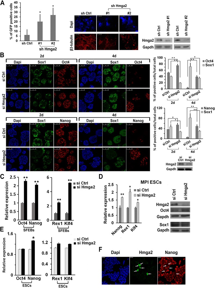
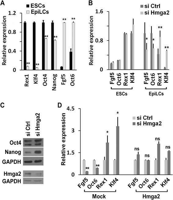
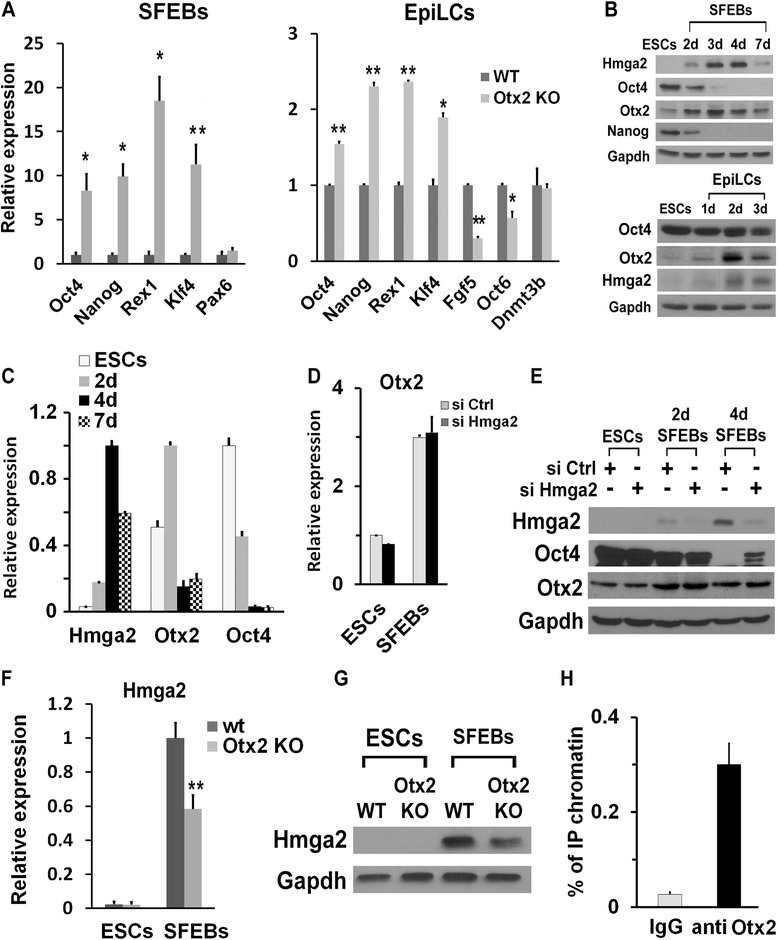
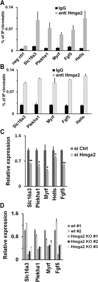
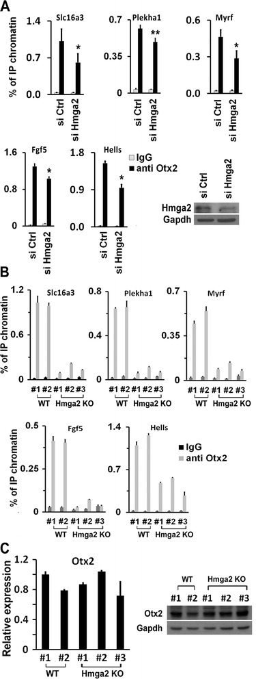
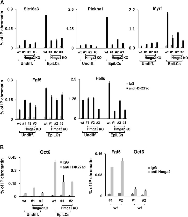
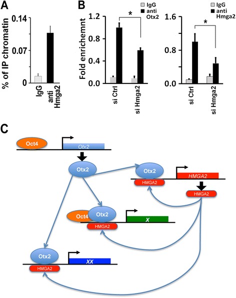
Results: Here, we demonstrate that Hmga2 accumulates soon after the induction of ESC differentiation. Its suppression hampers the exit of ESCs from the pluripotent ground state and their differentiation into EpiLCs. Mechanistically, Hmga2 controls the differentiation process by cooperating with Otx2 in the pioneering of new enhancers. In Hmga2 null induced pluripotent stem cells we observe that Otx2 fails to regulate its target genes upon the induction of differentiation. Hmga2 associates to Otx2-bound loci in EpiLCs, and in Hmga2 KO cells Otx2 is unable to engage and activate the new enhancers, thus indicating that Hmga2 is required for the binding of Otx2 to its cis-elements. We find that this mechanism also operates on the Hmga2 gene, which is one of the targets of Otx2, thus indicating the existence of a positive feedback loop.

Conclusions: Our findings reveal a novel mechanism necessary for the exit of ESCs from the pluripotent ground state. Upon the induction of ESC differentiation, Otx2 alone or in combination with Oct4 engages new enhancers, which are silent in undifferentiated ESCs. The Hmga2 gene is activated by Otx2 and Hmga2 protein binds to the enhancers targeted by Otx2, thus facilitating the engagement and/or the stable association of Otx2. Therefore, our results demonstrate that Hmga2 is a key element of the regulatory network that governs the exit of ESCs from the pluripotent ground state.

Keywords: Embryonic stem cell differentiation; Enhancer activation; High-mobility group protein.


Figures

Fig. 1
Fig. 1
Hmga2 suppression interferes with embryonic stem cell (ESC) differentiation. a ESCs expressing GFP under the control of Nanog gene promoter were transfected with the control shRNA (sh Ctrl) and two different shRNAs targeting Hmga2, which induced a significant decrease of the Hmga2 protein. Knockdown cells were induced to differentiate into neurons. After 8 days of differentiation, cells were harvested and the number of GFP expressing cells was measured by FACS, subtracting the fluorescent background of parental differentiated E14Tg2a ESCs. The histogram shows the percentage of GFP-positive cells in the indicated samples as mean ± SEM (n = 6). *P <0.05. The efficiency of neuron formation was evaluated at 10 days of differentiation by staining the cells with a neuro-specific antibody (anti-β3-tubulin). Scale bars: 250 μm. b Analysis of the phenotype induced by Hmga2 suppression during neuroectodermal differentiation of ESCs. ESCs were transfected with the indicated siRNA, and induced to differentiate through serum-free embryoid body (SFEB) formation for 2 and 4 days. Sectioned SFEBs were then subjected to immunostaining to analyze the presence of stemness (Oct4, Nanog) and neuroectodermal (Sox1) markers. Scale bars: 100 μm. The histograms represent the percentage of Oct4-, Nanog-, and Sox1-positive cells counted in more than six independent fields. *P <0.05, **P <0.01. The efficiency of Hmga2 silencing by siRNA was measured by western blot. c qPCR analysis of the expression changes of Oct4, Nanog, Rex1, and Klf4 in differentiated ESCs upon suppression of Hmga2. Data represent mean of three independent experiments (n = 3) ± SEM. *P <0.05. **P <0.01. d MPI ESCs were transfected with a control siRNA or a specific Hmga2 siRNA and induced to differentiate into SFEBs. At 4 days of differentiation, SFEBs were collected and the expression of the indicated markers was analyzed by qPCR and western blot. Data represent mean of three independent experiments (n = 3) ± SEM. *P <0.05. e qPCR analysis of the expression of Oct4, Nanog, Rex1, and Klf4 in undifferentiated ESCs upon suppression of Hmga2. Data represent mean of independent experiments (Oct4, Rex1, Klf4, n = 3; Nanog, n = 4) ± SEM. *P <0.05. f Representative images of the double staining of undifferentiated ESCs with Nanog (red) and Hmga2 (green) antibodies. The arrows indicate cells expressing Hmga2, which corresponds to cells expressing the lowest level of Nanog. Scale bars: 20 μm
Fig. 2
Fig. 2
Hmga2 suppression interferes with the transition of embryonic stem cells (ESCs) into epiblast-like stem cells (EpiLCs). a qPCR analysis to compare the expression levels of the indicated genes in ESCs versus EpiLCs. Data are reported as means (n = 3) ± SEM of biological replicates. b Comparison of the gene expression changes of ESC (Rex1 and Klf4) and EpiLC (Fgf5 and Oct6) markers in ESCs and EpiLCs upon Hmga2 silencing. Data are reported as means (n = 3) ± SEM of biological replicates (in the case of Fgf5 and Oct6 in EpiLC n = 4). c Western blot analysis to measure the expression level of Oct4, Nanog, and Hmga2 in EpiLCs derived from ESCs transfected with the indicated siRNAs. d Gene expression changes as a consequence of Hmga2 knockdown in EpiLCs transfected with GFP (mock) or Hmga2 under the control of the β-actin gene promoter. qPCR data are reported as mean of relative mRNA expression ± SEM of biological replicates (Fgf5 and Rex1, n = 4; Oct6 and Klf4, n = 3). *P <0.05, **P <0.01, ns: not significant
Fig. 3
Fig. 3
Induced pluripotent stem cells (iPSCs) derived from Hmga2 knockout (KO) embryonic fibroblasts (MEFs) are unable to properly differentiate. a iPSC clones derived from wildtype (wt; #1 and #2) and Hmga2 KO (#1, #2, #3) MEFs and grown under 2i + leukemia inhibitory factor (LIF) conditions are induced to differentiate into neuroectoderm through serum-free embryoid body (SFEB) formation for 4 days. Sectioned SFEBs were stained for neuroectodermal (Sox1) and stemness (Oct4) markers. Scale bars: 100 μm. b Western blot analysis to evaluate the expression of the indicated proteins in 4 day-differentiated SFEBs derived from different wt and Hmga2 KO iPSC clones and qPCR analysis of the indicated mRNAs. qPCR data are reported as means of relative mRNA expression ± SEM of two biological replicates for each of the indicated wt or Hmga2 KO iPSC clones. c The expression of the endodermal marker Gata 4 was evaluated by qPCR in wt and KO Hmga2 iPSCs after 4 days of differentiation as SFEBs. qPCR data represent mean ± SD (n = 2) for each of the indicated wt or Hmga2 KO iPSC clones. d Different clones of wt and Hmga2 KO iPSCs were induced to differentiate into cardiomyocytes through the hanging drops method. Histogram reports the percentage of embryoid bodies showing rhythmically contracting areas (beating hearts) on day 13 of differentiation. e Immunofluorescence analysis showing the extent of the areas positive for the cardiac ventricular marker myosin Mlc2v or the stemness marker Oct4 in wt and Hmga2 KO iPSCs differentiated for 13 days through the hanging drop method. f The expression of the stemness markers was measured by qPCR analysis in wt and Hmga2 KO iPSCs differentiated for 13 days through the hanging drop method. qPCR data represent mean ± SD (n = 2) for each of the indicated wt or Hmga2 KO iPSC clones. g The expression of Oct4 and Nanog was analyzed by western blot in wt and Hmga2 KO clones induced to differentiate into EpiLCs. The relative expression of the indicated genes was measured by qPCR analysis in EpiLCs obtained from wt and Hmga2 KO iPSC clones. qPCR data represent means of independent experiments (n = 2; in the case of Oct6 n = 3) ± SD for each of the indicated wt or Hmga2 KO iPSC clones. h The relative expression of Pax6, Sox1, and Gata4 was measured by qPCR analysis in SFEBs obtained from wt and Hmga2 KO iPSC clones, stably transfected with an Hmga2 or GFP (Mock) under the control of β-actin gene promoter. qPCR data represent the mean of three independent experiments (n = 3) ± SEM for each of the indicated wt or Hmga2 KO iPSC clones. *P <0.05. The expression levels of endogenous Hmga2 (>) and of the transfected form (#) are reported in the western blot. i The relative expression of Fgf5 and Oct6 was measured by qPCR analysis in EpiLCs obtained from wt and Hmga2 KO iPSC clones, stably transfected with Hmga2 or GFP (Mock) under the control of β-actin gene promoter. qPCR data represent mean ± SEM of three independent experiments (n = 3) for each of the indicated wt or Hmga2 KO iPSC clones. *P <0.05. j Wt and Hmga2 KO iPSCs, transfected or not with Hmga2 cDNA, were induced to differentiate into cardiomyocytes through the hanging drops method. The histogram reports the percentage of embryoid bodies showing rhythmically contracting areas (beating hearts) on day 13 of differentiation
Fig. 4
Fig. 4
Otx2 controls Hmga2 expression in epiblast-like stem cells (EpiLCs). a Phenotypic analysis of Otx2 knockout (KO) and wildtype (wt) differentiated embryonic stem cells (ESCs). The indicated markers were analyzed by qPCR at 4 days of differentiation as serum-free embryoid bodies (SFEBs) or into EpiLCs. qPCR data represent the means of independent biological replicates (Oct4, n = 4; Nanog, Rex1, and Pax6, n = 3; Klf4, n = 5) ± SEM. *P <0.05, **P <0.01. b Western blot analysis of undifferentiated and differentiated ESCs. Expression levels of Hmga2, Oct4, and Otx2 were measured in undifferentiated ESCs, during neuroectodermal differentiation through SFEBs and in EpiLCs. c Expression profile of Otx2, Oct4, and Hmga2 mRNAs at the indicated time points during ESC differentiation analyzed by qPCR. Data represent mean ± SD from two independent biological replicates (n = 2). d Otx2 mRNA levels in undifferentiated ESCs and in SFEBs at day 4 transfected with control or Hmga2 siRNAs. Data represent mean ± SD from two independent biological replicates (n = 2). e Oct4 and Otx2 expression in SFEBs at 2 and 4 days of differentiation derived from ESCs transfected with the indicated siRNAs. f Wt and Otx2 KO ESCs were induced to differentiate into SFEBs for 4 days. The expression of Hmga2 was analyzed by qPCR. Data represent mean of three independent experiments (n = 3) ± SEM. **P <0.01. g Hmga2 protein levels were measured in wt and Otx2 KO undifferentiated ESCs and 4-day differentiated SFEBs. h Otx2 association with the genomic region upstream Hmga2. ChIP-qPCR analysis of Otx2 binding to the genomic region upstream the transcriptional start site of Hmga2 (–9 kb) in EpiLCs. qPCR data represent mean of four independent experiments (n = 4) ± SEM
Fig. 5
Fig. 5
Hmga2 is involved in the regulation of Otx2 target genes upon the exit of embryonic stem cells (ESCs) from the pluripotent ground state. a ChIP-qPCR analysis of the association of Hmga2 to gene targets of Otx2 in wildtype (wt) epiblast-like stem cells (EpiLCs). The negative control (neg ctrl) corresponds to the region 10.1 kb upstream of the Hmga2 transcriptional start site. qPCR data represent means of three independent experiments (n = 3) ± SEM. b ChIP-qPCR analysis of the association of Hmga2 to gene targets of Otx2 in wt induced pluripotent stem cells (iPSCs) induced to differentiate. qPCR data represent means of two independent experiments (n = 2) ± SD. c The relative level of the indicated mRNAs was measured by qPCR in EpiLCs derived from ESCs transfected with si Ctrl and si Hmga2. qPCR data represent mean of three independent experiments (n = 3) ± SEM. *P <0.05, **P <0.01. d qPCR analysis of the expression of the indicated genes in wt and Hmga2 KO iPSC clones differentiated into EpiLCs. qPCR data represent means of two independent experiments (n = 2) ± SD for each of the indicated wt or Hmga2 KO iPSC clones
Fig. 6
Fig. 6
Hmga2 is necessary for the pioneering activity of Otx2 in the engagement of new enhancers during the transition from embryonic stem cells (ESCs) into epiblast-like stem cells (EpiLCs). a Otx2 association with the indicated genes upon Hmga2 silencing. ESCs transfected with si Ctrl or si Hmga2 were induced to differentiate into EpiLCs and the Otx2 binding to the enhancers of indicated genes was measured by ChIP-qPCR. Data are presented as mean ± SEM of independent biological replicates (Plekha1, FGF5, Hells, n = 3; Slc16a3, Myrf, n = 4). *P <0.05, **P <0.01. b ChIP-qPCR analysis of the binding of Otx2 to the enhancers of the indicated genes in EpiLCs derived from wt and Hmga2 KO induced pluripotent stem cell (iPSC) clones. qPCR data represent mean of two independent experiments (n = 2) ± SD for each of the indicated wt or Hmga2 KO iPSC clones. c Otx2 mRNA and protein expression levels in wt and KO iPSCs induced to differentiate into EpiLCs. qPCR data are means of three independent experiments (n = 3) ± SD
Fig. 7
Fig. 7
Enhancer activation in wildtype (wt) and Hmga2 knockout (KO) cells. a The activation of the indicated enhancers was analyzed by measuring the levels of H3K27ac by ChIP-qPCR in undifferentiated and differentiated wt and Hmga2 KO induced pluripotent stem cells (iPSCs). qPCR data represent means of two independent experiments (n = 2) ± SD for each of the indicated wt or Hmga2 KO iPSC clones. b The levels of H3K27ac on Oct6 enhancer were measured by ChIP-qPCR in undifferentiated and differentiated wt and Hmga2 KO iPSCs. The right panel reports the ChIP of Hmga2 that demonstrates the absence of binding to the Oct6 enhancer in EpiLCs. qPCR data represent means of two independent experiments (n = 2) ± SD for two wt iPSC clones
Fig. 8
Fig. 8
Hmga2 assists Otx2 in the activation of the Hmga2 gene. a The association of Hmga2 to the genomic region upstream of its own gene was measured by ChIP-qPCR in epiblast-like stem cells (EpiLCs). qPCR data represent mean of independent biological replicates (n = 5) ± SEM. b Otx2 binding to the genomic region upstream of Hmga2 gene (left panel) was measured by ChIP-qPCR in EpiLCs derived from embryonic stem cells transfected with the indicated siRNAs. Data are presented as means of independent experiments (n = 4) ± SEM. *P <0.05. The right panels shows the Hmga2 binding to its own enhancer in EpiLCs upon suppression of Hmga2. Data are presented as means of independent experiments (n = 3) ± SEM. * P <0.05. c Model illustrating the cooperation of Otx2 and Hmga2 in transcriptional control during the exit from the pluripotent ground state

Similar articles

Cited by

References

    1. Nichols J, Smith A. Pluripotency in the embryo and in culture. Cold Spring Har Perspect Biol. 2012;4:a008128. - PMC - PubMed
    1. Wray J, Kalkan T, Smith AG. The ground state of pluripotency. Biochem Soc Trans. 2010;38:1027–1032. doi: 10.1042/BST0381027. - DOI - PubMed
    1. Yang SH, Kalkan T, Morrisroe C, Smith A, Sharrocks AD. A genome-wide RNAi screen reveals MAP kinase phosphatases as key ERK pathway regulators during embryonic stem cell differentiation. PLoS Genet. 2012;8:e1003112. doi: 10.1371/journal.pgen.1003112. - DOI - PMC - PubMed
    1. Betschinger J, Nichols J, Dietmann S, Corrin PD, Paddison PJ, Smith A. Exit from pluripotency is gated by intracellular redistribution of the bHLH transcription factor Tfe3. Cell. 2013;153:335–347. doi: 10.1016/j.cell.2013.03.012. - DOI - PMC - PubMed
    1. Davies OR, Lin CY, Radzisheuskaya A, Zhou X, Taube J, Blin G, et al. Tcf15 primes pluripotent cells for differentiation. Cell Rep. 2013;3:472–484. doi: 10.1016/j.celrep.2013.01.017. - DOI - PMC - PubMed

Publication types

LinkOut - more resources