

Intravital and Kidney Slice Imaging of Podocyte Membrane Dynamics
- PMID: 27036737
- PMCID: PMC5084896
- DOI: 10.1681/ASN.2015121303
Intravital and Kidney Slice Imaging of Podocyte Membrane Dynamics
Abstract
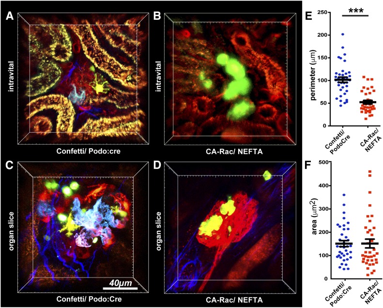
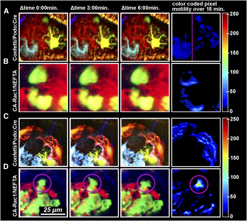
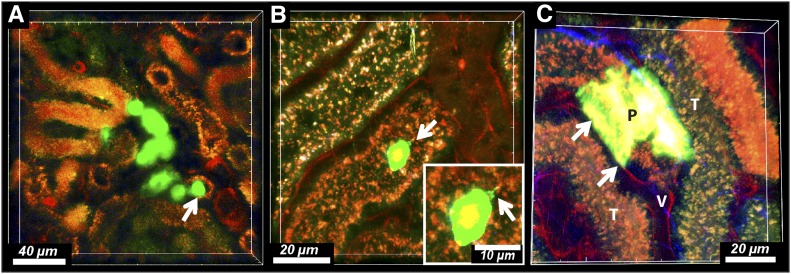
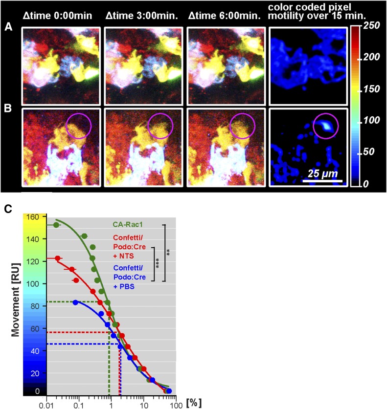
In glomerular disease, podocyte injury results in a dramatic change in cell morphology known as foot process effacement. Remodeling of the actin cytoskeleton through the activity of small GTPases was identified as a key mechanism in effacement, with increased membrane activity and motility in vitro However, whether podocytes are stationary or actively moving cells in vivo remains debated. Using intravital and kidney slice two-photon imaging of the three-dimensional structure of mouse podocytes, we found that uninjured podocytes remained nonmotile and maintained a canopy-shaped structure over time. On expression of constitutively active Rac1, however, podocytes changed shape by retracting processes and clearly exhibited domains of increased membrane activity. Constitutive activation of Rac1 also led to podocyte detachment from the glomerular basement membrane, and we detected detached podocytes crawling on the surface of the tubular epithelium and occasionally, in contact with peritubular capillaries. Podocyte membrane activity also increased in the inflammatory environment of immune complex-mediated GN. Our results provide evidence that podocytes transition from a static to a dynamic state in vivo, shedding new light on mechanisms in foot process effacement.
Keywords: cytoskeleton; glomerular disease; podocyte.
Copyright © 2016 by the American Society of Nephrology.
Figures
Similar articles
-
Light microscopic visualization of podocyte ultrastructure demonstrates oscillating glomerular contractions.Am J Pathol. 2013 Feb;182(2):332-8. doi: 10.1016/j.ajpath.2012.11.002. Epub 2012 Dec 13. Am J Pathol. 2013. PMID: 23246153
-
Nephrin Tyrosine Phosphorylation Is Required to Stabilize and Restore Podocyte Foot Process Architecture.J Am Soc Nephrol. 2016 Aug;27(8):2422-35. doi: 10.1681/ASN.2015091048. Epub 2016 Jan 22. J Am Soc Nephrol. 2016. PMID: 26802179 Free PMC article.
-
Acute podocyte injury is not a stimulus for podocytes to migrate along the glomerular basement membrane in zebrafish larvae.Sci Rep. 2017 Mar 2;7:43655. doi: 10.1038/srep43655. Sci Rep. 2017. PMID: 28252672 Free PMC article.
-
The role of podocytes in glomerular pathobiology.Clin Exp Nephrol. 2003 Dec;7(4):255-9. doi: 10.1007/s10157-003-0259-6. Clin Exp Nephrol. 2003. PMID: 14712353 Review.
-
Stretch, tension and adhesion - adaptive mechanisms of the actin cytoskeleton in podocytes.Eur J Cell Biol. 2006 Apr;85(3-4):229-34. doi: 10.1016/j.ejcb.2005.09.006. Epub 2005 Oct 18. Eur J Cell Biol. 2006. PMID: 16546566 Review.
Cited by
-
Stressed podocytes-mechanical forces, sensors, signaling and response.Pflugers Arch. 2017 Aug;469(7-8):937-949. doi: 10.1007/s00424-017-2025-8. Epub 2017 Jul 7. Pflugers Arch. 2017. PMID: 28687864 Review.
-
Protein tyrosine phosphatase 1B is a regulator of alpha-actinin4 in the glomerular podocyte.Biochim Biophys Acta Mol Cell Res. 2024 Jan;1871(1):119590. doi: 10.1016/j.bbamcr.2023.119590. Epub 2023 Sep 18. Biochim Biophys Acta Mol Cell Res. 2024. PMID: 37730132 Free PMC article.
-
New Insights into Podocyte Biology in Glomerular Health and Disease.J Am Soc Nephrol. 2017 Jun;28(6):1707-1715. doi: 10.1681/ASN.2017010027. Epub 2017 Apr 12. J Am Soc Nephrol. 2017. PMID: 28404664 Free PMC article. Review.
-
Proteinuric Kidney Diseases: A Podocyte's Slit Diaphragm and Cytoskeleton Approach.Front Med (Lausanne). 2018 Sep 11;5:221. doi: 10.3389/fmed.2018.00221. eCollection 2018. Front Med (Lausanne). 2018. PMID: 30255020 Free PMC article. Review.
-
Novel fluorescence techniques to quantitate renal cell biology.Methods Cell Biol. 2019;154:85-107. doi: 10.1016/bs.mcb.2019.04.013. Epub 2019 May 17. Methods Cell Biol. 2019. PMID: 31493823 Free PMC article.
References
-
- Kaplan JM, Kim SH, North KN, Rennke H, Correia LA, Tong HQ, Mathis BJ, Rodríguez-Pérez JC, Allen PG, Beggs AH, Pollak MR: Mutations in ACTN4, encoding alpha-actinin-4, cause familial focal segmental glomerulosclerosis. Nat Genet 24: 251–256, 2000 - PubMed
-
- Kim JM, Wu H, Green G, Winkler CA, Kopp JB, Miner JH, Unanue ER, Shaw AS: CD2-associated protein haploinsufficiency is linked to glomerular disease susceptibility. Science 300: 1298–1300, 2003 - PubMed
MeSH terms
Grants and funding
LinkOut - more resources
Full Text Sources
Other Literature Sources
Molecular Biology Databases
Research Materials
