. 2016 Nov;27(11):3447-3458.
doi: 10.1681/ASN.2015101100. Epub 2016 Apr 1.

Mucin-1 Increases Renal TRPV5 Activity In Vitro, and Urinary Level Associates with Calcium Nephrolithiasis in Patients

Affiliations

Mucin-1 Increases Renal TRPV5 Activity In Vitro, and Urinary Level Associates with Calcium Nephrolithiasis in Patients

Mingzhu Nie et al. J Am Soc Nephrol. 2016 Nov.

Abstract

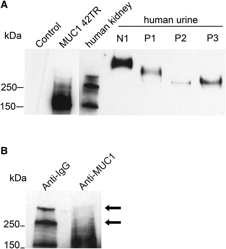
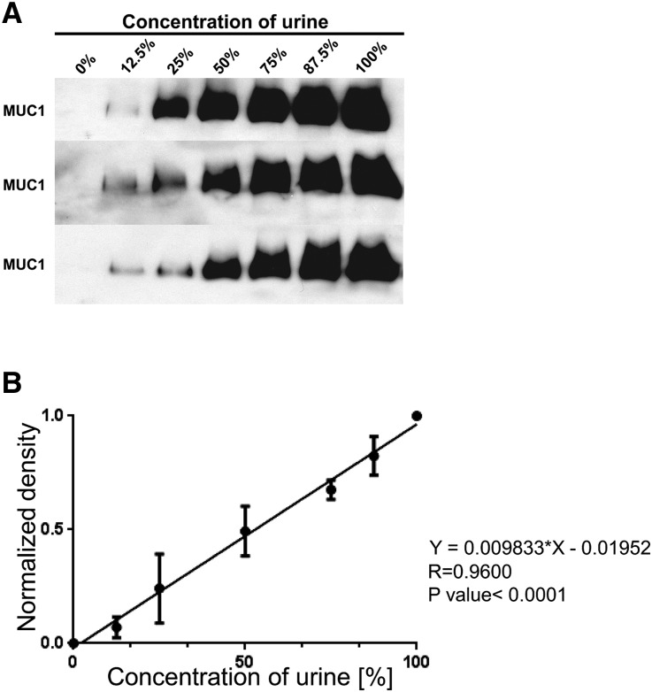
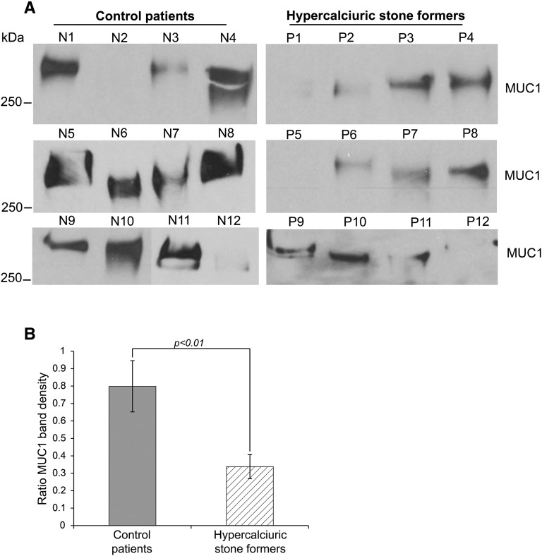
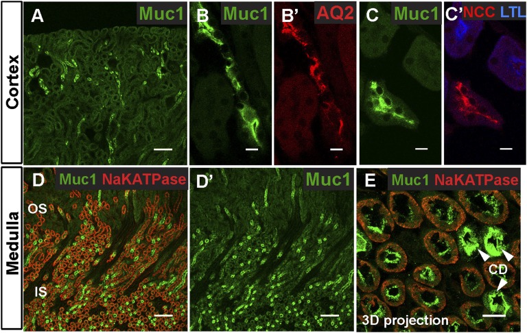
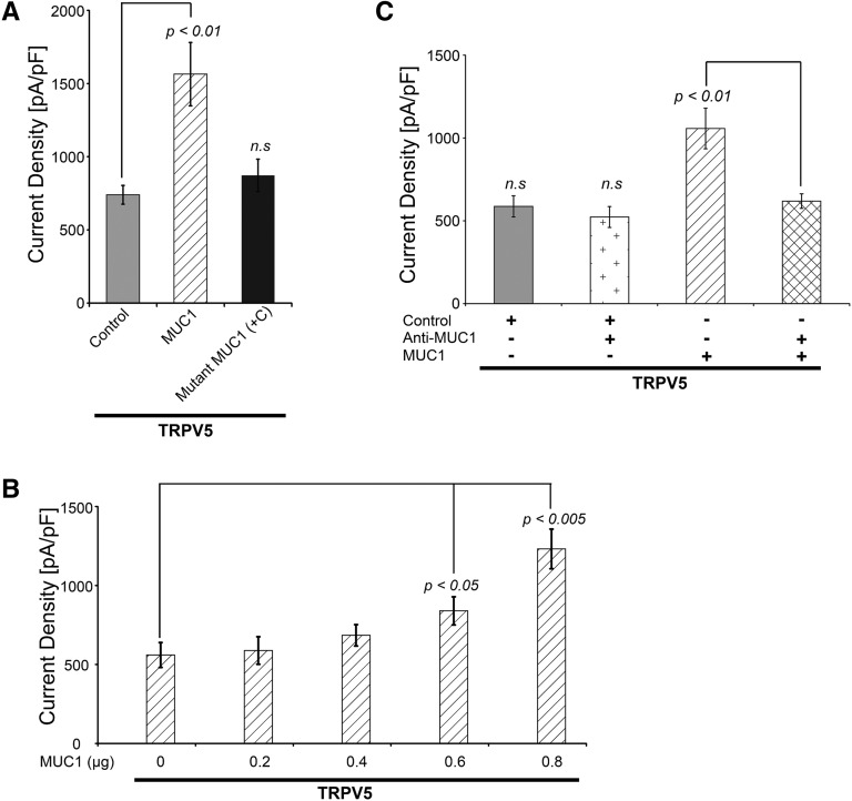
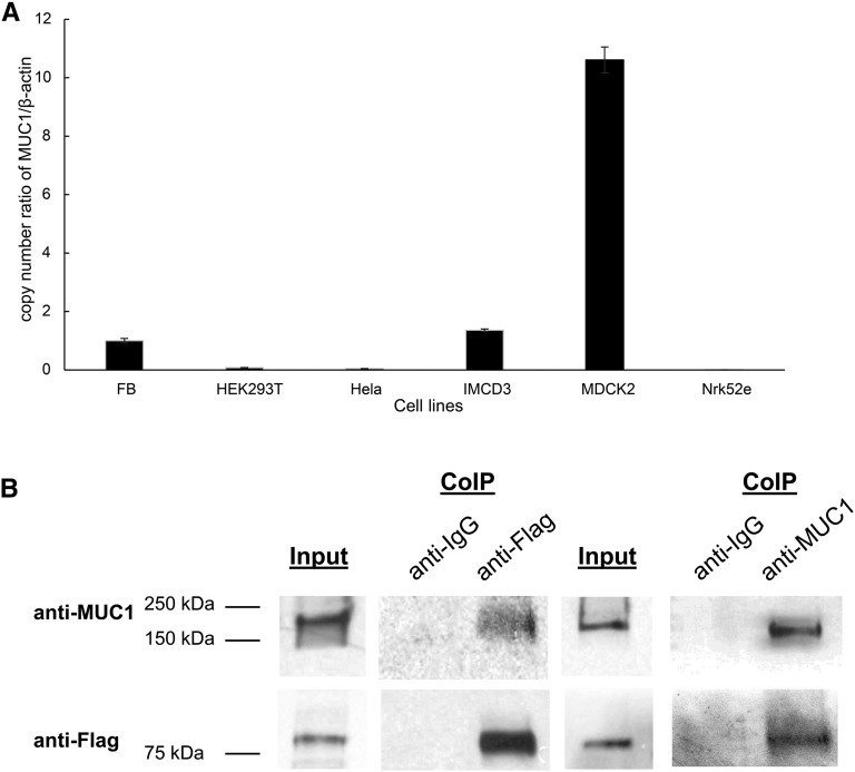
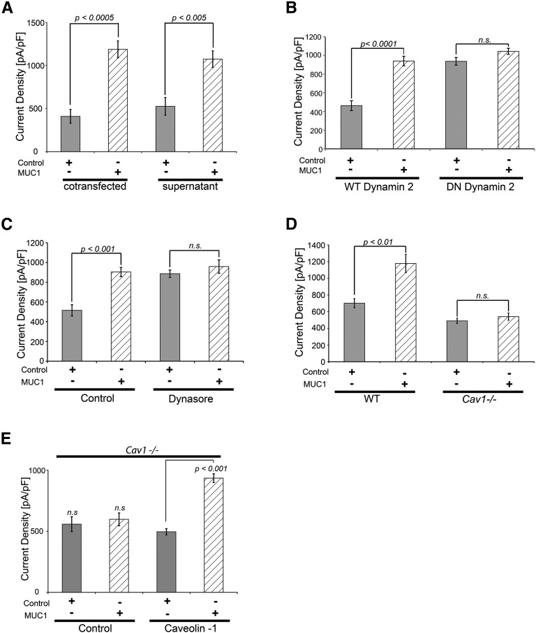
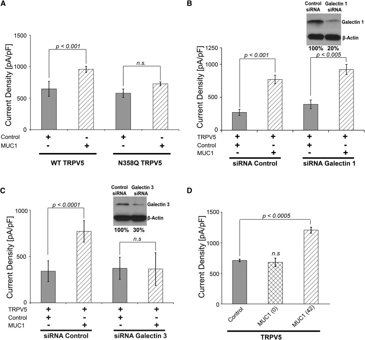
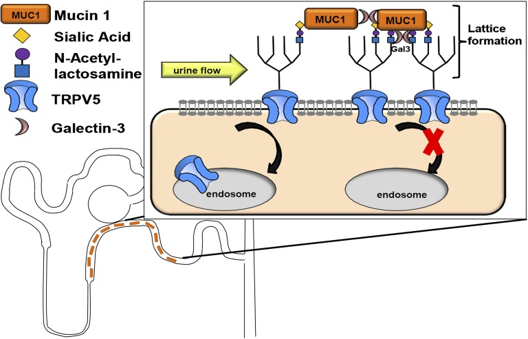
Hypercalciuria is a major risk factor for nephrolithiasis. We previously reported that Uromodulin (UMOD) protects against nephrolithiasis by upregulating the renal calcium channel TRPV5. This channel is crucial for calcium reabsorption in the distal convoluted tubule (DCT). Recently, mutations in the gene encoding Mucin-1 (MUC1) were found to cause autosomal dominant tubulointerstitial kidney disease, the same disease caused by UMOD mutations. Because of the similarities between UMOD and MUC1 regarding associated disease phenotype, protein structure, and function as a cellular barrier, we examined whether urinary MUC1 also enhances TRPV5 channel activity and protects against nephrolithiasis. We established a semiquantitative assay for detecting MUC1 in human urine and found that, compared with controls (n=12), patients (n=12) with hypercalciuric nephrolithiasis had significantly decreased levels of urinary MUC1. Immunofluorescence showed MUC1 in the thick ascending limb, DCT, and collecting duct. Applying whole-cell patch-clamp recording of HEK cells, we found that wild-type but not disease mutant MUC1 increased TRPV5 activity by impairing dynamin-2- and caveolin-1-mediated endocytosis of TRPV5. Coimmunoprecipitation confirmed a physical interaction between TRPV5 and MUC1. However, MUC1 did not increase the activity of N-glycan-deficient TRPV5. MUC1 is characterized by variable number tandem repeats (VNTRs) that bind the lectin galectin-3; galectin-3 siRNA but not galectin-1 siRNA prevented MUC1-induced upregulation of TRPV5 activity. Additionally, MUC1 lacking VNTRs did not increase TRPV5 activity. Our results suggest that MUC1 forms a lattice with the N-glycan of TRPV5 via galectin-3, which impairs TRPV5 endocytosis and increases urinary calcium reabsorption.

Keywords: calcium; electrophysiology; endocytosis; hypercalciuria; ion channel; kidney stones.


Figures

Figure 1.
Figure 1.
MUC1 is expressed in human kidney and secreted in urine. (A) Compared with HEK293 cell lysate (control; lane 1), we identified a clear MUC1 band in cell lysate from HEK293 cells transfected with an MUC1 plasmid containing 42 TRs, yielding a protein size of approximately 200 kD by Western blot. In lysate from human kidneys, we detected three different bands at approximately 170, 260, and >300 kD. Shown is MUC1 expression in human urine from a healthy control (N1) and three patients with hypercalciuric nephrolithiasis (P1–P3). (B) Immunodepletion of human kidney lysate with MUC1 or control IgG shows the MUC1 antibody specificity (arrows).
Figure 2.
Figure 2.
Urinary MUC1 concentration can be analyzed semiquantitatively by Western blot. (A) Three separate MUC1 dilution experiments with increasingly less diluted urine from the same control individual resulted in comparable results regarding MUC1 band density. (B) A linear function between the urine concentration and the normalized MUC1 band density (diluted MUC1 band/100% MUC1 band density) was found in the three different dilution experiments.
Figure 3.
Figure 3.
Urinary MUC1 is significantly decreased in calcium stone formers versus controls. (A) The 24-hour urine samples from 12 controls and 12 hypercalciuric stone formers were compared semiquantitatively regarding urine MUC1 band density. (B) The urinary MUC1 band densities of control individuals and patients were analyzed. To compare multiple urine samples, even on different SDS gels, we determined an MUC1 band ratio by dividing the patient’s urinary MUC1 band density by the MUC1 band density of a urine sample used repeatedly as standard on every SDS gel. Significantly lower urine MUC1 band ratios were found in hypercalciuric stone formers (ratio of MUC1 band density: 0.8±0.15 for the control group versus 0.34±0.07 for stone formers; P<0.01).
Figure 4.
Figure 4.
Muc1 is expressed along different nephron segments in mouse kidney. (A, B, B′, C, and C′) Muc1 immunostaining in the renal cortex. A shows the tubules at low magnification. Muc1 (green) localizes to (B and B′) tubules with Aquaporin 2 (AQ2; red; a marker of the CDs) and (C and C′) tubules with NCC (red; a marker of DCTs) but not with L. tetragonolobus lectin (LTL; blue; a marker of proximal tubules). (D, D′, and E) Within the outer and inner stripes of the outer medulla, Muc1 (green) is highly expressed in CDs (arrowheads in E; AQ2 is not shown here). Higher magnification of a three-dimensional (3D) projection of a z stack reveals that low levels of Muc1 also localize to medullary tubules containing basolateral Na+/K+ ATPase (red), which depict TALs. Scale bars, 100 μm in A, D, and D′; 10 μm in B, B′, C, C′, and E.
Figure 5.
Figure 5.
MUC1 increases TRPV5 whole–cell current density in a dose-dependent fashion. (A) In HEK293 cells, coexpression with WT MUC1 increased the TRPV5 current density (1565±217 pA/pF; P<0.01) compared with control (740±64 pA/pF) and ADTKD–causing frameshift mutant (+C) MUC1 (872±111 pA/pF; NS; n=5 for each group). (B) Cotransfection of increasing amounts of MUC1 plasmid resulted in increasingly higher TRPV5 current density (840±89 pA/pF with 0.6 μg MUC1 versus 560±79 pA/pF with 0 μg MUC1, P<0.05; n=5 for each group). (C) MUC1 antibody significantly decreased TRPV5 current density in TRPV5 and MUC1 cotransfected cells (1057±122 pA/pF for MUC1 versus 618±44 pA/pF for MUC1 and anti-MUC1; P<0.01). Anti-MUC1 alone had no effect on TRPV5 current density (587±64 pA/pF for control versus 523±66 pA/pF for control and anti-MUC1; NS; n=5 for each group).
Figure 6.
Figure 6.
MUC1 is abundantly expressed and binds physically to TRPV5. (A) MUC1 mRNA is endogenously expressed in multiple cell lines. We identified MUC1 mRNA in a range of different cell lines from a variety of species using quantitative RT-PCR. Endogenous MUC1 was strongly detected in MDCK2, IMCD3, and fibroblast (FB) cells, and to a lower degree, also detected in HEK293T cells. A very small amount of MUC1 mRNA was found in HeLa and Nrk52e cells. (B) MUC1 binds TRPV5 channel. HEK293 cells were transiently transfected with MUC1 and TRPV5-Flag. The antibody used for immunoprecipitation is shown above each panel (coimmunoprecipitation [CoIP]). Immunoprecipitated proteins were identified using Western blotting (WB) and specific antibodies as shown on the left. Cell lysate is shown at the left of each immunoprecipitation experiment. MUC1 is detected with precipitation of the MUC1-TRPV5 complex using anti-Flag antibody (lane 3), and TRPV5 is detected using anti-MUC1 antibody for precipitation (lane 6).
Figure 7.
Figure 7.
MUC1 upregulates TRPV5 from the extracellular space and impairs dynamin-2– and Cav1–dependent TRPV5 endocytosis. (A) MUC1 increases TRPV5 current density from the extracellular space. Cells cotransfected with MUC1 (410±80 pA/pF and control versus 1186±97 pA/pF and MUC1; P<0.0005) as well as TRPV5 transfected cells exposed to MUC1-containing supernatant show increased TRPV5 current density (526±102 pA/pF and control supernatant versus 1072±96 pA/pF and MUC1 supernatant; P<0.005), confirming extracellular upregulation of TRPV5 by MUC1. (B) MUC1 increases TRPV5 activity by impairing dynamin-2–dependent endocytosis of the channel. Whereas cells transfected with WT dynamin-2 showed the expected upregulation of TRPV5 current density with MUC1 (for WT dynamin-2: 462±53 pA/pF and control versus 940±51 pA/pF and MUC1; P<0.0001), cells transfected with dominant negative dynamin-2 displayed increased TRPV5 current density at baseline, indicating constitutive TRPV5 endocytosis by dynamin-2. In these cells, no additional increase of TRPV5 activity was noticed with MUC1 (for dominant negative dynamin-2: 937±41 pA/pF and control versus 1043±33 pA/pF and MUC1), indicating that TRPV5 upregulation by MUC1 occurs by impairing dynamin-2–dependent endocytosis. (C) Confirmation of MUC1 upregulation of TRPV5 by impairing dynamin–dependent TRPV5 endocytosis using dynasore. Using the dynamin GTPase inhibitor dynasore, which blocks dynamin-dependent endocytosis, we confirmed the lack of MUC1 effect on TRPV5 current density (no dynasore: 515±57 pA/pF and control versus 904±47 pA/pF and MUC1; P<0.001; plus dynasore: 887±37 pA/pF and control versus 959±68 pA/pF and MUC1; NS). (D) MUC1 increases TRPV5 activity by impairing Cav1-dependent endocytosis of the channel. Whereas WT fibroblasts showed the expected upregulation of TRPV5 current density with MUC1 (WT cells: 701±37 pA/pF and control versus 1178±109 pA/pF and MUC1; P<0.01), Cav1-deficient cells (Cav1−/−) displayed no further increase of TRPV5 activity when cotransfected with MUC1 (Cav1−/− cells: 490±31 pA/pF and control versus 541±43 pA/pF and MUC1; NS), indicating that TRPV5 upregulation by MUC1 occurs by impairing Cav1-dependent endocytosis. (E) TRPV5 upregulation by MUC1 in Cav1−/− cells is rescued by cotransfection of Cav1. Forced overexpression of Cav1 but not control plasmid restored the ability of TRPV5 upregulation by MUC1 (no Cav1: 558±61 pA/pF and control versus 598±53 pA/pF and MUC1; NS; with Cav1: 496±25 pA/pF and control versus 935±36 pA/pF and MUC1; P<0.001; n=5 for each group).
Figure 8.
Figure 8.
MUC1 upregulation of TRPV5 requires the TRPV5 N-glycan and galectin-3. (A) The N-glycan of TRPV5 is required for upregulation by MUC1. WT TRPV5 responded to MUC1 cotransfection (WT TRPV5: 648±45 pA/pF and control versus 958±14 pA/pF and MUC1; P<0.001), whereas N-glycan–deficient (N358Q) TRPV5 did not react to MUC1 stimulation (N358Q TRPV5: 578±90 pA/pF and control versus 728±64 pA/pF and MUC1; NS). (B) Galectin-1 is not required for TRPV5 upregulation by MUC1. Knockdown of galectin-1 using siRNA did not impair the response of TRPV5 current density to MUC1 (control siRNA: 270±46 pA/pF and control versus 769±65 pA/pF and MUC1; P<0.001; galectin-1 siRNA: 393±65 pA/pF and control versus 920±77 pA/pF and MUC1; P<0.005). Using Western blotting, knockdown of galectin-1 is shown in the upper right panel. Quantification of the galectin-1 knockdown by densitometry shows a reduction of 80%. (C) Knockdown of galectin-3 abrogates upregulation of TRPV5 current density by MUC1 (control siRNA: 341±37 pA/pF and control versus 770±52 pA/pF and MUC1; P<0.0001; galectin-3 siRNA: 372±53 pA/pF and control versus 364±72 pA/pF and MUC1; NS). Using Western blotting, knockdown of galectin-3 is shown in the upper right panel. Quantification of the galectin-3 knockdown by densitometry shows a reduction of 70% (D) MUC1 lacking VNTR is not able to upregulate TRPV5 current density (711±23 pA/pF and control versus 681±68 pA/pF and zero-TR MUC1; NS) in contrast to MUC1 containing 42 TRs (1207±49 pA/pF and 42-TR MUC1; P<0.0005; n=5 for each group).
Figure 9.
Figure 9.
Model of TRPV5 channel upregulation by urinary MUC1. MUC1 is secreted in the ultrafiltrate along the TAL and the distal nephron. In the DCT and connecting tubule, urinary MUC1 binds to the N-glycan of TRPV5 (shown in the boxed area). Two different scenarios are possible. Either galectin-3 binds to the TRPV5 N-glycan, and MUC1 stabilizes the binding by binding galectin-3 via the O-glycans of the MUC1 VNTR, or MUC1 may bind directly to TRPV5 N-glycans, and galectin-3 is needed for aggregation of multiple MUC1 molecules via the O-glycans of the MUC1 VNTR. In both situations, galectin-3 enables lattice formation of TRPV5 because of MUC1, which impairs TRPV5 endocytosis and increases TRPV5 cell surface abundance (shown in the box).

Comment in

Similar articles

Cited by

References

    1. Sakhaee K, Maalouf NM, Kumar R, Pasch A, Moe OW: Nephrolithiasis-associated bone disease: Pathogenesis and treatment options. Kidney Int 79: 393–403, 2011 - PMC - PubMed
    1. Kumar V, Lieske JC: Protein regulation of intrarenal crystallization. Curr Opin Nephrol Hypertens 15: 374–380, 2006 - PubMed
    1. Moe OW, Pearle MS, Sakhaee K: Pharmacotherapy of urolithiasis: Evidence from clinical trials. Kidney Int 79: 385–392, 2011 - PMC - PubMed
    1. Frick KK, Bushinsky DA: Molecular mechanisms of primary hypercalciuria. J Am Soc Nephrol 14: 1082–1095, 2003 - PubMed
    1. Costanzo LS, Windhager EE, Ellison DH: Calcium and sodium transport by the distal convoluted tubule of the rat. 1978. J Am Soc Nephrol 11: 1562–1580, 2000 - PubMed