. 2016 Mar 31:4:32.
doi: 10.1186/s40478-016-0300-0.

A local insult of okadaic acid in wild-type mice induces tau phosphorylation and protein aggregation in anatomically distinct brain regions

Affiliations

A local insult of okadaic acid in wild-type mice induces tau phosphorylation and protein aggregation in anatomically distinct brain regions

Siân Baker et al. Acta Neuropathol Commun. .

Abstract

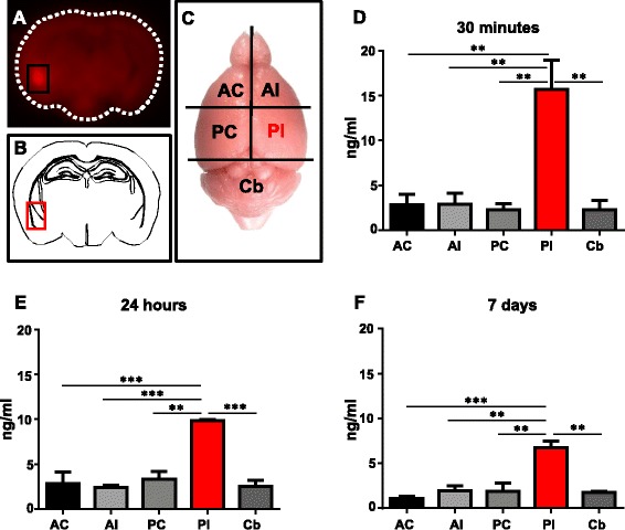
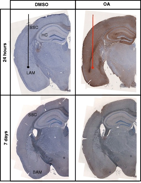
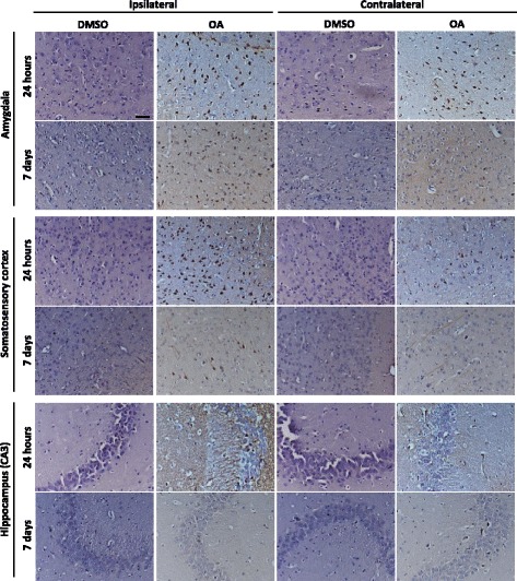
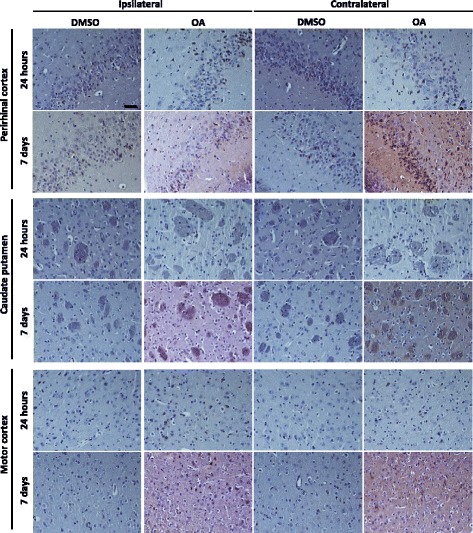
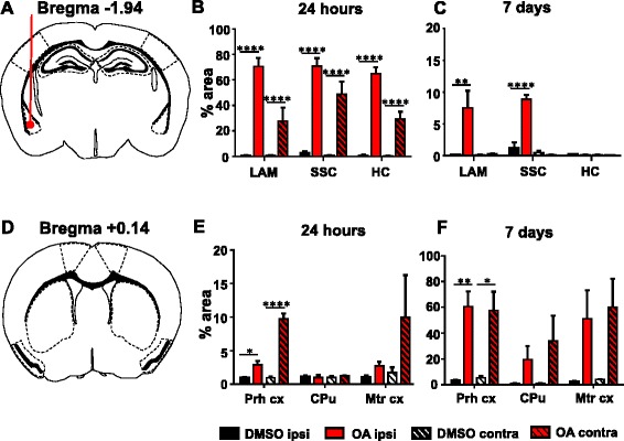
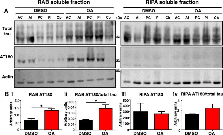
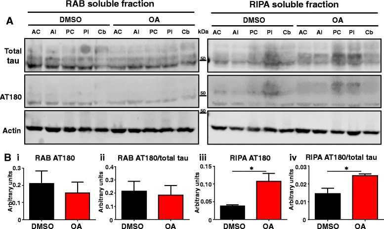
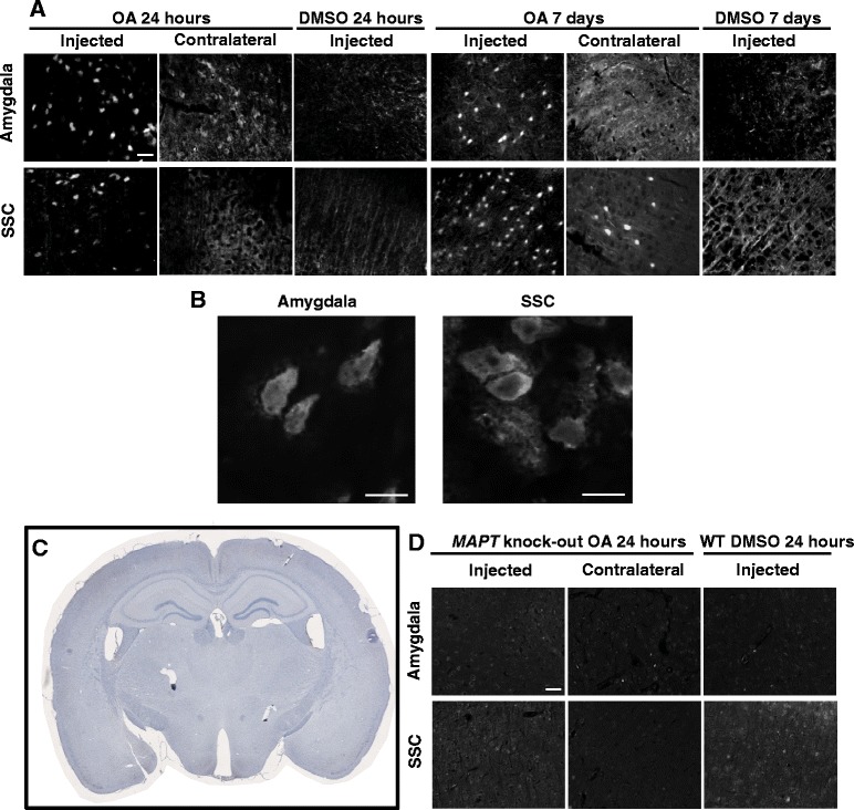
In Alzheimer's disease (AD), the distribution and density of neurofibrillary tangles, a histological hallmark comprised predominately of phosphorylated tau protein, follows a distinct pattern through anatomically connected brain regions. Studies in transgenic mice engineered to regionally confine tau expression have suggested spreading of tau within neural networks. Furthermore, injection of protein lysates isolated from brains of transgenic mice or patients with tauopathies, including AD, were shown to behave like seeds, accelerating tau pathology and tangle formation in predisposed mice. However, it remains unclear how the initiation of primary aggregation events occurs and what triggers further dissemination throughout the neural system. To consolidate these findings, we pursued an alternative approach to assess the spreading of endogenous phosphorylated tau. To generate endogenous seeds, 130 nl of 100 μM protein phosphatase 2A inhibitor okadaic acid (OA) was injected unilaterally into the amygdala of 8-month-old C57Bl/6 wild-type mice. OA was detected in brain tissue by ELISA, and found to be restricted to the injected hemispheric quadrant, where it remained detectable a week post-injection. OA injection induced tau phosphorylation that was observed not only at the injection site but also in anatomically distinct areas across both hemispheres, including the cortex and hippocampus 24 h post-injection. An increase in insoluble tau was also observed in both hemispheres of injected brains by 7 days. Furthermore, thioflavin-S detected protein aggregation at the injection site and in the cortex of both injected and contralateral hemispheres. OA injection induced no thioflavin-positivity in tau knock-out mice. The data demonstrates that a local OA insult can rapidly initiate changes in protein phosphorylation, solubility and aggregation at anatomically distant sites. This model suggests that tau phosphorylation can be both a primary response to an insult, and a secondary response communicated to non-exposed brains regions. The study highlights the use of OA to assist in understanding the initiation of tau spreading in vivo.

Keywords: Alzheimer’s disease; Okadaic acid; Phosphorylation; Protein phosphatase 2A; Spreading; Tau; Tauopathy.


Figures

Fig. 1
Fig. 1
Brain dissection to confirm injection site. a Mice are unilaterally injected at the co-ordinates for the lateral amygdala with Evans blue dye and culled 30 min later. 1 mm thick brain sections are imaged at 700 nm where injected dye fluoresces red. b Comparison to neuroanatomy at Bregma −1.94 mm confirms injection location to the lateral amygdala. c Mice brains are dissected into the anterior contralateral (AC), anterior ipsilateral (AI), posterior contralateral (PC), posterior ipsilateral (PI) and cerebellum (Cb) for analysis by ELISA and western blotting. The PI fraction contains the lateral amygdala injection site. d-f OA concentration in brain tissue is quantified by ELISA. Significant levels of OA are detected in the PI fraction alone at all time points, with no significant difference observed between any of the non-injected fractions, with concentrations comparable to homogenised tissue controls (Additional file 1: Figure S1). The PI fraction maintains significantly elevated levels of OA at 24 h and 7 days after injection, although the concentration is reduced. (n = 3, **p < 0.01, ***p < 0.001)
Fig. 2
Fig. 2
Characterisation of tau pathology. Representative coronal sections from the injection site are stained for phospho-tau with AT180 and counterstained with haematoxylin for cell bodies. Tissue injected with DMSO have relatively low levels of phospho-tau immunoreactivity. At 24 h phospho-tau is abundant at the injection site in the lateral amygdala (LAM) and can be seen throughout the layers of the cortex (CX) and hippocampus (HC) in OA injected. At 7 days phospho-tau persists primarily in the basal amygdala (BAM) and somatosensory cortex (SSC) in the OA injected mice
Fig. 3
Fig. 3
Propagation of tau phosphorylation occurs across hemispheres at 24 h at remains up to 7 days. Representative images of AT180 immunoreactivty are taken from Bregma – 1.94 at the coronal level of injection. Tau phosphorylation at AT180 is observed in both the injected and contralateral hemisphere at 24 h post OA injection. Positive neurones and neuropil staining is abundant in both the lateral amygdala and hippocampus. Furthermore positive cell bodies and processes are clearly defined in the somatosensory cortex, although qualitatively more are seen in the injected hemisphere. By 7 days phospho-tau positive neurones are predominately observed in the injected hemisphere in fewer numbers than at 24 h. Phosphorylation does not persist in the hippocampus at 7 days. Scale bar = 50 μm
Fig. 4
Fig. 4
Tau phosphorylation propagates to areas anterior to the injection site. Representative images of AT180 immunoreactivty are taken from brain regions 2.08 mm anterior to the injection. At 24 h there is relatively little tau phosphorylation detectable in anterior regions, although a few positive cell bodies in the perirhinal cortex are noted. At 7 days post OA injection tau phosphorylation is pronounced in the perirhinal cortex in both the injected and contralateral hemisphere. In the striatum phospho-tau immunoreactivity is seen confined to within axon bundles. Only a small proportion of phospho-tau positive neurones are observed in the motor cortex. Scale bar = 50 μM
Fig. 5
Fig. 5
Propagation of tau pathology to anatomically distinct brain regions. Regions of interest for analysis are taken from a lateral amygdala (LA), somatosensory cortex (SSC) and hippocampus (HC) in sections at the level of injection and d perirhinal cortex (Prh cx), caudate putamen (CPu) and motor cortex (Mtr cx) from sections anterior to the injection site at Bregma + 0.14 mm. Quantification of percentage area analysis at 24 h b, e shows significant increase in phospho-tau immunoreactivity in both the ipsilateral (ipsi) and contralateral (contra) hemispheres of OA-injected mice. By 7 days significant changes are predominately noted in the ipsilateral hemisphere (c) with the exception of the perirhinal cortex (f). (n = 4–5, *P < 0.05, **P < 0.01, ****P < 0.0001)
Fig. 6
Fig. 6
Soluble tau is hyperphosphorylated at 24 h. a Representative western blots for analysis of tau expression for total tau by DAKO and the phospho-tau by AT180. b i) Quantification using the sum of all fractions shows a significant increase in total AT180 at 24 h in OA injected mice when compared to DMSO injected controls. ii) The proportion of tau phosphorylated at AT180 is significantly increased by OA. No significant changes are observed in the insoluble, RIPA fraction (iii-iv). (n = 3, *p < 0.05)
Fig. 7
Fig. 7
Insoluble tau is hyperphosphorylated at 7 days. a Representative western blots for analysis of tau expression for total tau by DAKO and the phospho-tau by AT180. b i) Quantification using the sum of all fractions shows no significant change in total or phospho-tau in the soluble fraction. (iii) AT180 significantly increases by 7 days in OA injected mice in the insoluble fraction compared to DMSO injected controls. The ratio of phosphorylated tau as noted by the AT180/total tau is significantly increased in the RIPA (iv) fractions but is unchanged in RAB fractions (ii). (n = 3, *p < 0.05)
Fig. 8
Fig. 8
Insoluble proteins form NFT-like aggregates rapidly after OA injection at both the injection site and distal brain regions. a β-pleated sheet protein structures are identified by thioflavin-S. Intracellular aggregates are observed at both the injection site and distal sites in the somatosensory cortex at 24 h, which persist 7 days post injection. Scale bar = 50 μM. b High magnification of thioflavin-S positive cells in the amygdala and somatosensory cortex. Scale bar = 10 μm. c Coronal sections taken from OA injected MAPT KO show no phospho-tau immunoreactivity. d No thioflavin positive structures are observed in MAPT KO mice with background staining comparable to the wild-type controls at 24 h. Scale bar = 50 μm

References

    1. Querfurth HW, LaFerla FM. Alzheimer’s disease. N Engl J Med. 2010;362:329–44. doi: 10.1056/NEJMra0909142. - DOI - PubMed
    1. Braak H, Alafuzoff I, Arzberger T, Kretzschmar H, Del Tredici K. Staging of Alzheimer disease-associated neurofibrillary pathology using paraffin sections and immunocytochemistry. Acta Neuropathol. 2006;112:389–404. doi: 10.1007/s00401-006-0127-z. - DOI - PMC - PubMed
    1. Montine TJ, Phelps CH, Beach TG, Bigio EH, Cairns NJ, Dickson DW, et al. National Institute on Aging-Alzheimer’s Association guidelines for the neuropathologic assessment of Alzheimer’s disease: a practical approach. Acta Neuropathol. 2012;123:1–11. doi: 10.1007/s00401-011-0910-3. - DOI - PMC - PubMed
    1. Hardy J, Revesz T. The spread of neurodegenerative disease. N Engl J Med. 2012;366:2126–8. doi: 10.1056/NEJMcibr1202401. - DOI - PubMed
    1. Calafate S, Buist A, Miskiewicz K, Vijayan V, Daneels G, de Strooper B, et al. Synaptic contacts enhance cell-to-cell tau pathology propagation. Cell Rep. 2015;11:1176–83. doi: 10.1016/j.celrep.2015.04.043. - DOI - PubMed

Publication types

LinkOut - more resources