

Review
doi: 10.1124/pr.115.010652.
Mechanisms of Vascular Smooth Muscle Contraction and the Basis for Pharmacologic Treatment of Smooth Muscle Disorders
Affiliations
- PMID: 27037223
- PMCID: PMC4819215
- DOI: 10.1124/pr.115.010652
Item in Clipboard
Review
Mechanisms of Vascular Smooth Muscle Contraction and the Basis for Pharmacologic Treatment of Smooth Muscle Disorders
Pharmacol Rev.
2016 Apr.
Abstract
The smooth muscle cell directly drives the contraction of the vascular wall and hence regulates the size of the blood vessel lumen. We review here the current understanding of the molecular mechanisms by which agonists, therapeutics, and diseases regulate contractility of the vascular smooth muscle cell and we place this within the context of whole body function. We also discuss the implications for personalized medicine and highlight specific potential target molecules that may provide opportunities for the future development of new therapeutics to regulate vascular function.
Copyright © 2016 by The American Society for Pharmacology and Experimental Therapeutics.
Figures
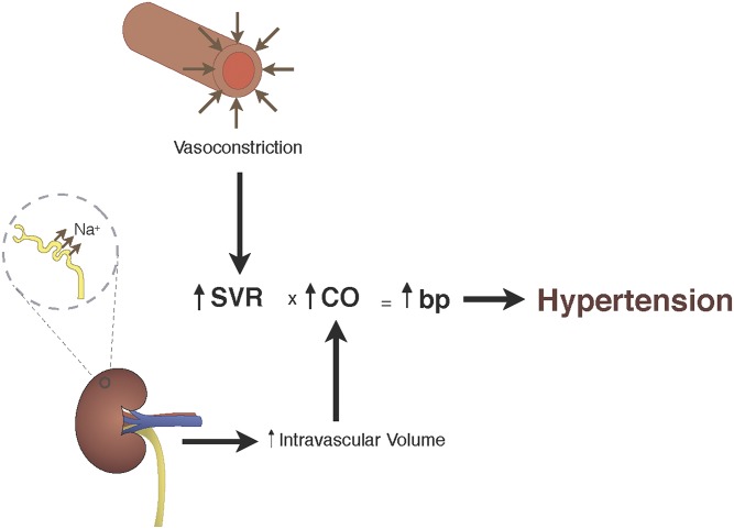
SVR versus kidney: Blood pressure is the product of systemic vascular resistance and cardiac output (BP = SVR × CO). Changes in Na+ reabsorption will increase or decrease intravascular volume and result in an increase or decrease cardiac output, which will alter blood pressure. Similarly, alterations in vascular tone can either increase or decrease SVR, which leads to an increase or decrease in blood pressure (see text for details).
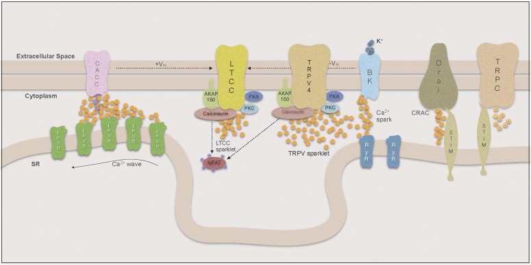
Compartmentalization of Ca signaling.
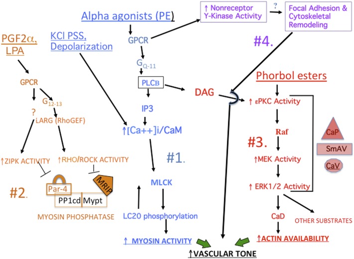
Overview of pathways regulating vascular tone. See text for details. For additional detailed pathways, see subsequent figures.
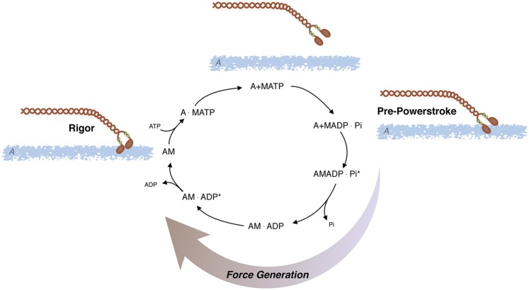
AMATPase: Actomyosin ATPase cycle; ATP is hydrolyzed by myosin (M) and the subsequent interaction of myosin with actin (A) produces force and/or displacement (see text for details).
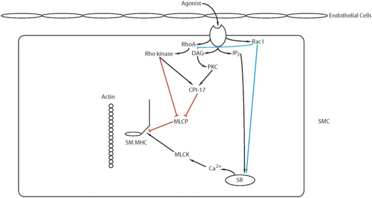
Ca2+ sensitization: Agonist activation of G-protein coupled receptors activates several signaling pathways (IP3, RhoA/Rho kinase, PKC, RAC1) that modulate Ca2+ release from the SR and/or lead to Ca2+ sensitization of the contractile filaments (see text for details).
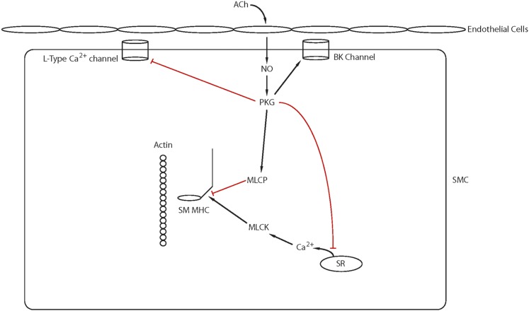
Ca2+ desensitization: ACh stimulation of muscarinic receptors on the vascular endothelium leads to the production of NO, and NO diffuses into smooth muscle cells to activate guanylate cyclase. The NO/cGMP signaling pathway relaxes smooth muscle by both decreasing intracellular Ca2+ and activating MLC phosphatase, which results in Ca2+ desensitization of the contractile filaments (see text for details).
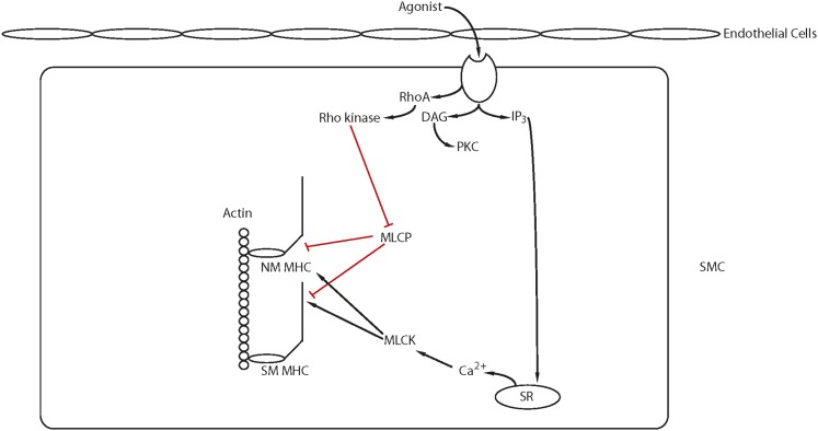
Force maintenance involving either nonmuscle myosin or smooth muscle myosin: There are several proposed mechanisms for force maintenance in smooth muscle including the interaction of actin with either smooth muscle or NM myosin (latch crossbridge) as well as changes in the cytoskeleton (see text for details and see also Fig. 3).
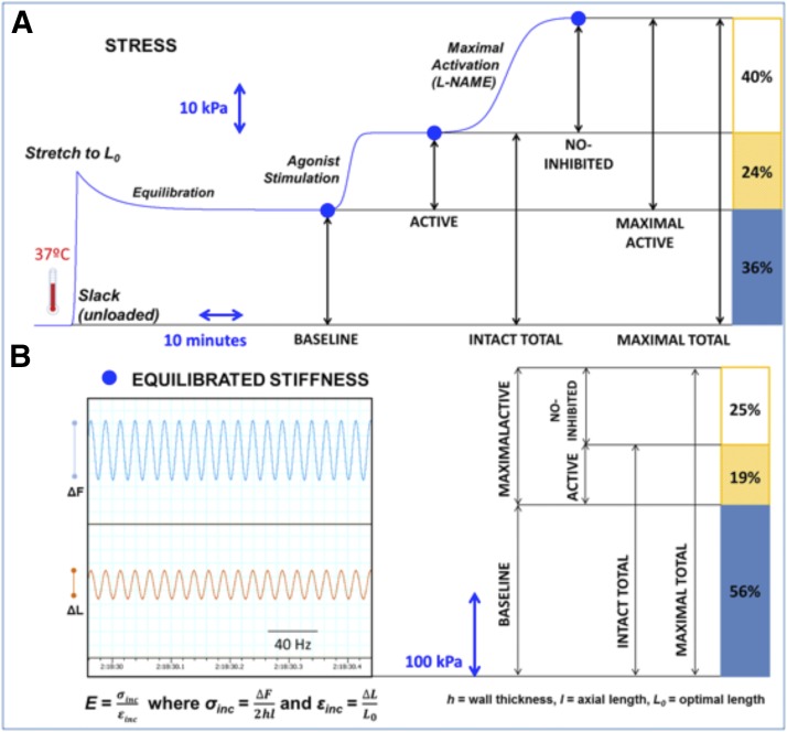
Separability of contractile components to the generation of aortic stiffness. Modified from Gao et al. (2014b).
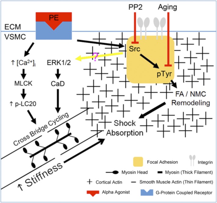
A diagrammatic model of how cytoskeletal remodeling could provide plasticity and an important “shock absorber” for the cardiovascular system (modified from Gao et al., 2014b.)
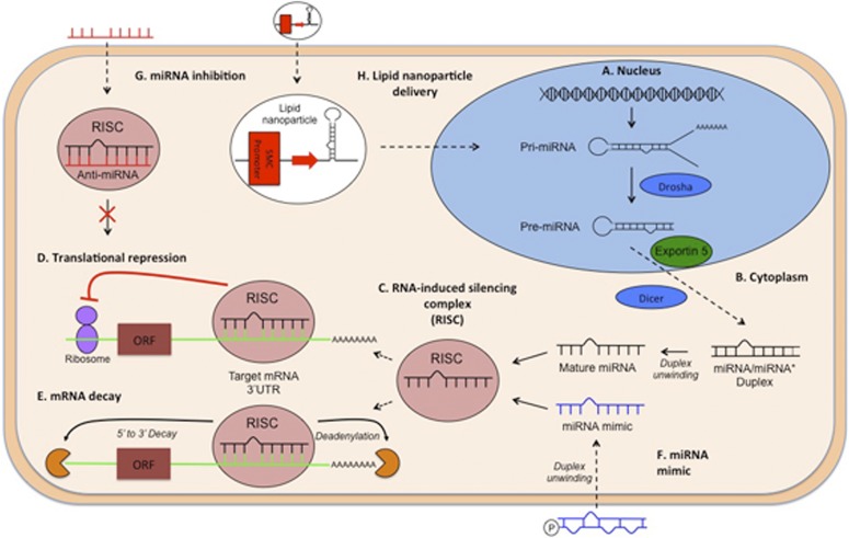
Summary of the main mechanisms of miR biogenesis and function and avenues for pharmacological intervention. (A) MiRs are first transcribed in the nucleus, primarily from introns located in both coding and noncoding DNA, into long primary transcripts termed pri-miRNAs, then are processed in the nucleus by the RNAse III enzyme Drosha into a shorter (∼70 nucleotide) hairpin duplex termed pre-miRs. (B) After export to the cytoplasm by the dsRNA-binding protein Exportin 5, pre-microRNAs are processed by an RNAse III enzyme, Dicer, into a short (21–25 nucleotide) duplex. (C) The miR duplex is unwound, primarily leading to preferential incorporation of a single strand onto Argonaute protein to form the RNA-induced silencing complex (RISC). A short sequence (6–8 nucleotides) at the 5′ end of the miR, known as the "seed" sequence, targets the RISC to complimentary sequences in the 3′ UTR of target mRNAs. (D) Translational repression, through the blocking of translation initiation or recruitment of translational blocking proteins, and/or (E) mRNA decay, via 5′ to 3′ decay and deadenylation of the poly (A) tail. (F) miR mimics are designed as RNA duplexes composed of a passenger strand, chemically modified (e.g., phosphate addition) to permit entry to the cell and subsequent unwinding after entry (i.e., by containing several nucleotide mismatches), and a guide strand, which consists of an identical sequence to the endogenous miR. (G) An anti-miR is designed to be complimentary to the endogenous miR of interest, thus inhibiting target mRNA binding, which therefore increases translation of the target mRNA. (H) A lipid nanoparticle delivery system can induce expression of a miR in specific cell types [e.g., in smooth muscle cells by expressing the miR under the influence of a smooth muscle cell (SMC) promoter].
References
-
- Abassi ZA, Gurbanov K, Mulroney SE, Potlog C, Opgenorth TJ, Hoffman A, Haramati A, Winaver J. (1997) Impaired nitric oxide-mediated renal vasodilation in rats with experimental heart failure: role of angiotensin II. Circulation 96:3655–3664. - PubMed
-
- Alahyan M, Webb MR, Marston SB, El-Mezgueldi M. (2006) The mechanism of smooth muscle caldesmon-tropomyosin inhibition of the elementary steps of the actomyosin ATPase. J Biol Chem 281:19433–19448. - PubMed
Publication types
MeSH terms
Substances
LinkOut - more resources
Full Text Sources
Other Literature Sources
Medical
