. 2016 Nov;26(11):1222-1234.
doi: 10.1093/glycob/cww042. Epub 2016 Apr 1.

Novel aspects of sialoglycan recognition by the Siglec-like domains of streptococcal SRR glycoproteins

Affiliations

Novel aspects of sialoglycan recognition by the Siglec-like domains of streptococcal SRR glycoproteins

Barbara A Bensing et al. Glycobiology. 2016 Nov.

Abstract

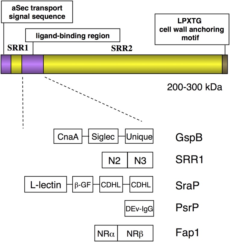
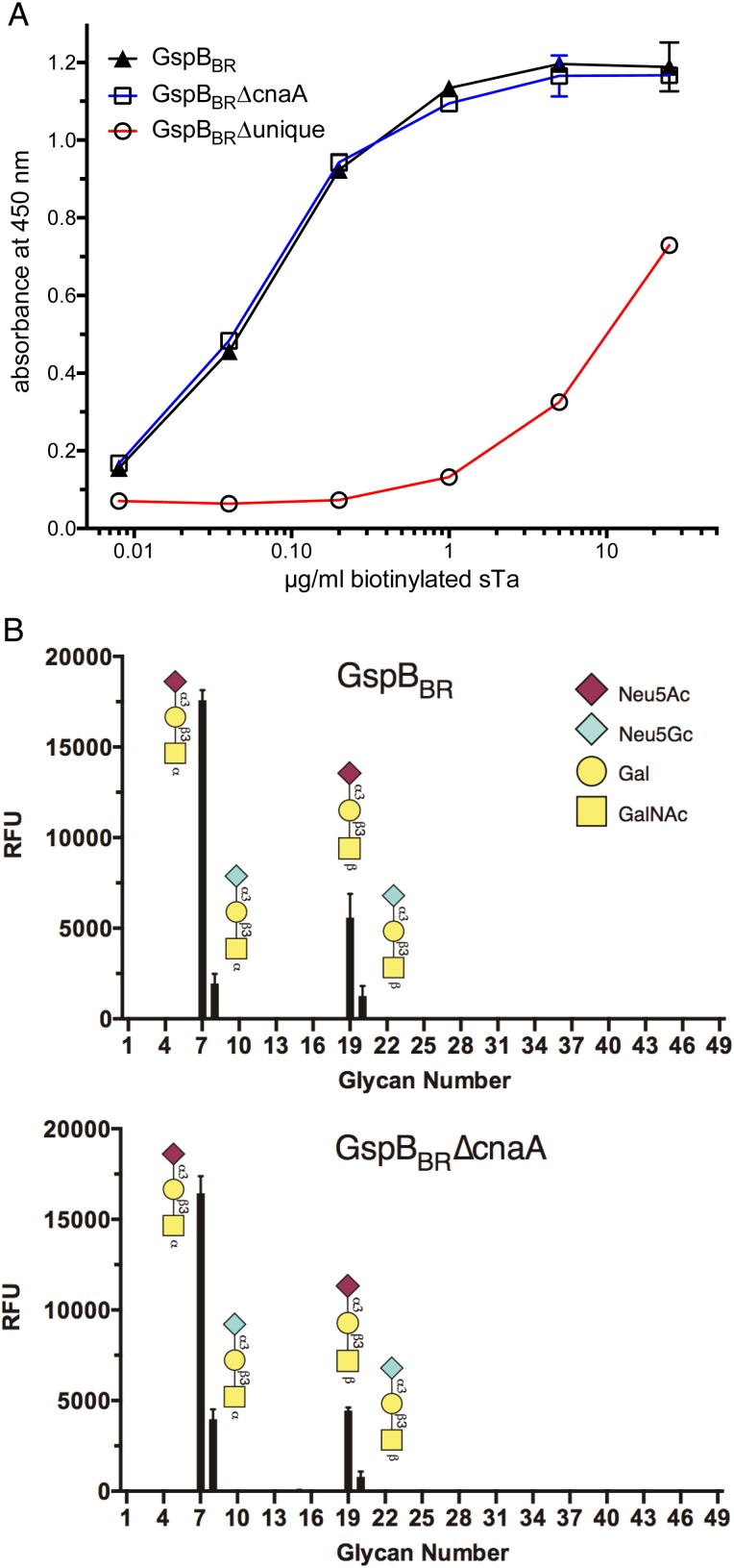
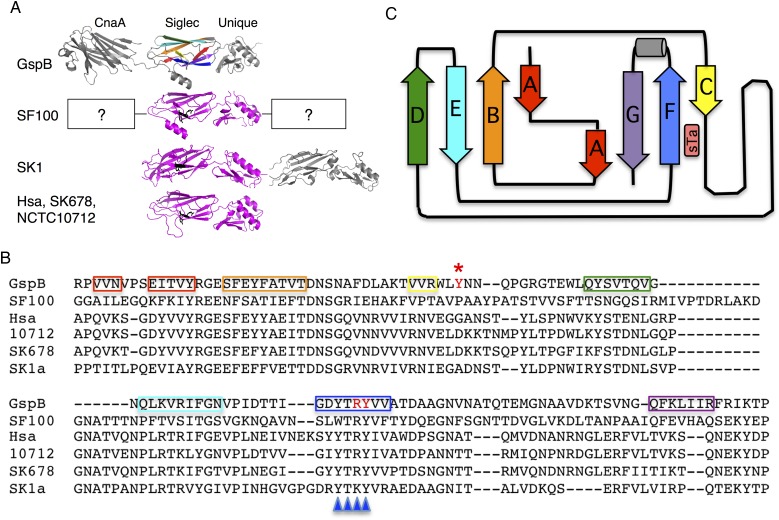
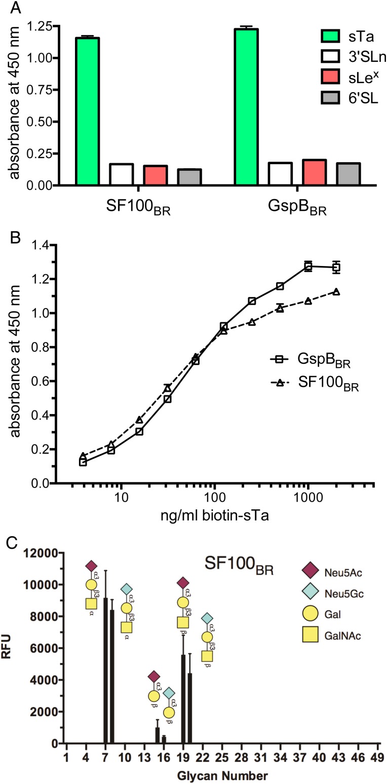
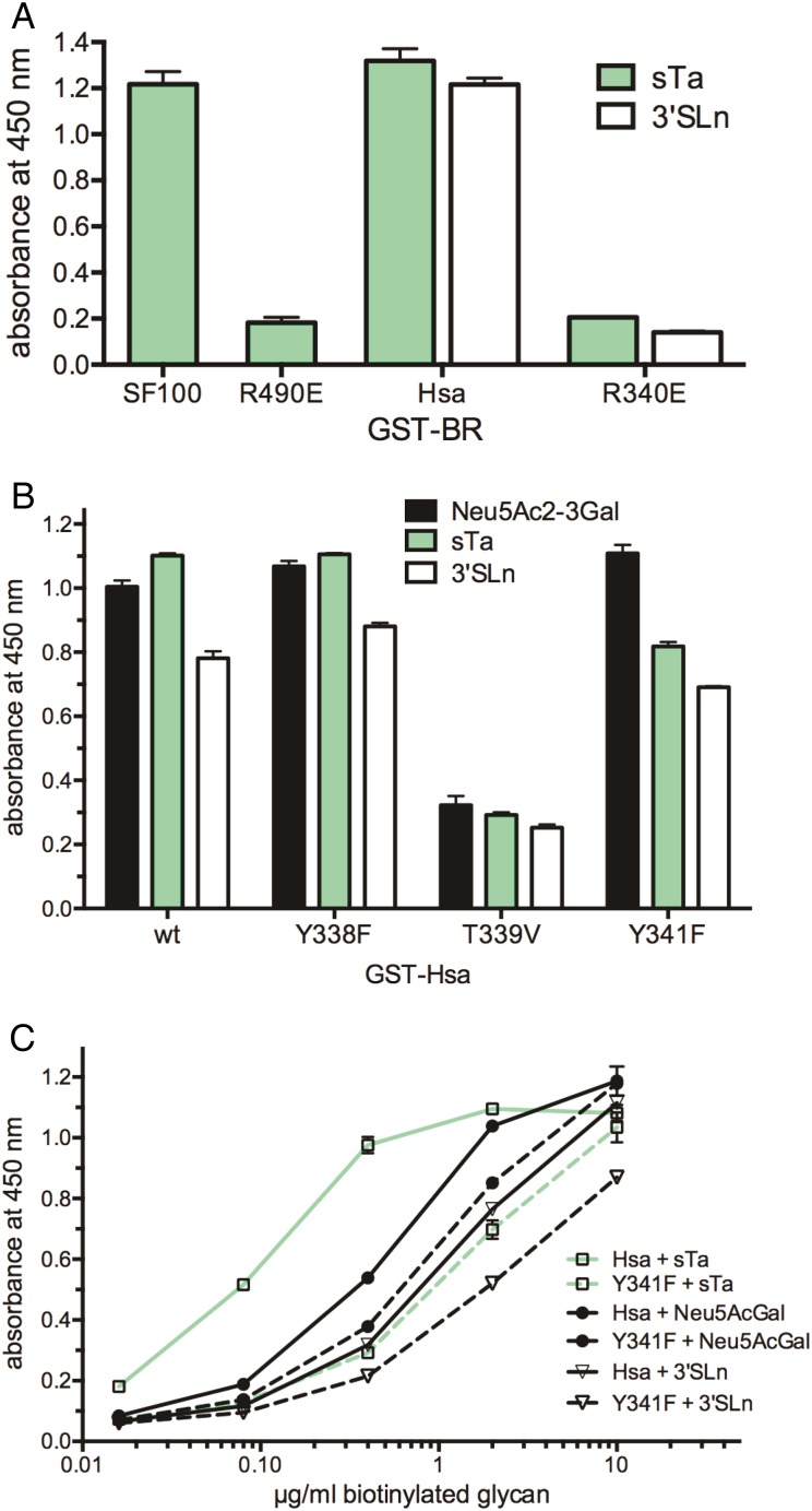
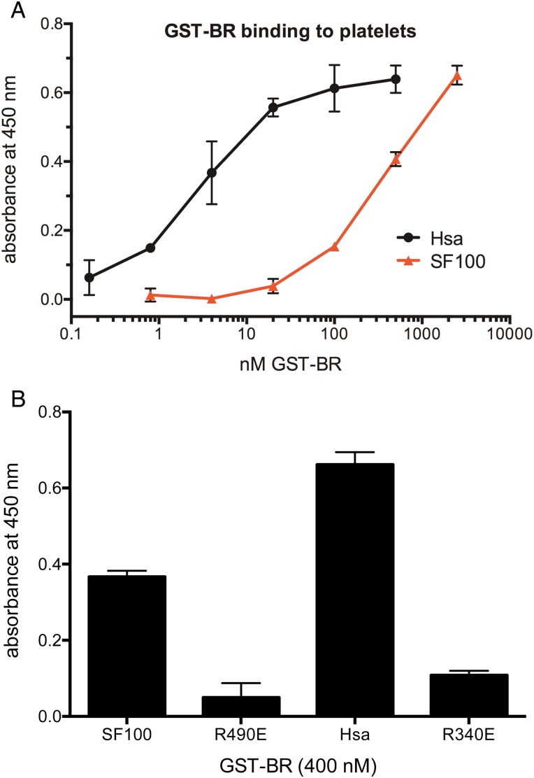
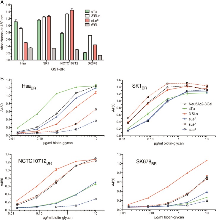
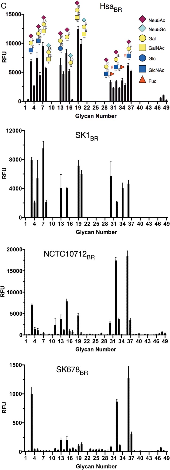
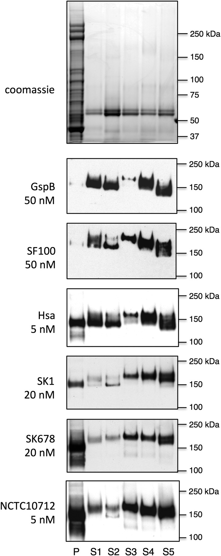
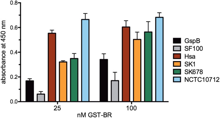
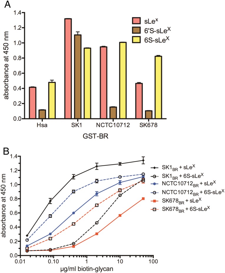
Serine-rich repeat glycoproteins are adhesins expressed by commensal and pathogenic Gram-positive bacteria. A subset of these adhesins, expressed by oral streptococci, binds sialylated glycans decorating human salivary mucin MG2/MUC7, and platelet glycoprotein GPIb. Specific sialoglycan targets were previously identified for the ligand-binding regions (BRs) of GspB and Hsa, two serine-rich repeat glycoproteins expressed by Streptococcus gordonii While GspB selectively binds sialyl-T antigen, Hsa displays broader specificity. Here we examine the binding properties of four additional BRs from Streptococcus sanguinis or Streptococcus mitis and characterize the molecular determinants of ligand selectivity and affinity. Each BR has two domains that are essential for sialoglycan binding by GspB. One domain is structurally similar to the glycan-binding module of mammalian Siglecs (sialic acid-binding immunoglobulin-like lectins), including an arginine residue that is critical for glycan recognition, and that resides within a novel, conserved YTRY motif. Despite low sequence similarity to GspB, one of the BRs selectively binds sialyl-T antigen. Although the other three BRs are highly similar to Hsa, each displayed a unique ligand repertoire, including differential recognition of sialyl Lewis antigens and sulfated glycans. These differences in glycan selectivity were closely associated with differential binding to salivary and platelet glycoproteins. Specificity of sialoglycan adherence is likely an evolving trait that may influence the propensity of streptococci expressing Siglec-like adhesins to cause infective endocarditis.

Keywords: MUC7; Siglec; endocarditis; platelet GPIb; sialyl-T antigen.


Figures

Fig. 1.
Fig. 1.
Diagram of SRR glycoproteins and structural variability of the BRs. The upper portion of the figure indicates the conserved domain organization for the family of adhesins and the lower portion indicates the structural diversity of the BRs. The S. gordonii GspB domains are described in the text. The Streptococcus agalactiae SRR1 has two MSCRAMM folds, designated N2 and N3 (Seo et al. 2013). The Staphylococcus aureus SraP has a legume lectin-like module (l-lectin) that adjoins a ubiquitin-like β-grasp fold and a pair of cadherin-like modules (Yang et al. 2014). The S. pneumoniae PsrP has a DE-variant Ig fold (Schulte et al. 2014). The Streptococcus parasanguinis Fap1 has a pair of novel domains designated non-repeat α and non-repeat β (Ramboarina et al. 2010). This figure is available in black and white in print and in color at Glycobiology online.
Fig. 2.
Fig. 2.
Binding by GspB ΔcnaA and Δunique domain variants. (A) GST-BRs (500 nM) were immobilized in 96-well plates, and biotinylated sTa was supplied at the indicated concentrations. Binding is reported as the mean ± standard deviation, with n = 3. (B) Binding of GST-GspBBR (upper) and the ΔcnaA domain variant (lower) to an array of immobilized glycans, which includes compounds present on salivary and platelet glycoproteins (n = 4). This figure is available in black and white in print and in color at Glycobiology online.
Fig. 3.
Fig. 3.
Comparison of additional Siglec-like BR sequences. (A) BR domain composition. The β strands A through G of the GspB Siglec domain are indicated in color. A, red; B, orange; C, yellow; D, green; E, cyan; F, blue; G, violet. The predicted structures of the SF100, SK1a and SK678 Siglec and Unique domains are shown in magenta, with the F-strand arginine and tyrosine residues indicated as black sticks. The SF100BR includes two domains of unknown structure, indicated with question marks. The SK1BR has two Siglec and two Unique domains; the Siglec domain of the second pair (SK1b, shown in grey) lacks the conserved YTRY motif. (B) Alignment of the Siglec domain sequences. GspB residues that are essential for sTa binding (Y443, R484 and Y485) are indicated in red, and Y443 is denoted by a red asterisk. The β-strand residues are boxed and colored as above. The YTRY motif is indicated with blue triangles. (C) Ribbon diagram of the V-set Ig fold of GspB, with strands colored as indicated above (adapted from Pyburn et al. 2011).
Fig. 4.
Fig. 4.
Ligand binding by the S. mitis SF100BR. (A) Binding of selected biotinylated glycans (10 µg/mL) to immobilized GST-BRs. Binding is reported as the mean ± standard deviation, with n = 4. (B) Binding of biotinylated sTa to immobilized GST-BRs. Binding is reported as the mean ± standard deviation, with n = 2. (C) Binding of GST-SF100BR to the array of immobilized glycans (n = 4). This figure is available in black and white in print and in color at Glycobiology online.
Fig. 5.
Fig. 5.
Contribution of the F-strand motif residues to sialoglycan binding by the SF100 and Hsa BRs. (A) Binding of biotinylated sTa or 3′SLn (10 µg/mL) to immobilized GST-BRs. R490E and R340E are the F-strand arginine mutants of GST-SF100BR or GST-HsaBR, respectively. Binding is reported as the mean ± standard deviation, with n = 4. (B) Binding of selected biotinylated glycans (5 µg/mL) to immobilized wild-type or mutant GST-HsaBR. Binding is reported as the mean ± standard deviation, with n = 3. (C) Binding of biotinylated glycans to immobilized GST-HsaBR wild type or the Y341F variant. Binding is reported as the mean ± standard deviation, with n = 2. This figure is available in black and white in print and in color at Glycobiology online.
Fig. 6.
Fig. 6.
The F-strand Arg is critical for platelet binding by Hsa and SF100 BRs. (A) Binding of GST-BRs to immobilized platelets. Binding is reported as the mean ± standard deviation, with n = 4. (B): Comparative binding of wild-type and mutant BRs (400 nM) to immobilized platelets. Binding is reported as the mean ± standard deviation, with n = 6. This figure is available in black and white in print and in color at Glycobiology online.
Fig. 7.
Fig. 7.
Ligand binding by the Hsa-related SK1, NCTC10712 and SK678 BRs. (A) Binding of selected biotinylated glycans (10 µg/mL) to immobilized GST-BRs. Binding is reported as the mean ± standard deviation, with n = 3. (B) Binding of biotinylated glycans to immobilized GST-BRs. Binding is reported as the mean ± standard deviation, with n = 2. (C) Binding of GST-BRs to the array of immobilized glycans (n = 4). This figure is available in black and white in print and in color at Glycobiology online.
Fig. 7.
Fig. 7.
Ligand binding by the Hsa-related SK1, NCTC10712 and SK678 BRs. (A) Binding of selected biotinylated glycans (10 µg/mL) to immobilized GST-BRs. Binding is reported as the mean ± standard deviation, with n = 3. (B) Binding of biotinylated glycans to immobilized GST-BRs. Binding is reported as the mean ± standard deviation, with n = 2. (C) Binding of GST-BRs to the array of immobilized glycans (n = 4). This figure is available in black and white in print and in color at Glycobiology online.
Fig. 8.
Fig. 8.
Far-western blot of six GST-BRs against salivary and platelet glycoproteins. Lanes contain a lysate of fresh, washed platelets from a single donor (P), or saliva from five different donors (S1 through S5). Glycoproteins were separated by electrophoresis through a 3–8% polyacrylamide gradient, and then stained (top panel) or transferred to nitrocellulose and then probed with GST-BRs as indicated. No signals were detected outside of the cropped region, aside from a high MW band in the platelet sample. The 140–150 kDa BR-reactive bands were previously determined to contain predominantly GPIbα or MG2/MUC7 (Takamatsu et al. 2005, 2006). The precise amount of GPIbα and MG2/MUC7 within the samples is undetermined, but remained constant across the different BRs tested.
Fig. 9.
Fig. 9.
Quantitative binding to immobilized human platelets. GST-BRs (25 or 100 nM) were applied to monolayers of fixed platelets. Binding is reported as the mean ± standard deviation, with n = 4. This figure is available in black and white in print and in color at Glycobiology online.
Fig. 10.
Fig. 10.
Binding of the Hsa-like BRs to sulfated forms of sialyl Lewis X. (A) Binding of biotinylated sLeX, 6′S-sLeX or 6S-sLeX (10 µg/mL) to immobilized GST-BRs. Binding is reported as the mean ± standard deviation, with n = 3. (B) Binding of biotinylated sLeX or 6S-sLeX to immobilized GST-BRs. Binding is reported as the mean ± standard deviation, with n = 2. This figure is available in black and white in print and in color at Glycobiology online.

References

    1. Alphey MS, Attrill H, Crocker PR, van Aalten DM. 2003. High resolution crystal structures of Siglec-7. Insights into ligand specificity in the Siglec family. J Biol Chem. 278:3372–3377. - PubMed
    1. Angata T. 2006. Molecular diversity and evolution of the Siglec family of cell-surface lectins. Mol Divers. 10:555–566. - PubMed
    1. Attrill H, Takazawa H, Witt S, Kelm S, Isecke R, Brossmer R, Ando T, Ishida H, Kiso M, Crocker PR et al. 2006. The structure of siglec-7 in complex with sialosides: Leads for rational structure-based inhibitor design. Biochem J. 397:271–278. - PMC - PubMed
    1. Bensing BA, Lopez JA, Sullam PM. 2004. The Streptococcus gordonii surface proteins GspB and Hsa mediate binding to sialylated carbohydrate epitopes on the platelet membrane glycoprotein Ibalpha. Infect Immun. 72:6528–6537. - PMC - PubMed
    1. Bensing BA, Loukachevitch LV, McCulloch KM, Yu H, Vann KR, Wawrzak Z, Anderson S, Chen X, Sullam PM, Iverson TM. 2016. Structural basis for Sialoglycan binding by the Streptococcus sanguinis SrpA adhesin. J Biol Chem. 291:7230–7240. - PMC - PubMed

Publication types

MeSH terms