Review
. 2016 Apr 1;8(4):a005900.
doi: 10.1101/cshperspect.a005900.

Signals and Receptors

Affiliations
Review

Signals and Receptors

Carl-Henrik Heldin et al. Cold Spring Harb Perspect Biol. .

Abstract

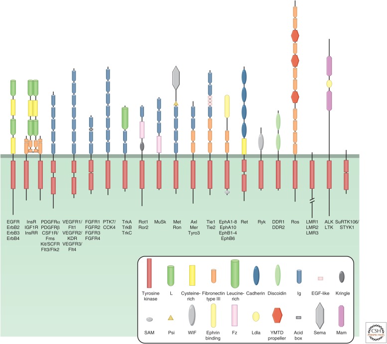
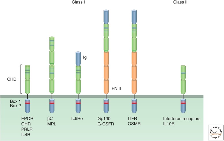
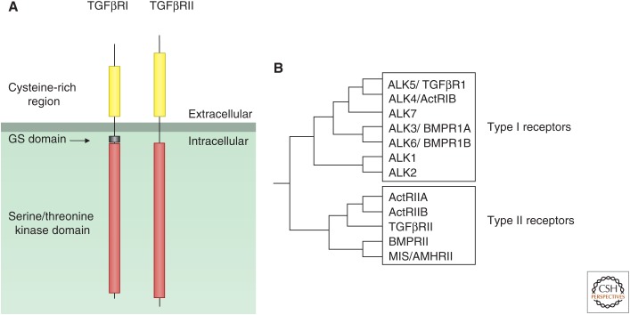
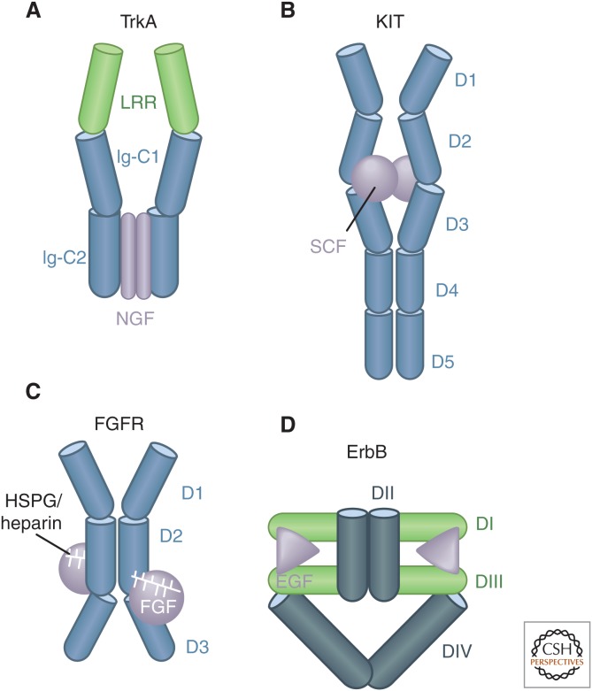
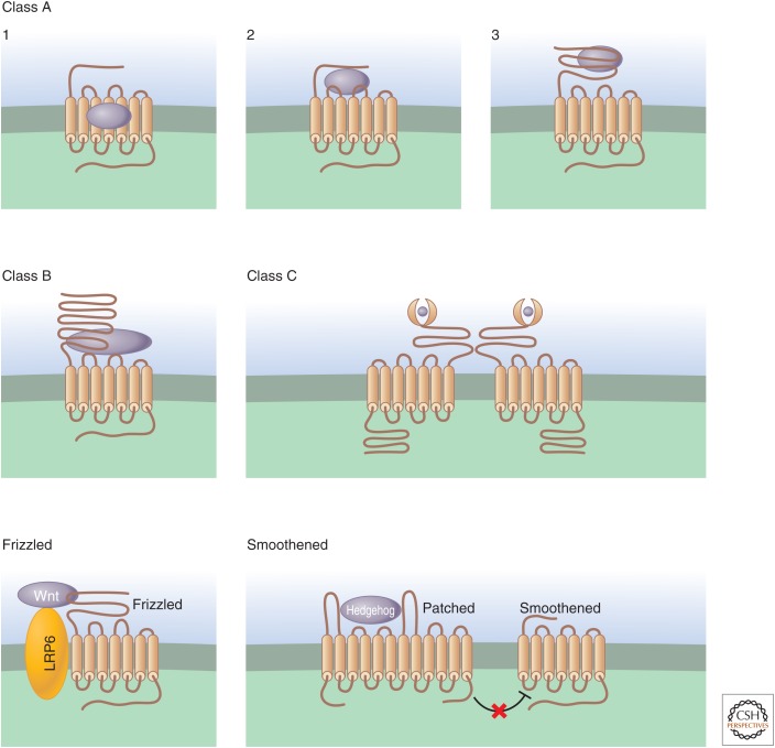
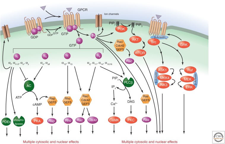
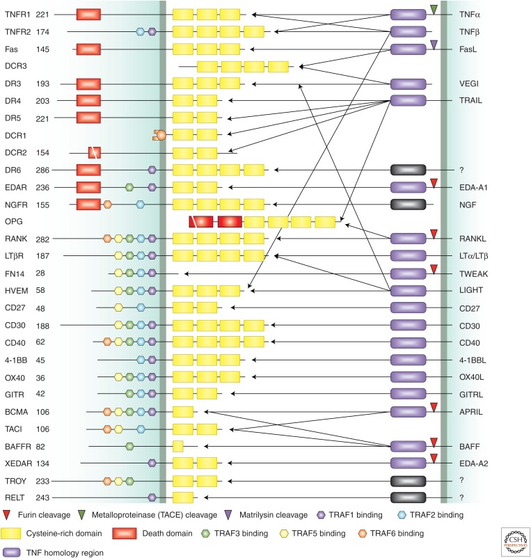
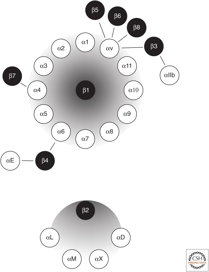
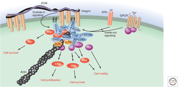
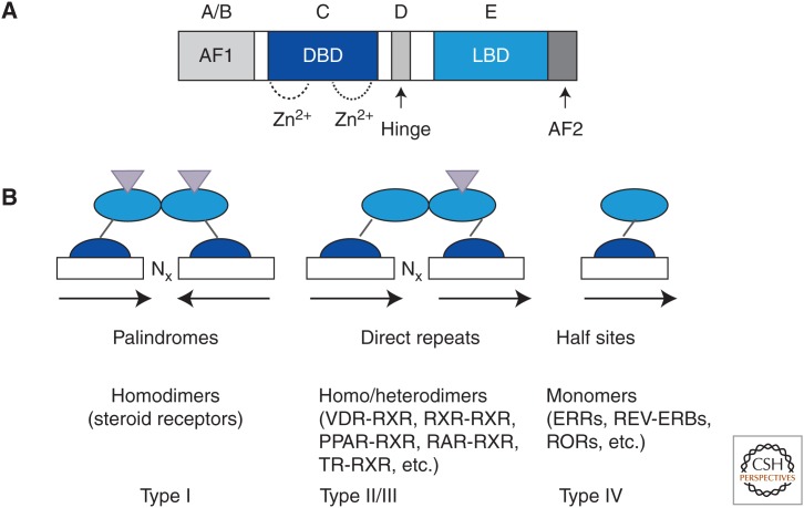
Communication between cells in a multicellular organism occurs by the production of ligands (proteins, peptides, fatty acids, steroids, gases, and other low-molecular-weight compounds) that are either secreted by cells or presented on their surface, and act on receptors on, or in, other target cells. Such signals control cell growth, migration, survival, and differentiation. Signaling receptors can be single-span plasma membrane receptors associated with tyrosine or serine/threonine kinase activities, proteins with seven transmembrane domains, or intracellular receptors. Ligand-activated receptors convey signals into the cell by activating signaling pathways that ultimately affect cytosolic machineries or nuclear transcriptional programs or by directly translocating to the nucleus to regulate transcription.


Figures

Figure 1.
Figure 1.
Receptor tyrosine kinase (RTK) families. The 20 subfamilies of human RTKs and their characteristic structural domains are shown. The individual members of each family are listed below. (From Lemmon and Schlessinger 2010; adapted, with permission.)
Figure 2.
Figure 2.
Cytokine receptor families. The structural features of the five subfamilies of class I and class II cytokine receptors are depicted. The characteristic cytokine homology domains (CHDs) with their four cysteine residues (blue lines) and a WW motif (green line), as well as the box 1 and box 2 regions (red bands), Ig-like domains, and FNIII domains are shown.
Figure 3.
Figure 3.
Serine/threonine kinase receptors. (A) The structural features of type I (TGFβRI) and type II (TGFβRII) serine/threonine kinase receptors. (B) The different members of the type I and type II receptor subfamilies and their evolutionary relations. Act, Activin; ALK, activin-receptor-like kinase.
Figure 4.
Figure 4.
Schematic illustration of different modes of dimerization of RTKs. (A) Some dimeric ligands, such as nerve growth factor (NGF), bind to receptors in a symmetric manner, but the receptors do not contact each other. (B) Other dimeric ligands, such as stem cell factor (SCF), also bind to RTKs in a symmetric manner, but the receptor dimer is in addition stabilized by direct receptor–receptor interactions. (C) In the case of fibroblast growth factor (FGF), a ternary complex involving the ligand, the receptor, and heparin/heparin sulfate stabilizes the receptor dimer. (D) In the case of members of the epidermal growth factor (EGF) receptor family such as ErbB, ligand binding induces a conformational change in the extracellular domain of the receptor that promotes direct receptor–receptor interactions. (From Lemmon and Schlessinger 2010; adapted, with permission.)
Figure 5.
Figure 5.
GPCRs use distinct structural features for ligand recognition. GPCRs have been classified based on their sequence similarities and ligand-binding properties. In class A GPCRs, the largest group, the ligand-binding site is deep within the transmembrane domains in subfamily 1. It involves interactions with the amino terminus, the extracellular loops, and the transmembrane domains in subfamily 2. The ligand-binding site is in the long extracellular domain in subfamily 3. Class B GPCRs are activated by high-molecular-weight hormones, which bind to the ligand-binding site within the long amino-terminal region, as well as some of the extracellular loops. Class C GPCRs are characterized by a very long amino terminus that shares some sequence similarity with periplasmic bacterial proteins; activation involves obligatory dimerization. The Frizzled family of receptors contains the Frizzled and “Smoothened” subfamilies, which are structurally distinct and have complex mechanisms of agonist activation. Wnt binds to and activates Frizzled through an interaction with a cysteine-rich amino-terminal region, whereas low-density lipoprotein-receptor-related protein 5 (LRP5) or LRP6 (single-transmembrane-span proteins) acts as a coreceptor (Lim and Nusse 2013). When Hedgehog binds to Patched, a negative regulatory effect of Patched on Smoothened activity is relieved, and Smoothened regulates both G-protein-dependent and -independent signals (Ingham 2012).
Figure 6.
Figure 6.
Regulation of classical second messenger systems and Ras and Rho GTPases by GPCRs. Agonist-activated GPCRs promote the dissociation of GDP bound to the α subunit pf heterotrimeric G proteins and its replacement by GTP. Gα and Gβγ subunits can then activate numerous downstream effectors. The 16 human G protein α subunits can be divided into the four subfamilies shown, and a single GPCR can couple to one or more families of Gα subunits. Downstream effectors regulated by their targets include a variety of second messenger systems, as well as members of the Ras and Rho families of small GTP-binding proteins, which, in turn, control the activity of multiple MAPKs, including ERK, JNK, p38, and ERK5. G-protein-dependent activation of these by GPCRs and β-arrestin-mediated G-protein-independent activation of ERK and JNK can have multiple effects in the cytosol. MAPKs also translocate to the nucleus, where they regulate gene expression. Activation of the PI3K–Akt and mTOR pathways plays a central role in the regulation of cell metabolism, migration, growth, and survival by GPCRs.
Figure 7.
Figure 7.
The tumor necrosis factor (TNF) receptor family. The structural features of the members of the TNF receptor family (left) and their ligands (right) are shown. Cysteine-rich domains, death domains, and interaction motifs for various members of the TRAF family are indicated. Cleavage sites for various proteases involved in processing of the ligands are also shown. (From Aggarwal 2003; adapted, with permission.)
Figure 8.
Figure 8.
The integrin family of cell adhesion receptors. Integrins are composed of a heterodimer of two transmembrane α and β subunits. The 18 α subunits and eight β subunits can form at least 24 heterodimeric complexes displaying distinct binding specificity and signaling capacity. (Adapted from Hynes 2002.)
Figure 9.
Figure 9.
Integrin-based cell adhesion and signaling. Integrin engagement at cell-matrix adhesions or interaction with a repertoire of cell-surface ligands results in the rapid assembly of a multifunctional protein network (Geiger and Yamada 2011) containing many cytoskeletal, adaptor, and signaling proteins. This contributes to cell adhesion and activates multiple signaling events. The adhesive properties of integrins are, in turn, regulated by a variety of signaling pathways; this is known as inside-out signaling.
Figure 10.
Figure 10.
General structure and binding of nuclear receptors. (A) Domain organization of a typical nuclear receptor. (B) Three modes of signal transduction: as monomers, heterodimers, and homodimers. (From Sonoda et al. 2008: modified, with permission, © Elsevier.)

References

    1. Aggarwal BB. 2003. Signalling pathways of the TNF superfamily: A double-edged sword. Nat Rev Immunol 3: 745–756. - PubMed
    1. Amit I, Citri A, Shay T, Lu Y, Katz M, Zhang F, Tarcic G, Siwak D, Lahad J, Jacob-Hirsch J, et al. 2007. A module of negative feedback regulators defines growth factor signaling. Nat Genet 39: 503–512. - PubMed
    1. Anastasiadis PZ, Reynolds AB. 2001. Regulation of Rho GTPases by p120-catenin. Curr Opin Cell Biol 13: 604. - PubMed
    1. Ancot F, Foveau B, Lefebvre J, Leroy C, Tulasne D. 2009. Proteolytic cleavages give receptor tyrosine kinases the gift of ubiquity. Oncogene 28: 2185–2195. - PubMed
    1. Andl CD, Mizushima T, Nakagawa H, Oyama K, Harada H, Chruma K, Herlyn M, Rustgi AK. 2003. Epidermal growth factor receptor mediates increased cell proliferation, migration, and aggregation in esophageal keratinocytes in vitro and in vivo. J Biol Chem 278: 1824–1830. - PubMed

MeSH terms