. 2016 Apr 2:16:32.
doi: 10.1186/s12896-016-0262-0.

Depleting extracellular vesicles from fetal bovine serum alters proliferation and differentiation of skeletal muscle cells in vitro

Affiliations

Depleting extracellular vesicles from fetal bovine serum alters proliferation and differentiation of skeletal muscle cells in vitro

Hala Aswad et al. BMC Biotechnol. .

Abstract

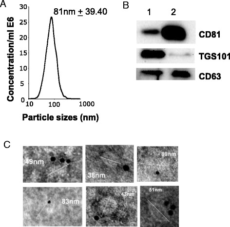
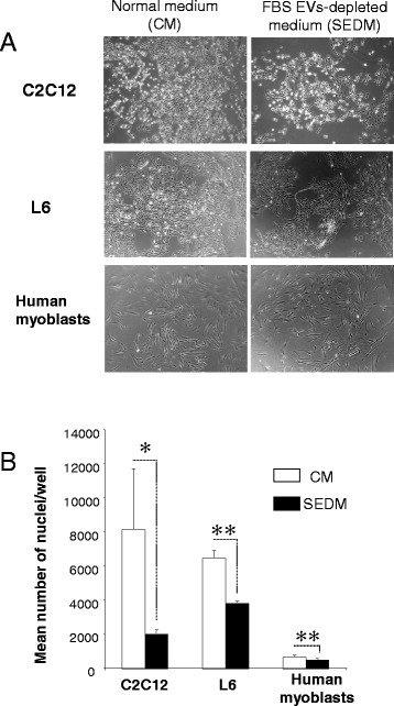
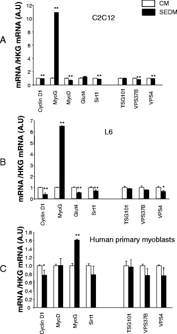
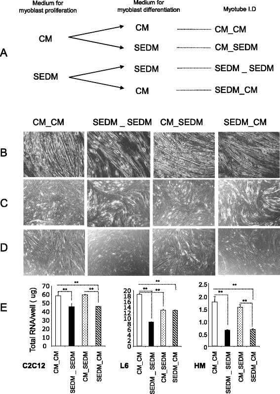
Background: Fetal bovine serum (FBS) contains a wide range of growth factors, hormones, vitamins, amino acids, fatty acids and trace elements required for cell growth. It was shown that animal sera contain also extracellular vesicles (EVs) with important biological properties; thus we wondered whether EVs present in FBS would influence muscle cell phenotype. EVs were removed from sera by ultracentrifugation (18 h). C2C12, L6 and human primary myoblasts, were grown either in classical media (CM) or in EVs-depleted media. Differentiation was induced by replacing the culture medium either with CM or EV-depleted media. qRT-PCR of relevant genes and miRNA involved in proliferation, differentiation, energy metabolism and EVs formation and secretion were performed.

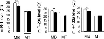
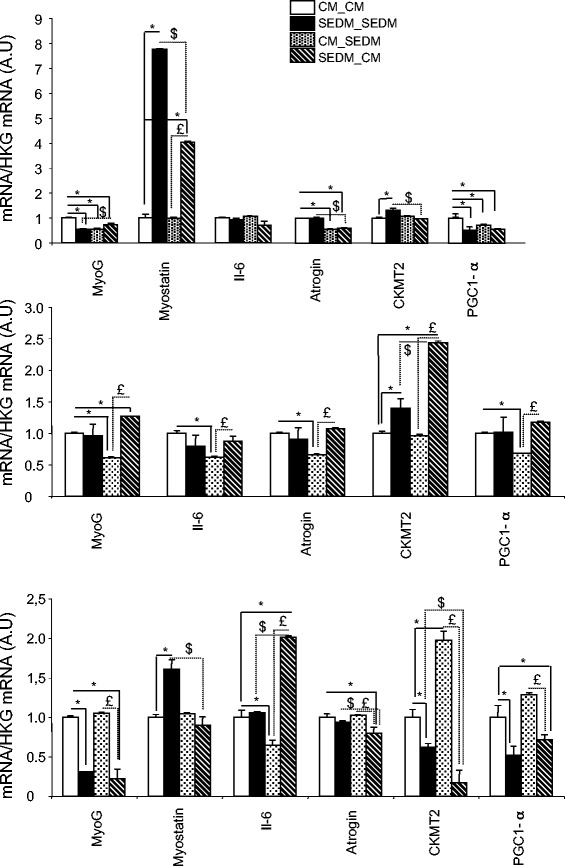
Results: Growth of myoblasts in EV-free media during proliferation produces the most unfavorable situation for proper myotube formation, when considering C212 and human myoblasts. Removing EVs from serum committed myoblasts to differentiate precociously (induction of myogenin and decreased expression of myomiR involved in myogenesis). C2C12 and human myoblasts, grown constantly in EV-depleted media during proliferation and differentiation, formed less myotubes than in CM. They had a reduced level of myogenin and a strong increase in myostatin expression, a negative regulator of muscle cell differentiation that affects myotube size. This situation was not reversed when confluent myoblasts were switched to CM for differentiation. Like C2C12 and human cells, L6 formed less myotubes in EVs-depleted media. However, as they do not express myostatin, L6 myotubes were larger and expressed higher level of CKTM2 compared to myotubes grown in CM suggesting that they had reached a higher level of differentiation.

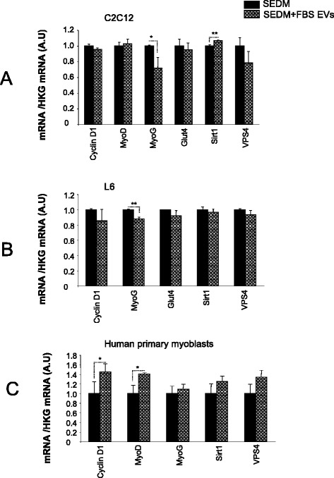
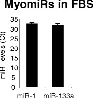
Conclusions: Researchers studying the role of muscle EVs in culture conditions should consider that depleting EVs from serum alters the phenotype of muscle cells. Interestingly, the cross-talk between myoblasts and myotubes during myogenesis (Forterre 2014, PLoS One. 2014 Jan 2;9(1):e84153) can be recapitulate by using FBS-EVs as well. This implies that EVs can transfer specific signals to cells from unrelated species and that part of serum EV composition is evolutionarily conserved (e.g.; myomiR are detected in FBS-EVs). EVs in body fluids could have an unsuspected function during embryogenesis and in regulation of cellular processes such as hypertrophy and hyperplasia.

Keywords: Bovine serum; C2C12; Extracellular vesicles; Gene expression; Human myoblasts; L6; Muscle cell; Proliferation; miRNAs.


Figures

Fig. 1
Fig. 1
Fetal bovine serum (FBS) contain exosome-like vesicles. FBS-derived vesicles were pellet at 100,000 g for 18 h, resuspended in PBS and re-pelleted at 100,000 g for 70 min. a FBS-EV size distributions measured with Nanosight. b Immuno-blotting for enriched exosomal proteins in FBS (1); C2C12 released exosomes were used as positive controls (2). TSG101 = Tumor susceptibility gene 101, CD81 = Cluster of Differentiation 81 (tetraspanin). c TEM images of purified FBS-EVs. Nanovesicles are labeled with anti-CD81 gold particles to confirm their exosomal origin
Fig. 2
Fig. 2
FBS-derived vesicles are involved in myoblast proliferation. a light-microscopy (x4) of representative wells from C2C12, L6 or human primary myoblasts grown in 6-well plates either in normal proliferative medium or EV-depleted proliferative medium. b Mean number of nuclei per well
Fig. 3
Fig. 3
Removing FBS-derived vesicles from bovine serum affects gene expression. Quantitative RT-PCR of relevant genes for proliferation (Cyclin D1, Sirt1), differentiation (MyoD, MyoG), metabolism (Glut4) and vesicle trafficking and EV release (TSG101, VPS37B, VPS4). Data are expressed as fold compared to CM, normalized by house keeping gene (HKG). In white, cells grown in control media containing FBS-EVs (CM), in black, cells grown in CM depleted of FBS-EVs (SEDM). a C2C12 myoblasts; b L6 rat myoblasts; c human primary myoblasts
Fig. 4
Fig. 4
Removing FBS-derived vesicles from bovine serum affects myomiR expression in C2C12. Quantification of miR-1, miR-133a and miR-206 in C2C12, grown either in CM (white) or in EVs-depleted proliferation media (black). Data are expressed as Ct values
Fig. 5
Fig. 5
Removing EVs from culture medium sera affect myoblast differentiation. a Workflow showing myoblast growth conditions. CM = control media containing FBS-EVs, SEDM = media depleted of FBS-EVs. b-c-d Light-microscopy (x4) of representative wells showing myotubes after 8 days of differentiation; B = C2C12, C = L6 and D = human myotubes. e Mean quantities of total RNA/well (NanoDrop quantification) from the 3 types of myotubes after 8 days of differentiation
Fig. 6
Fig. 6
Removing FBS-derived vesicles from bovine serum affects gene expression in myotubes. Quantitative RT-PCR of relevant genes for differentiation (MyoG, CKMT2), myotube size (myostatin, atrogin), metabolism (PGC1-α) or encoding secreted proteins (Il-6). Data are expressed as fold compared to CM_CM, normalized by house keeping gene (HKG). * = p < 0.01 all conditions vs CM_CM, $ = p < 0.01 SEDM_SEDM vs SEDM_CM. £ = p < 0.01 CM_SEDM vs SEDM_CM
Fig. 7
Fig. 7
Effect of FBS-EVs on myoblasts gene expression quantified by qRT-PCR. Data are expressed as fold compared to EV-depleted condition, normalized by house keeping gene (HKG). In black, cells grown in serum depleted of its EVs (SEDM), in grey, cells grown in SEDM supplemented with FBS EVs. a C2C12 myoblasts; b L6 rat myoblasts; c human primary myoblasts
Fig. 8
Fig. 8
FBS-EVs contain miRNA involved in myogenesis (myomiRs). Data are expressed as Ct values

Similar articles

Cited by

References

    1. Carrel A, Ebeling AH. Age and multiplication of fibroblasts. J Exp Med. 1921;34:599–623. doi: 10.1084/jem.34.6.599. - DOI - PMC - PubMed
    1. Treadwell PE, Ross JD. Characterization of human cells: variation in growth rate, volume, morphology and growth efficiency in media supplemented with human serum or bovine fetal serum. Exp Cell Res. 1963;29:356–79. doi: 10.1016/0014-4827(63)90390-2. - DOI - PubMed
    1. Kirikae T, Tamura H, Hashizume M, Kirikae F, Uemura Y, Tanaka S, Yokochi T, Nakano M. Endotoxin contamination in fetal bovine serum and its influence on tumor necrosis factor production by macrophage-like cells J774.1 cultured in the presence of the serum. Int J Immunopharmacol. 1997;19:255–62. doi: 10.1016/S0192-0561(97)00066-0. - DOI - PubMed
    1. Khodabukus A, Baar K. The effect of serum origin on tissue engineered skeletal muscle function. J Cell Biochem. 2014;115:2198–207. doi: 10.1002/jcb.24938. - DOI - PubMed
    1. Camussi G, Deregibus MC, Bruno S, Cantaluppi V, Biancone L. Exosomes/microvesicles as a mechanism of cell-to-cell communication. Kidney Int. 2010;78:838–48. doi: 10.1038/ki.2010.278. - DOI - PubMed

Publication types

LinkOut - more resources