. 2016 Apr 7;165(2):382-95.
doi: 10.1016/j.cell.2016.02.046. Epub 2016 Mar 31.

The Antagonistic Gene Paralogs Upf3a and Upf3b Govern Nonsense-Mediated RNA Decay

Affiliations

The Antagonistic Gene Paralogs Upf3a and Upf3b Govern Nonsense-Mediated RNA Decay

Eleen Y Shum et al. Cell. .

Abstract

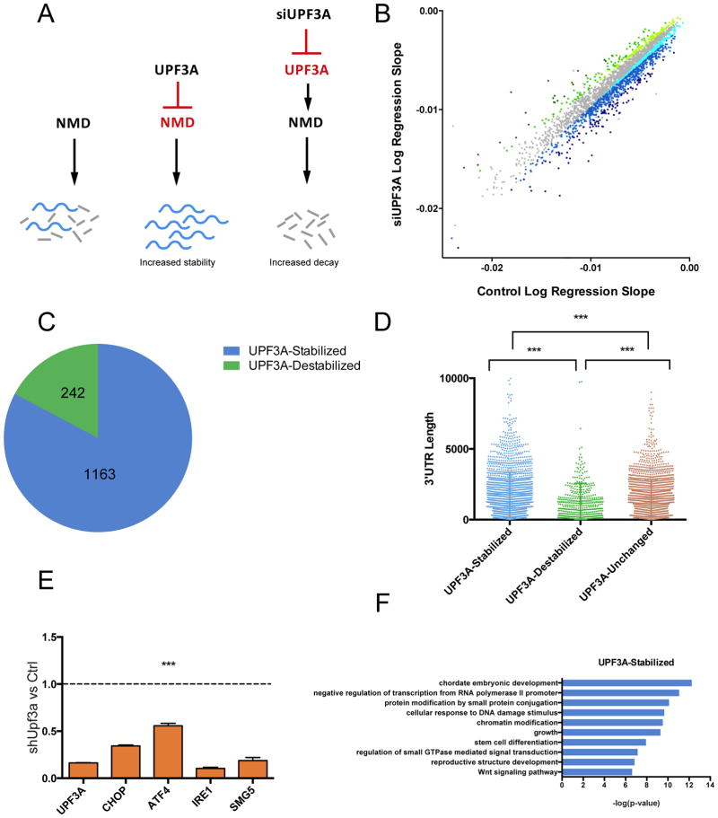
Gene duplication is a major evolutionary force driving adaptation and speciation, as it allows for the acquisition of new functions and can augment or diversify existing functions. Here, we report a gene duplication event that yielded another outcome--the generation of antagonistic functions. One product of this duplication event--UPF3B--is critical for the nonsense-mediated RNA decay (NMD) pathway, while its autosomal counterpart--UPF3A--encodes an enigmatic protein previously shown to have trace NMD activity. Using loss-of-function approaches in vitro and in vivo, we discovered that UPF3A acts primarily as a potent NMD inhibitor that stabilizes hundreds of transcripts. Evidence suggests that UPF3A acquired repressor activity through simple impairment of a critical domain, a rapid mechanism that may have been widely used in evolution. Mice conditionally lacking UPF3A exhibit "hyper" NMD and display defects in embryogenesis and gametogenesis. Our results support a model in which UPF3A serves as a molecular rheostat that directs developmental events.


Figures

Figure 1
Figure 1. UPF3A is a NMD inhibitor
(A) qPCR analysis of NMD substrates in mouse P19 cells transfected with a Upf3a shRNA or a negative control (Ctrl) shRNA construct, the latter of which was given a value of “1.” (B) NMD substrate half-lives determined by qPCR analysis. P19 cells were transfected with the constructs described in panel A, followed by ActD treatment for the indicated times. (C) Depletion of Upf3a leads to destabilization of a NMD reporter. P19 cells were transfected with the constructs described in panel A and analyzed by the NMD reporter described in the Materials and Methods. (D) qPCR analysis of NMD substrates in mEFs electroporated with either Upf3a shRNA (shUpf3a) or a control shRNA (Ctrl). (E) qPCR analysis of NMD substrate mRNAs in Upf3b-null mNSCs transfected as in panel A. Graphs are represented as mean and standard error (SEM) of replicates. *P<0.05; **P<0.01; ***P<0.001; ****P<0.0001
Figure 2
Figure 2. Genome-wide Half-Life Analysis of UPF3A-Regulated mRNAs
(A) Model: UPF3A stabilizes NMD target transcripts by inhibiting NMD. Depletion of UPF3A stimulates NMD. (B) Scatter plot of RNA half-life slope of UPF3A-depleted (UPF3A siRNA transfected) vs. Control (control siRNA transfected) P19 cells (R>0.7). UPF3A-stabilized and -destabilized transcripts are blue and green, respectively. Transcripts not exhibiting altered stability are gray. Darker shades of blue and green convey progressively increasing regulation. (C) Proportion of significantly UPF3A-stabilized and –destabilized transcripts (destabilized and stabilized, respectively, in response to UPF3A depletion (R>0.7) in the transfected cells described in panel B. (D) The distribution of 3′ UTR length in UPF3A-destabilized and -stabilized transcripts in the transfected cells described in panel B. ***P<0.001 (unpaired Student’s t-test). (E) qPCR analysis of NMD substrates in stably UPF3A-depleted (shUPF3A) HeLa cells vs Control (shLuc) HeLa cells. (F) GO analysis of functional categories overrepresented (p<0.05) in the UPF3A- stabilized mRNAs defined in panel C.
Figure 3
Figure 3. Evolution and Functional Analysis of UPF3A
(A) Two copies of upf3 appear to have emerged at the dawn of vertebrates. See Figure S3A for a phylogenetic tree showing the evolution of these paralogs. (B, F) Functional analysis of human UPF3A (hUPF3A). P19 cells were transiently transfected with shRNAs (mouse Upf3a [shUpf3a] or negative control [Ctrl] shRNAs) and the hUPF3A expression constructs (WT or the mutants depicted in panel E) or empty vector (EV). NMD magnitude was assessed using the NMD reporter described in Figure 1C. (C) qPCR analysis of NMD substrates in HeLa cells, comparing expression in cells transiently transfected with UPF3A expression vector or empty vector, the latter of which was set to “1.” (D) Model: UPF3B promotes NMD by bridging the EJC with UPF1 and UPF2. (E) Schematic of hUPF3A constructs used in panel F. Both of the mutant UPF3A proteins were previously shown to be expressed at levels similar to that of the wild-type UPF3A protein (Kunz et al., 2006). (G) Amino-acid similarity between mouse and human UPF3A/UPF3B. Values below 70% are shown in red. *P<0.05; ***P<0.001
Figure 4
Figure 4. Mechanism of UPF3A Action
(A) Model: UPF3A sequesters UPF2 away from the NMD machinery in order to silence NMD. (B) Schematic of different hUPF3A and hUPF3B constructs used in panels C and D. These UPF3 mutants were previously shown to be expressed at levels similar to that of the corresponding wild-type UPF3 proteins (Kunz et al., 2006). (C, D) A single amino acid in the EJC-interaction domain is critical for NMD repressor activity. Experiments were performed as in Figure 3B with the constructs shown in Figure 4B. (E) Schematic of an mRNA reporter with 6 MS-binding sites or a control reporter lacking the MS-binding sites (Ctrl). (F) Left: Northern blot analysis of HeLa cells co-transfected with the indicated expression vectors and siRNAs, along with both the reporters described in panel E. Right: Quantification of the mRNA reporter level were normalized to the levels of the Ctrl reporter mRNA in 3 independent experiments. (G) Evidence that UPF3A inhibits UPF1-EJC interactions. Left: Co-IP analysis of UPF1 with the EJC components, MLN51 and MAGOH, in UPF3A-depleted P19 cells (shUpf3a) or P19 cells transfected with a negative-control shRNA (Ctrl). Right: Quantification of MLN51- and MAGOH-UPF1 interactions in UPF3A-depleted cells relative to control cells (n=3). *P<0.05
Figure 5
Figure 5. Upf3a is Required for Early Embryogenesis
(A) Upf3a conditional knockout scheme and location of primers used for the detection of the floxed and knockout alleles at the Upf3a locus. (B) Genomic PCR analysis of tails from mice with the indicated genotypes. The data show successful insertion of the targeted allele harboring loxP sites. (C) Genotypes of the progeny from Upf3a+/− breeding pairs at the indicated embryonic and postnatal time points. (D) E3.5 embryos isolated from superovulated Upf3a+/− female mice bred with Upf3a+/− male mice. The embryos were manually flushed out of fallopian tubes of superovulated mice and the distribution of genotypes in different morphological groups is shown. (E–F) Immunofluorescence analysis of UPF3A (red) and nuclear LAMIN (green) expression in Panel E shows mouse blastocysts and Panel F shows a mouse 1-cell embryo with polar body. DAPI staining (blue) shows the position of nuclei. *P<0.05; ****P<0.0001
Figure 6
Figure 6. UPF3A Expression Pattern
(A–B) Expression pattern of Upf3a and Upf3b mRNA, as determined by qPCR analysis. Panel A shows adult mouse tissues and Panel B shows germ cell subsets. Values are relative to that in epididymis, which was given a value of “1.” (C) Immunohistochemical analysis of UPF3A and UPF3B protein expression in adult mouse testes. Pre-immune IgG serves as a negative control. L, leptotene spermatocyte; P, pachytene spermatocyte; S, spermatid. (D) H&E staining of Upf3a-cKO and littermate control testis at different postnatal time points. Most seminiferous tubules in the mutant had delayed spermatogenesis at P28, as evidenced by the presence of pachytene spermatocytes (P) and round spermatids (RS) near the lumen. Wild-type tubules typically have elongated spermatids (ES) at the lumen. Green arrows points to vacuoles, which were commonly present in mutant testes.
Figure 7
Figure 7. Loss of UPF3A in Male Germ Cells Elevates NMD and Perturbs Spermatogenesis
(A) qPCR analysis of markers representing different germ cell stages in 4-week-old Upf3a-cKO vs. control (Ctrl) littermate mice (n=2–3 per genotype). “1” represents expression level normalized to Ctrl level. (B) Spermatocytes per tubule, as quantified by γ-H2AX expression pattern in testis cross-sections (see text) from 4-week-old Upf3a-cKO vs. control littermate mice (n=2–3 per genotype). (C) FACS analysis of Hoescht 33342-stained cells from 4-week-old Upf3a-cKO vs. littermate control seminiferous tubules. The data are plotted as Hoescht-Blue (DNA content) versus Hoescht-Red (DNA condensation) and the numbers indicate percentage of gated live cells. Population A, leptotene spermatocytes; population B, zygotene-pachytene-diplotene spermatocytes; and population C, round and elongated spermatids. (D) FACS analysis of population C from panel C, showing strong deficit in both round spermatids (RS) and elongated spermatids (ES), which are distinguished by size (forward scatter [FSC]). (E–G) qPCR analysis of 4-week-old Upf3a-cKO vs. littermate control mice. Panel E shows dramatically reduced levels of spermatocyte markers (Stra8, Spo11, and Sypc3) in purified 4N spermatocytes from cKO mice. The 4N spermatocytes were purified as in panel C. Panel F shows expression of known NMD substrates in spermatocytes isolated as in panel E. Panel G shows expression of known NMD substrates in the olfactory epithelium of Krt5-Cre;Upf3afl/fl mice. n=2–3 per genotype for all 3 panels. “1” represents expression level normalized to Ctrl level. (H) Model: Cell A has high levels of UPF3B and consequently high NMD. Cell B has high levels of UPF3A and thus has low NMD, allowing accumulation of NMD substrates. *P<0.05; **P<0.01; ***P<0.001

Comment in

Similar articles

Cited by

References

    1. Bastos H, Lassalle B, Chicheportiche A, Riou L, Testart J, Allemand I, Fouchet P. Flow cytometric characterization of viable meiotic and postmeiotic cells by Hoechst 33342 in mouse spermatogenesis 2005 - PubMed
    1. Boelz S, Neu-Yilik G, Gehring NH, Hentze MW, Kulozik AE. A chemiluminescence-based reporter system to monitor nonsense-mediated mRNA decay 2006 - PubMed
    1. Bruno IG, Karam R, Huang L, Bhardwaj A, Lou CH, Shum EY, … Wilkinson MF. Identification of a microRNA that activates gene expression by repressing nonsense-mediated RNA decay 2011 - PMC - PubMed
    1. Buchwald G, Ebert J, Basquin C, Sauliere J, Jayachandran U, Bono F, Le Hir H, Conti E. Insights into the recruitment of the NMD machinery from the crystal structure of a core EJC-UPF3b complex 2010 - PMC - PubMed
    1. Chan W-K, Huang L, Gudikote JP, Chang YF, Imam JS, MacLean JA, 2nd, Wilkinson MF. An alternative branch of the nonsense-mediated decay pathway 2007 - PMC - PubMed

Publication types