

Altered cellular redox status, sirtuin abundance and clock gene expression in a mouse model of developmentally primed NASH
- PMID: 27040510
- PMCID: PMC4874946
- DOI: 10.1016/j.bbalip.2016.03.026
Altered cellular redox status, sirtuin abundance and clock gene expression in a mouse model of developmentally primed NASH
Abstract
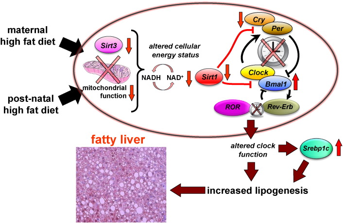
Background: We have previously shown that high fat (HF) feeding during pregnancy primes the development of non-alcoholic steatohepatits (NASH) in the adult offspring. However, the underlying mechanisms are unclear.
Aims: Since the endogenous molecular clock can regulate hepatic lipid metabolism, we investigated whether exposure to a HF diet during development could alter hepatic clock gene expression and contribute to NASH onset in later life.
Methods: Female mice were fed either a control (C, 7%kcal fat) or HF (45%kcal fat) diet. Offspring were fed either a C or HF diet resulting in four offspring groups: C/C, C/HF, HF/C and HF/HF. NAFLD progression, cellular redox status, sirtuin expression (Sirt1, Sirt3), and the expression of core clock genes (Clock, Bmal1, Per2, Cry2) and clock-controlled genes involved in lipid metabolism (Rev-Erbα, Rev-Erbβ, RORα, and Srebp1c) were measured in offspring livers.
Results: Offspring fed a HF diet developed NAFLD. However HF fed offspring of mothers fed a HF diet developed NASH, coupled with significantly reduced NAD(+)/NADH (p<0.05, HF/HF vs C/C), Sirt1 (p<0.001, HF/HF vs C/C), Sirt3 (p<0.01, HF/HF vs C/C), perturbed clock gene expression, and elevated expression of genes involved lipid metabolism, such as Srebp1c (p<0.05, C/HF and HF/HF vs C/C).
Conclusion: Our results suggest that exposure to excess dietary fat during early and post-natal life increases the susceptibility to develop NASH in adulthood, involving altered cellular redox status, reduced sirtuin abundance, and desynchronized clock gene expression.
Keywords: Aging; Circadian; Development; Fatty liver; High fat; Maternal diet.
Copyright © 2016 The Authors. Published by Elsevier B.V. All rights reserved.
Figures


Similar articles
-
Circadian Rhythm Alteration of the Core Clock Genes and the Lipid Metabolism Genes Induced by High-Fat Diet (HFD) in the Liver Tissue of the Chinese Soft-Shelled Turtle (Trionyx sinensis).Genes (Basel). 2024 Jan 25;15(2):157. doi: 10.3390/genes15020157. Genes (Basel). 2024. PMID: 38397147 Free PMC article.
-
Effect of H₂S on the circadian rhythm of mouse hepatocytes.Lipids Health Dis. 2012 Feb 8;11:23. doi: 10.1186/1476-511X-11-23. Lipids Health Dis. 2012. PMID: 22316301 Free PMC article.
-
A Slow-Digesting Carbohydrate Diet during Rat Pregnancy Protects Offspring from Non-Alcoholic Fatty Liver Disease Risk through the Modulation of the Carbohydrate-Response Element and Sterol Regulatory Element Binding Proteins.Nutrients. 2019 Apr 14;11(4):844. doi: 10.3390/nu11040844. Nutrients. 2019. PMID: 31013988 Free PMC article.
-
Dissecting the Rev-erbα Cistrome and the Mechanisms Controlling Circadian Transcription in Liver.Cold Spring Harb Symp Quant Biol. 2015;80:233-8. doi: 10.1101/sqb.2015.80.027508. Epub 2015 Sep 14. Cold Spring Harb Symp Quant Biol. 2015. PMID: 26370410 Review.
-
The Clock-NAD+ -Sirtuin connection in nonalcoholic fatty liver disease.J Cell Physiol. 2022 Aug;237(8):3164-3180. doi: 10.1002/jcp.30772. Epub 2022 May 26. J Cell Physiol. 2022. PMID: 35616339 Review.
Cited by
-
Effects of maternal branched-chain amino acid and alanine supplementation on growth and biomarkers of protein metabolism in dams fed a low-protein diet and their offspring.Amino Acids. 2022 Jul;54(7):977-988. doi: 10.1007/s00726-022-03157-1. Epub 2022 Mar 30. Amino Acids. 2022. PMID: 35353249
-
Interaction between stress responses and circadian metabolism in metabolic disease.Liver Res. 2017 Sep;1(3):156-162. doi: 10.1016/j.livres.2017.11.002. Liver Res. 2017. PMID: 29430321 Free PMC article.
-
Influence of mental stress and environmental toxins on circadian clocks: Implications for redox regulation of the heart and cardioprotection.Br J Pharmacol. 2020 Dec;177(23):5393-5412. doi: 10.1111/bph.14949. Epub 2020 Feb 4. Br J Pharmacol. 2020. PMID: 31833063 Free PMC article. Review.
-
Redox Regulatory Changes of Circadian Rhythm by the Environmental Risk Factors Traffic Noise and Air Pollution.Antioxid Redox Signal. 2022 Oct;37(10-12):679-703. doi: 10.1089/ars.2021.0272. Epub 2022 Apr 18. Antioxid Redox Signal. 2022. PMID: 35088601 Free PMC article. Review.
-
SIRT1-dependent mechanisms and effects of resveratrol for amelioration of muscle wasting in NASH mice.BMJ Open Gastroenterol. 2020 May;7(1):e000381. doi: 10.1136/bmjgast-2020-000381. BMJ Open Gastroenterol. 2020. PMID: 32371503 Free PMC article.
References
-
- Adams L.A., Angulo P. Recent concepts in non-alcoholic fatty liver disease. Diabet. Med. 2005;22:1129–1133. - PubMed
-
- Ozcan U., Cao Q., Yilmaz E., Lee A.H., Iwakoshi N.N., Ozdelen E., Tuncman G., Gorgun C., Glimcher L.H., Hotamisligil G.S. Endoplasmic reticulum stress links obesity, insulin action, and type 2 diabetes. Science. 2004;306:457–461. - PubMed
-
- Shneider B.L., Gonzalez-Peralta R., Roberts E.A. Controversies in the management of pediatric liver disease: hepatitis B, C and NAFLD: summary of a single topic conference. Hepatology. 2006;44:1344–1354. - PubMed
Publication types
MeSH terms
Substances
Grants and funding
LinkOut - more resources
Full Text Sources
Other Literature Sources
Medical
Molecular Biology Databases
Research Materials
Miscellaneous
