. 2016 Jul;1858(7 Pt A):1436-42.
doi: 10.1016/j.bbamem.2016.03.027. Epub 2016 Mar 31.

Effect of the achondroplasia mutation on FGFR3 dimerization and FGFR3 structural response to fgf1 and fgf2: A quantitative FRET study in osmotically derived plasma membrane vesicles

Affiliations

Effect of the achondroplasia mutation on FGFR3 dimerization and FGFR3 structural response to fgf1 and fgf2: A quantitative FRET study in osmotically derived plasma membrane vesicles

Sarvenaz Sarabipour et al. Biochim Biophys Acta. 2016 Jul.

Abstract

The G380R mutation in the transmembrane domain of FGFR3 is a germline mutation responsible for most cases of Achondroplasia, a common form of human dwarfism. Here we use quantitative Fӧster Resonance Energy Transfer (FRET) and osmotically derived plasma membrane vesicles to study the effect of the achondroplasia mutation on the early stages of FGFR3 signaling in response to the ligands fgf1 and fgf2. Using a methodology that allows us to capture structural changes on the cytoplasmic side of the membrane in response to ligand binding to the extracellular domain of FGFR3, we observe no measurable effects of the G380R mutation on FGFR3 ligand-bound dimer configurations. Instead, the most notable effect of the achondroplasia mutation is increased propensity for FGFR3 dimerization in the absence of ligand. This work reveals new information about the molecular events that underlie the achondroplasia phenotype, and highlights differences in FGFR3 activation due to different single amino-acid pathogenic mutations.

Keywords: Achondroplasia; Dimer stability; Dimerization; Fibroblast growth factor receptor 3; Receptor tyrosine kinases; skeletal disorders.


Figures

Fig. 1
Fig. 1
A vesicle, co-expressing FGFR3 G380R EC + TM-YFP and FGFR3 G380R EC + TM-mCherry, imaged and analyzed in the FRET, acceptor, and donor channels. Images were acquired with a Nikon laser scanning confocal microscope. The images are analyzed with a Matlab code that has been discussed in detail in a previous publication [3]. The intensity across the membrane (open blue symbols) is fit to a Gaussian (solid line) after background correction [30]. The residual from the fit is also shown. (For interpretation of the references to color in this figure legend, the reader is referred to the web version of this article.)
Fig. 2
Fig. 2
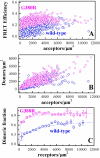
(A) FRET data for FGFR3 G380R EC + TM construct (magenta circles) are compared to previously published data for the wild-type (blue diamonds) [22]. The FRET efficiency is shown as a function of the acceptor concentration. Each data point represents a single vesicle. (B) Donor versus acceptor concentrations in each vesicle. (C) Dimeric fraction as a function of total receptor concentration, calculated as described in Supplemental Information. The solid lines are the best fits to the data. (For interpretation of the references to color in this figure legend, the reader is referred to the web version of this article.)
Fig. 3
Fig. 3
FRET efficiencies, donor concentrations and acceptor concentrations, measured for the G380R EC + TM FGFR3, in the presence of 5 μg/ml fgf1 (black) or fgf2 (olive). For comparison, we also show the G380R EC + TM FGFR3 data in the absence of ligand (open magenta circles). Each data point represents a single vesicle. In each case, 300 to 500 individual plasma membrane derived vesicles were imaged 1 hour after adding the ligand. FRET does not depend on the receptor concentration, as expected in the case of saturating ligand. The Intrinsic FRET for each vesicle was determined according to equation (11) in Supplementary Information. (For interpretation of the references to color in this figure legend, the reader is referred to the web version of this article.)
Fig. 4
Fig. 4
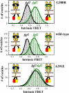
Histograms of measured Intrinsic FRET in single vesicles for the FGFR3 G380 EC + TM construct (top panel), compared to published data for the wild-type (middle panel) and the A391E mutant (bottom panel) [22]. Also shown is a graphical representation of the changes in the average distance between the fluorescent proteins, based on changes in Intrinsic FRET (not to scale).

Similar articles

Cited by

References

    1. Horton WA, Hall JG, Hecht JT. Achondroplasia. Lancet. 2007;370:162–172. - PubMed
    1. Foldynova-Trantirkova S, Wilcox WR, Krejci P. Sixteen Years and Counting: the current understanding of fibroblast growth factor receptor 3 (FGFR3) signaling in skeletal dysplasias. Hum. Mutat. 2012;33:29–41. - PMC - PubMed
    1. Shiang R, Thompson LM, Zhu Y-Z, Church DM, Fielder TJ, Bocian M, Winokur ST, Wasmuth JJ. Mutations in the transmembrane domain of FGFR3 cause the most common genetic form of dwarfism, achondroplasia. Cell. 1994;78:335–342. - PubMed
    1. Deng C, Wynshaw-Boris A, Zhou F, Kuo A, Leder P. Fibroblast growth factor receptor 3 is a negative regulator of bone growth. Cell. 1996;84:911–921. - PubMed
    1. Cohen MM. Some chondrodysplasias with short limbs: molecular perspectives. Am. J. Med. Genet. 2002;112:304–313. - PubMed

Publication types

MeSH terms