. 2016 Apr 7;98(4):772-81.
doi: 10.1016/j.ajhg.2016.01.016. Epub 2016 Mar 31.

Recessive Inactivating Mutations in TBCK, Encoding a Rab GTPase-Activating Protein, Cause Severe Infantile Syndromic Encephalopathy

Affiliations

Recessive Inactivating Mutations in TBCK, Encoding a Rab GTPase-Activating Protein, Cause Severe Infantile Syndromic Encephalopathy

Jessica X Chong et al. Am J Hum Genet. .

Abstract

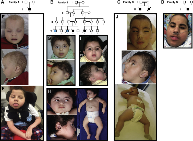
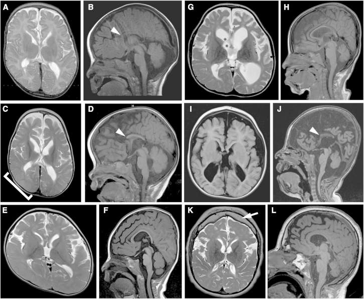
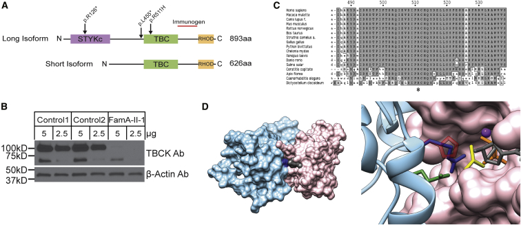
Infantile encephalopathies are a group of clinically and biologically heterogeneous disorders for which the genetic basis remains largely unknown. Here, we report a syndromic neonatal encephalopathy characterized by profound developmental disability, severe hypotonia, seizures, diminished respiratory drive requiring mechanical ventilation, brain atrophy, dysgenesis of the corpus callosum, cerebellar vermis hypoplasia, and facial dysmorphism. Biallelic inactivating mutations in TBCK (TBC1-domain-containing kinase) were independently identified by whole-exome sequencing as the cause of this condition in four unrelated families. Matching these families was facilitated by the sharing of phenotypic profiles and WES data in a recently released web-based tool (Geno2MP) that links phenotypic information to rare variants in families with Mendelian traits. TBCK is a putative GTPase-activating protein (GAP) for small GTPases of the Rab family and has been shown to control cell growth and proliferation, actin-cytoskeleton dynamics, and mTOR signaling. Two of the three mutations (c.376C>T [p.Arg126(∗)] and c.1363A>T [p.Lys455(∗)]) are predicted to truncate the protein, and loss of the major TBCK isoform was confirmed in primary fibroblasts from one affected individual. The third mutation, c.1532G>A (p.Arg511His), alters a conserved residue within the TBC1 domain. Structural analysis implicated Arg511 as a required residue for Rab-GAP function, and in silico homology modeling predicted impaired GAP function in the corresponding mutant. These results suggest that loss of Rab-GAP activity is the underlying mechanism of disease. In contrast to other disorders caused by dysregulated mTOR signaling associated with focal or global brain overgrowth, impaired TBCK function results in progressive loss of brain volume.

Keywords: GTPase-activating protein; Mendelian disease; exome sequencing; infantile encephalopathy; mTOR signaling.


Figures

Figure 1
Figure 1
Pedigrees and Pictures of Individuals with TBCK-Related Encephalopathy (A–D) Pedigrees for families A–D (A–D, respectively), in whom loss-of-function mutations in TBCK segregate with disease. Solid black fill indicates individuals affected by TBCK-related encephalopathy. Light blue fill in family B indicates individuals affected by a hematologic phenotype without neurodevelopmental features; neither is homozygous for p.Lys455. WES was performed on individuals marked with asterisks. (E–J) Images show the similar facial features and hypotonia in individual A-II-1 at 25 months (E) and 13 years (F), individual B-IV-4 at 17 months (G) and 4 years, 3 months (H), individual B-IV-6 at 18 months (I), individual C-II-1 at 21 months (J), and individual D-II-1 at 14 years. See Table 1 for detailed clinical information on each affected individual and Figure 2 and Table S3 for imaging information.
Figure 2
Figure 2
Brain-Imaging Features in Individuals with TBCK-Related Encephalopathy (A–D) Axial T2-weighted (A and C) and sagittal T1-weighted images (B and D) show progressive loss of gray- and white-matter volume (most severe in the frontal lobes), demonstrated by increasing ventriculomegaly and extra-axial spaces (cortical and cerebellar) in individual A-II-1 between 22 days of age (A and B) and 29 months of age (C and D). Also present are swelling of the right parieto-occipital lobe, presumably due to a metabolic stroke (bracket in C), a diffusely thin corpus callosum with absent rostrum (arrowheads in B and D), and mild cerebellar vermis hypoplasia with a relatively spared brainstem. (E and F) Axial T2-weighted (E) and sagittal T1-weighted (F) images show ventriculomegaly, prominent extra-axial spaces, a diffusely thin but complete corpus callosum, and mild cerebellar vermis hypoplasia in individual B-IV-4 at 18 months of age. Right plagiocephaly is also present. (G and H) Axial T2-weighted (G) and sagittal T1-weighted (H) images show marked ventriculomegaly, prominent extra-axial spaces, a diffusely thin but complete corpus callosum, and mild cerebellar vermis hypoplasia in individual B-IV-6 at 6 years of age. Synechiae are also present in the right frontal horn (asterisks in G). (I and J) Axial T1-weighted (I) and sagittal T1-weighted (J) images show ventriculomegaly, prominent extra-axial spaces, a diffusely thin corpus callosum with an absent rostrum and anterior body (arrowhead in J), and mild cerebellar vermis hypoplasia in individual C-II-1 at 21 months of age. (K and L) Axial T2-weighted (K) and sagittal T1-weighted (L) images show mild ventriculomegaly, prominent extra-axial spaces, a diffusely thin corpus callosum, mild cerebellar vermis hypoplasia with a relatively preserved brainstem, and a thick frontal bone (arrow in K) in individual D-II-1 at 14 years of age.
Figure 3
Figure 3
Biochemical and Structural Characterization of TBCK Mutations (A) TBCK encodes two isoforms containing a TBC1 domain (TBC) flanked by a rhodanese domain (RHOD) at the C terminus. The long isoform also contains a pseudokinase domain (STYKc) at the N terminus. The location of the identified variants is reported. (B) Western blot shows that TBCK amounts were dramatically lower in a cell line from individual A-II-1 than in two control cell lines. The TBCK monoclonal antibody (1/250, Sigma HPA039951) recognizes a C-terminal fragment of the protein (depicted as a red line in A). After stripping, β-actin (1/5,000, Sigma A5441) was used as a loading control. The predicted sizes of the TBCK long and short isoforms are 101 and 71 kDa, respectively. (C) Conservation of the catalytic arginine “finger” (Arg511 in TBCK) in TBCK orthologs. (D) Homology model of the TBCK TBC1 domain complexed with the Rab33 GTPase. In the overall structure of the complex (left), both proteins are shown in a surface representation; Rab is colored pink, GDP is gray, the TBC1 domain is light blue, and Arg511 is dark blue. In the enlarged view of the active site of the GTPase (right), GDP is reported in gray sticks, and the phosphates are colored orange. AlF3, which in the crystal structure mimics the transition state for GTP hydrolysis, is shown in yellow, and the Mg atom is shown as a purple sphere. The TBC1 domain is reported in ribbon representation, and the side chains of key residues are shown as sticks. The two catalytic “fingers” Arg511 (blue) and Gln546 (green) are shown together with the disease-associated His511, shown in semi-transparent red.

Similar articles

Cited by

References

    1. Lalani S.R., Zhang J., Schaaf C.P., Brown C.W., Magoulas P., Tsai A.C.-H., El-Gharbawy A., Wierenga K.J., Bartholomew D., Fong C.-T. Mutations in PURA cause profound neonatal hypotonia, seizures, and encephalopathy in 5q31.3 microdeletion syndrome. Am. J. Hum. Genet. 2014;95:579–583. - PMC - PubMed
    1. Hunt D., Leventer R.J., Simons C., Taft R., Swoboda K.J., Gawne-Cain M., Magee A.C., Turnpenny P.D., Baralle D., DDD study Whole exome sequencing in family trios reveals de novo mutations in PURA as a cause of severe neurodevelopmental delay and learning disability. J. Med. Genet. 2014;51:806–813. - PMC - PubMed
    1. Perez Y., Kadir R., Volodarsky M., Noyman I., Flusser H., Shorer Z., Gradstein L., Birnbaum R.Y., Birk O.S. UNC80 mutation causes a syndrome of hypotonia, severe intellectual disability, dyskinesia and dysmorphism, similar to that caused by mutations in its interacting cation channel NALCN. J. Med. Genet. 2015 - PubMed
    1. Stray-Pedersen A., Cobben J.-M., Prescott T.E., Lee S., Cang C., Aranda K., Ahmed S., Alders M., Gerstner T., Aslaksen K., Care4Rare Canada Consortium. Baylor-Hopkins Center for Mendelian Genomics Biallelic Mutations in UNC80 Cause Persistent Hypotonia, Encephalopathy, Growth Retardation, and Severe Intellectual Disability. Am. J. Hum. Genet. 2016;98:202–209. - PMC - PubMed
    1. Shamseldin H.E., Faqeih E., Alasmari A., Zaki M.S., Gleeson J.G., Alkuraya F.S. Mutations in UNC80, Encoding Part of the UNC79-UNC80-NALCN Channel Complex, Cause Autosomal-Recessive Severe Infantile Encephalopathy. Am. J. Hum. Genet. 2016;98:210–215. - PMC - PubMed

Publication types

MeSH terms

Substances