Review
. 2016 Jun;79(6):463-79.
doi: 10.1002/jemt.22648. Epub 2016 Apr 4.

Advanced light microscopy core facilities: Balancing service, science and career

Affiliations
Review

Advanced light microscopy core facilities: Balancing service, science and career

Elisa Ferrando-May et al. Microsc Res Tech. 2016 Jun.

Abstract

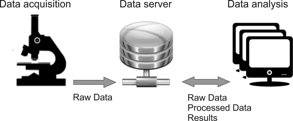
Core Facilities (CF) for advanced light microscopy (ALM) have become indispensable support units for research in the life sciences. Their organizational structure and technical characteristics are quite diverse, although the tasks they pursue and the services they offer are similar. Therefore, throughout Europe, scientists from ALM-CFs are forming networks to promote interactions and discuss best practice models. Here, we present recommendations for ALM-CF operations elaborated by the workgroups of the German network of ALM-CFs, German Bio-Imaging (GerBI). We address technical aspects of CF planning and instrument maintainance, give advice on the organization and management of an ALM-CF, propose a scheme for the training of CF users, and provide an overview of current resources for image processing and analysis. Further, we elaborate on the new challenges and opportunities for professional development and careers created by CFs. While some information specifically refers to the German academic system, most of the content of this article is of general interest for CFs in the life sciences. Microsc. Res. Tech. 79:463-479, 2016. © 2016 THE AUTHORS MICROSCOPY RESEARCH AND TECHNIQUE PUBLISHED BY WILEY PERIODICALS, INC.

Keywords: core facility administration; instrument performance tests; microscopy room requirements; user and staff training; user/staff/instrument ratio.


Figures

Figure 1
Figure 1
Development and current composition of the GerBI network. (A) Number of registrations per year. CF: ALM‐core facility, RG: microscopy research group. (B) Composition of GerBI: canonical core facilities (orange, 40), research groups (light orange, 11), and sites operating as both research group and facility (yellow, 9). The outer segment indicates the total number of users of registered CFs per year (status as of January 2016). [Color figure can be viewed in the online issue, which is available at wileyonlinelibrary.com.]
Figure 2
Figure 2
Characteristics of a representative group of German imaging CFs. (A) The graph displays the number of facility users per year (y‐axis) vs. the number of supporting staff (x‐axis). Each circle represents one CF. The size of the circle depends on the number of instruments, which is shown inside. The circle sectors indicate the proportion of high vs. medium vs. low level systems. The systems were defined as follows: High: Superresolution microscopy, fluorescence correlation imaging, MP and nonlinear imaging, light sheet imaging, laser capture microdissection; Medium: Point scanning and spinning disk confocal, total internal reflection microscopy, fluorescence resonance energy transfer; Low: Wide‐field, deconvolution, and stereo microscopes. The dashed line indicates the median user/staff ratio. (B) Enlarged view of the inset shown in (A). [Color figure can be viewed in the online issue, which is available at wileyonlinelibrary.com.]
Figure 3
Figure 3
User to staff ratio in German ALM‐CFs. Each green bar represents one facility. The blue line indicates the median. [Color figure can be viewed in the online issue, which is available at wileyonlinelibrary.com.]
Figure 4
Figure 4
Experiment pipeline of a standard approach within quantitative biology. The figure depicts the information flow from the (i) biological approach, (ii) data acquisition, (iii) image analysis up to (iv) data exploration including the interpretation of the underlying biological process.
Figure 5
Figure 5
Image raw data is written and stored on a centralized data server, ideally featuring an integrated image/data repository and preferably a data base, respectively. The server ensures user and data management and external access for data analysis workstations.

References

    1. Allan C, Burel JM, Moore J, Blackburn C, Linkert M, Loynton S, Macdonald D, Moore WJ, Neves C, Patterson A, Porter M, Tarkowska A, Loranger B, Avondo J, Lagerstedt I, Lianas L, Leo S, Hands K, Hay RT, Patwardhan A, Best C, Kleywegt GJ, Zanetti G, Swedlow JR. 2012. OMERO: Flexible, model‐driven data management for experimental biology. Nat Methods 9:245–253. - PMC - PubMed
    1. Anderson KI, Sanderson J, Peychl J. 2007. Design and Function of a Light‐Microscopy Facility. In: S.L. Shorte and F. Frischknecht, editors. Imaging Cellular and Molecular Biological Functions. Springer. pp. 93–113.
    1. Carpenter AE, Jones TR, Lamprecht MR, Clarke C, Kang IH, Friman O, Guertin DA, Chang JH, Lindquist RA, Moffat J, Golland P, Sabatini DM. 2006. CellProfiler: Image analysis software for identifying and quantifying cell phenotypes. Genome Biol 7:R100. - PMC - PubMed
    1. Carpenter AE, Kamentsky L, Eliceiri KW. 2012. A call for bioimaging software usability. Nat Methods 9:666–670. - PMC - PubMed
    1. Cole RW, Jinadasa T, Brown CM. 2011. Measuring and interpreting point spread functions to determine confocal microscope resolution and ensure quality control. Nat Protoc 6:1929–1941. - PubMed

LinkOut - more resources