RepA-WH1 prionoid: Clues from bacteria on factors governing phase transitions in amyloidogenesis
- PMID: 27040981
- PMCID: PMC4981189
- DOI: 10.1080/19336896.2015.1129479
RepA-WH1 prionoid: Clues from bacteria on factors governing phase transitions in amyloidogenesis
Abstract
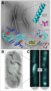
In bacterial plasmids, Rep proteins initiate DNA replication by undergoing a structural transformation coupled to dimer dissociation. Amyloidogenesis of the 'winged-helix' N-terminal domain of RepA (WH1) is triggered in vitro upon binding to plasmid-specific DNA sequences, and occurs at the bacterial nucleoid in vivo. Amyloid fibers are made of distorted RepA-WH1 monomers that assemble as single or double intertwined tubular protofilaments. RepA-WH1 causes in E. coli an amyloid proteinopathy, which is transmissible from mother to daughter cells, but not infectious, and enables conformational imprinting in vitro and in vivo; i.e. RepA-WH1 is a 'prionoid'. Microfluidics allow the assessment of the intracellular dynamics of RepA-WH1: bacterial lineages maintain two types (strains-like) of RepA-WH1 amyloids, either multiple compact cytotoxic particles or a single aggregate with the appearance of a fluidized hydrogel that it is mildly detrimental to growth. The Hsp70 chaperone DnaK governs the phase transition between both types of RepA-WH1 aggregates in vivo, thus modulating the vertical propagation of the prionoid. Engineering chimeras between the Sup35p/[PSI(+)] prion and RepA-WH1 generates [REP-PSI(+)], a synthetic prion exhibiting strong and weak phenotypic variants in yeast. These recent findings on a synthetic, self-contained bacterial prionoid illuminate central issues of protein amyloidogenesis.
Keywords: Hsp70 chaperone; RepA-WH1; amyloid polymorphism; amyloid proteinopathy; bacterial prionoid; phase transitions.
Figures

Comment on
- doi: 10.1016/j.str.2014.11.007
- doi: 10.1038/srep14669
- doi: 10.1111/mmi.12518
- doi: 10.3389/fmicb.2015.00311
Similar articles
-
Amyloidogenesis of bacterial prionoid RepA-WH1 recapitulates dimer to monomer transitions of RepA in DNA replication initiation.Structure. 2015 Jan 6;23(1):183-189. doi: 10.1016/j.str.2014.11.007. Epub 2014 Dec 24. Structure. 2015. PMID: 25543255
-
Pre-amyloid oligomers of the proteotoxic RepA-WH1 prionoid assemble at the bacterial nucleoid.Sci Rep. 2015 Oct 1;5:14669. doi: 10.1038/srep14669. Sci Rep. 2015. PMID: 26423724 Free PMC article.
-
Addressing Intracellular Amyloidosis in Bacteria with RepA-WH1, a Prion-Like Protein.Methods Mol Biol. 2018;1779:289-312. doi: 10.1007/978-1-4939-7816-8_18. Methods Mol Biol. 2018. PMID: 29886540
-
SynBio and the Boundaries between Functional and Pathogenic RepA-WH1 Bacterial Amyloids.mSystems. 2020 Jun 30;5(3):e00553-20. doi: 10.1128/mSystems.00553-20. mSystems. 2020. PMID: 32606029 Free PMC article. Review.
-
Twenty years of the pPS10 replicon: insights on the molecular mechanism for the activation of DNA replication in iteron-containing bacterial plasmids.Plasmid. 2004 Sep;52(2):69-83. doi: 10.1016/j.plasmid.2004.06.002. Plasmid. 2004. PMID: 15336485 Review.
Cited by
-
Structural analysis of a replication protein encoded by a plasmid isolated from a multiple sclerosis patient.Acta Crystallogr D Struct Biol. 2019 May 1;75(Pt 5):498-504. doi: 10.1107/S2059798319003991. Epub 2019 Apr 29. Acta Crystallogr D Struct Biol. 2019. PMID: 31063152 Free PMC article.
-
Enabling stop codon read-through translation in bacteria as a probe for amyloid aggregation.Sci Rep. 2017 Sep 19;7(1):11908. doi: 10.1038/s41598-017-12174-0. Sci Rep. 2017. PMID: 28928456 Free PMC article.
-
Membraneless organelles formed by liquid-liquid phase separation increase bacterial fitness.Sci Adv. 2021 Oct 22;7(43):eabh2929. doi: 10.1126/sciadv.abh2929. Epub 2021 Oct 20. Sci Adv. 2021. PMID: 34669478 Free PMC article.
-
Outlining Core Pathways of Amyloid Toxicity in Bacteria with the RepA-WH1 Prionoid.Front Microbiol. 2017 Apr 4;8:539. doi: 10.3389/fmicb.2017.00539. eCollection 2017. Front Microbiol. 2017. PMID: 28421043 Free PMC article.
References
-
- Otzen D. Functional amyloid: Turning swords into plowshares. Prion 2010; 4:256-64; PMID:20935497; http://dx.doi.org/10.4161/pri.4.4.13676 - DOI - PMC - PubMed
-
- Romero D, Kolter R. Functional amyloids in bacteria. Int Microbiol 2014; 17:65-73; PMID:26418850 - PubMed
-
- DePas WH, Chapman MR. Microbial manipulation of the amyloid fold. Res Microbiol 2012; 163:592-606; PMID:23108148; http://dx.doi.org/10.1016/j.resmic.2012.10.009 - DOI - PMC - PubMed
-
- Bieler S, Estrada L, Lagos R, Baeza M, Castilla J, Soto C. Amyloid formation modulates the biological activity of a bacterial protein, J Biol Chem 2005; 280:26880-5; PMID:15917245; http://dx.doi.org/10.1074/jbc.M502031200 - DOI - PubMed
-
- Liebman SW, Chernoff YO. Prions in yeast. Genetics 2012; 191:1041-72; PMID:22879407; http://dx.doi.org/10.1534/genetics.111.137760 - DOI - PMC - PubMed
Publication types
MeSH terms
Substances
LinkOut - more resources
Full Text Sources
Other Literature Sources
Molecular Biology Databases
