. 2016 Apr 20;90(2):388-99.
doi: 10.1016/j.neuron.2016.02.038. Epub 2016 Mar 31.

Morphological Substrates for Parallel Streams of Corticogeniculate Feedback Originating in Both V1 and V2 of the Macaque Monkey

Affiliations

Morphological Substrates for Parallel Streams of Corticogeniculate Feedback Originating in Both V1 and V2 of the Macaque Monkey

Farran Briggs et al. Neuron. .

Abstract

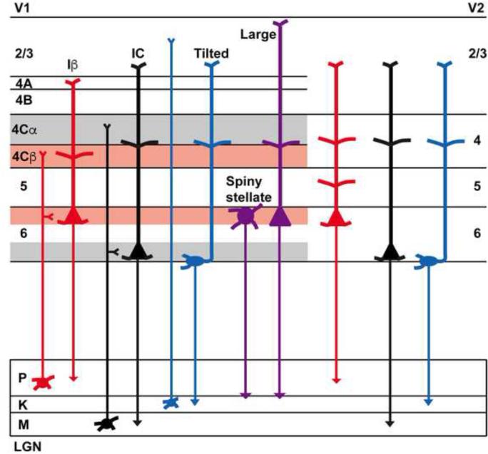
Corticothalamic circuits are essential for reciprocal information exchange between the thalamus and cerebral cortex. Nevertheless, the role of corticothalamic circuits in sensory processing remains a mystery. In the visual system, afferents from retina to the lateral geniculate nucleus (LGN) and from LGN to primary visual cortex (V1) are organized into functionally distinct parallel processing streams. Physiological evidence suggests corticogeniculate feedback may be organized into parallel streams; however, little is known about the diversity of corticogeniculate neurons, their local computations, or the structure-function relationship among corticogeniculate neurons. We used a virus-mediated approach to label and reconstruct the complete dendritic and local axonal arbors of identified corticogeniculate neurons in the macaque monkey. Our results reveal morphological substrates for parallel streams of corticogeniculate feedback based on distinct classes of neurons in V1 and V2. These results support the hypothesis that distinct populations of feedback neurons provide independent and unique information to the LGN.


Figures

Figure 1
Figure 1
Injection sites and expression of SADΔG-EGFP in CG neurons. A. Photographs of injections of virus in LGN in two monkeys indicating approximately the largest extent of each injection (see Supplemental Figure 1A for 3-D renderings of complete injection sites). Scale bars are 500 microns. B. Photograph of virus-labeled CG neurons in V1 and V2. Inset is slightly more magnified photograph of labeled CG neurons in V1 to illustrate the sublaminar expression pattern. Scale bars are 200 microns. V1 layers are labeled and indicated by dashed black lines in the inset. Here and throughout, layers 6A and 6B define upper and lower equal halves of layer 6. C. Photographs of 4 example V1 CG neurons. Scale bars are 100 microns. Layers are labeled and indicated by black dashed lines. D. Higher resolution photographs (showing the deep layers only) of less common morphological types of V1 CG neurons (neurons with large cell bodies [left] and spiny stellate neurons [middle, right]). Scale bars are 50 microns. Layers are labeled and indicated by dashed black lines. E. Photographs of 3 example V2 CG neurons. Scale bars are 100 microns. Layers are labeled and indicated by dashed black lines.
Figure 2
Figure 2
Classifying V1 CG cell types. A. Dendrogram of all V1 CG neurons illustrating the linkage distances, a measurement of clustering, of CG neurons in upper (red) and lower (blue) layer 6 as well as a distinct cluster of spiny stellate neurons within the upper cluster (purple box). B. Histogram of average percentage of BD (layer 6) and AD across V1 layers comparing upper (red) to lower (blue) CG neurons. Error bars represent SEMs. Asterisks illustrate significant differences (Bonferroni corrected to p < 0.007). C. Dendrogram of clustering of non-stellate upper V1 CG neurons into two classes: IB (dark red) and large (green). D. Dendrogram of clustering of lower V1 CG neurons into two classes: IC (blue) and tilted (orange). E. Cell body (CB) position as percent depth in layer 6 (0 is white matter border, 1 is layer 5/6 border) versus CB size (square microns) for all V1 CG neurons (labeled according to legend). F. Histogram of average CB position as percent depth in layer 6 for V1 CG neurons. Error bars represent SEMs, asterisk indicates that IC and tilted cells are deeper than upper CG neurons (p = 5.1×10⁻⁷). G. Histogram of average CB size (square microns) for V1 CG neurons. Error bars represent SEMs, asterisk indicates that large cells have larger cell bodies than all other V1 CG neurons (p = 2.2×10⁻⁹). H. Histogram of average AD angle (degrees relative to vertical) for all V1 CG neurons. Error bars represent SEMs, asterisk indicates that tilted cells have greater AD angles than all other V1 CG neurons (p = 4.6×10⁻⁸).
Figure 3
Figure 3
Example V1 CG neurons of each type. Reconstructions of V1 CG neurons including (left to right) Iβ, spiny stellate, large, IC, and tilted cells. Apical dendrites are illustrated in turquoise, basal dendrites are illustrated in black, axons are illustrated in purple, and cell bodies are illustrated in yellow. Grey lines illustrate laminar borders and layers are labeled for all reconstructions except where layers on neighboring reconstructions are aligned. Scale bars beneath each reconstruction represent 100 microns. For additional reconstructions of each cell type, see Supplemental Figure 3.
Figure 4
Figure 4
Dendritic organization of V1 CG neurons. A. Cell body (CB) position as percent depth in layer 6 versus index of BD in layer 6A vs. 6B for all V1 CG neurons (labeled according to the legend). Iβ, stellate, large cell indices are significantly different from IC, tilted (Iβ also different from large; p = 4×10-18). B. CB position versus percentage of AD in layer 5 for all V1 CG neurons. Iβ, large cells have significantly more AD in layer 5 than the other three cell types (p = 1×10⁻¹⁰). C. CB position versus percentage of AD in layer 2/3 for all but stellate V1 CG neurons. Iβ cells have significantly more AD in layer 2/3 than IC and tilted cells (p = 0.007). D. Percentage of AD in layer 4Cα versus layer 4Cβ for all but stellate V1 CG neurons. Iβ cells have significantly more AD in 4Cβ than 4Cα (p = 3.9×10⁻⁴).
Figure 5
Figure 5
Local axonal organization for V1 CG neurons. A. Histogram of average percentage of axon across the layers in V1 for upper (red) versus lower (blue) V1 CG neurons. Error bars represent SEMs, asterisks indicate significant differences (Bonferroni corrected to p < 0.007). B. Cell body (CB) position as percent depth in layer 6 versus index of axon in layer 6A vs. 6B. Indices for Iβ cells are significantly different than those for IC and tilted cells (p = 0.0004). C. Percentage of axon in layer 4Cα versus layer 4Cβ. Iβ cells have significantly more axon in 4Cβ than 4Cα (p = 3.3×10⁻⁵).
Figure 6
Figure 6
Classification and analysis of V2 CG neurons. A. Dendrogram of all V2 CG neurons illustrating linkage distance, a measure of clustering, of V2 CG neurons positioned in upper (red) and lower (blue) layer 6. B. Reconstructions of three V2 CG neurons representing upper (left) and lower (middle, right) groups. Conventions as in Figure 3; scale bars beneath each reconstruction represent 100 microns. C. Histogram of average percentage of BD (layer 6) and AD across V2 layers comparing upper (red) to lower (blue) V2 CG neurons. Error bars represent SEMs. Asterisks illustrate significant differences (Bonferroni corrected to p < 0.003). D. Cell body (CB) position as percent depth in layer 6 versus CB size (square microns) for V2 CG neurons. CB position and size are significantly different for upper and lower V2 CG neurons (p = 1.5×10⁻⁷ and p = 0.046, respectively). E. CB position versus indices of BD in layer 6A vs. 6B. Indices for upper and lower V2 CG neurons differ significantly (p = 6.4×10⁻⁸). F. CB position versus percentage of AD in layer 5. Upper V2 CG neurons had significantly more AD in layer 5 compared to lower V2 CG neurons (p = 0.002). G. CB position versus percentage of axon in layer 2/3. Lower V2 CG neurons had significantly more axon in layer 2/3 than upper V2 CG neurons (p = 0.03).
Figure 7
Figure 7
Schematic representation of CG cell classes in V1 and V2. Red, black, and blue colors represent predicted projections to LGN layers (to parvocellular, magnocellular, and koniocellular LGN layers, respectively) and purple illustrates potential projections of spiny stellate and large CG neurons in V1 to parvocellular and koniocellular layers.

References

    1. Ahmed B, Anderson JC, Douglas RJ, Martin KAC, Nelson JC. Polyneuronal innervation of spiny stellate neurons in cat visual cortex. J Comparative Neurology. 1994;341:39–49. - PubMed
    1. Bourassa J, Deschenes M. Corticothalamic projections from the primary visual cortex in rats: a single fiber study using biocytin as an anterograde tracer. Neuroscience. 1995;66:253–263. - PubMed
    1. Briggs F, Callaway EM. Layer-specific input to distinct cell types in layer 6 of monkey primary visual cortex. J Neurosci. 2001;21:3600–3608. - PMC - PubMed
    1. Briggs F, Usrey WM. Temporal properties of feedforward and feedback pathways between thalamus and visual cortex in the ferret. Thalamus and Related Systems. 2005;3:133–139. - PMC - PubMed
    1. Briggs F, Usrey WM. A fast, reciprocal pathway between the lateral geniculate nucleus and visual cortex in the macaque monkey. J Neurosci. 2007;27:5431–5436. - PMC - PubMed

Publication types