. 2016 Apr 20;90(2):374-87.
doi: 10.1016/j.neuron.2016.03.007. Epub 2016 Mar 31.

Inhibition, Not Excitation, Drives Rhythmic Whisking

Affiliations

Inhibition, Not Excitation, Drives Rhythmic Whisking

Martin Deschênes et al. Neuron. .

Abstract

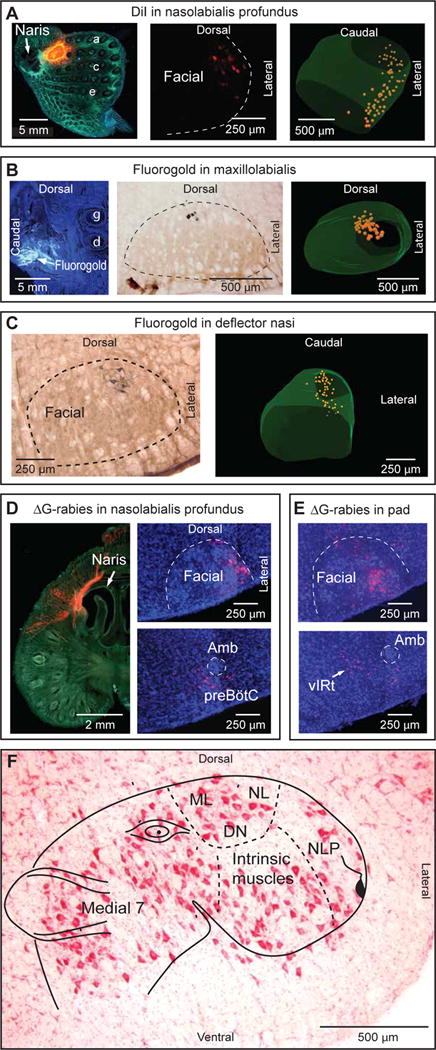
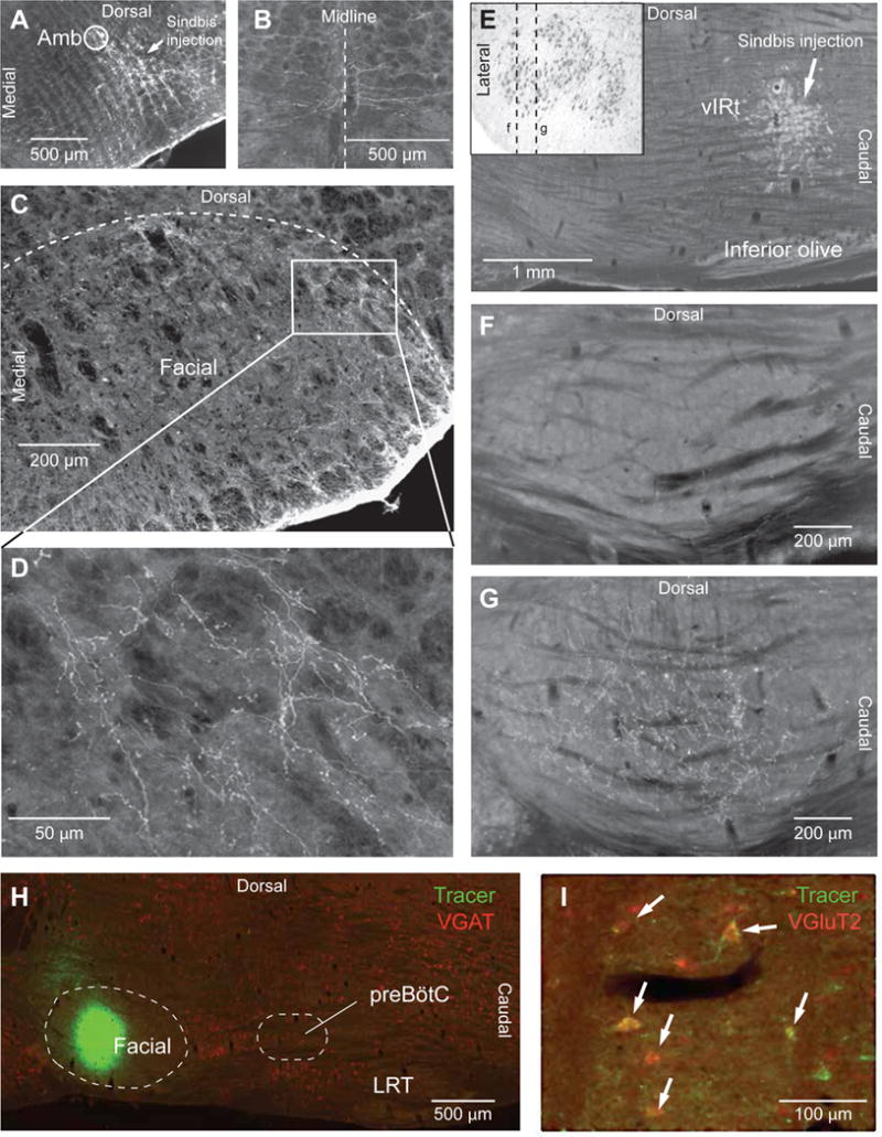
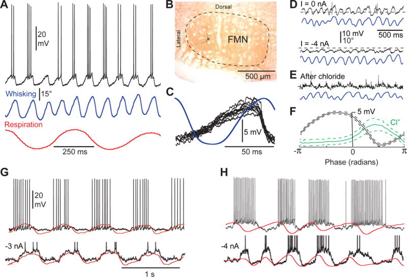
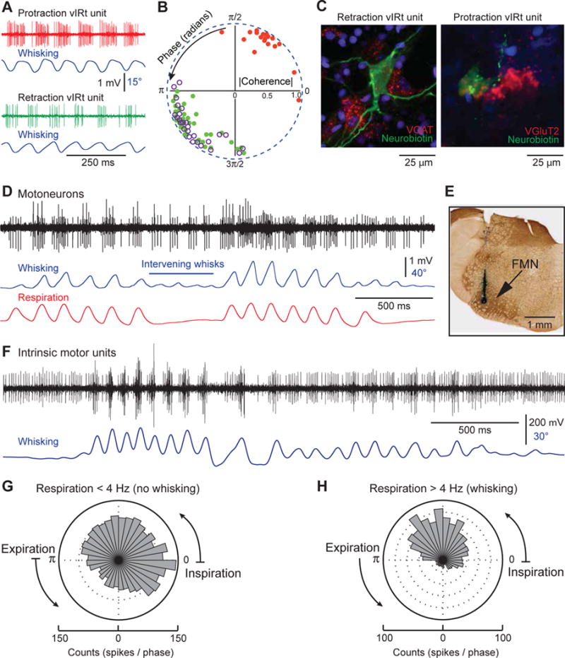
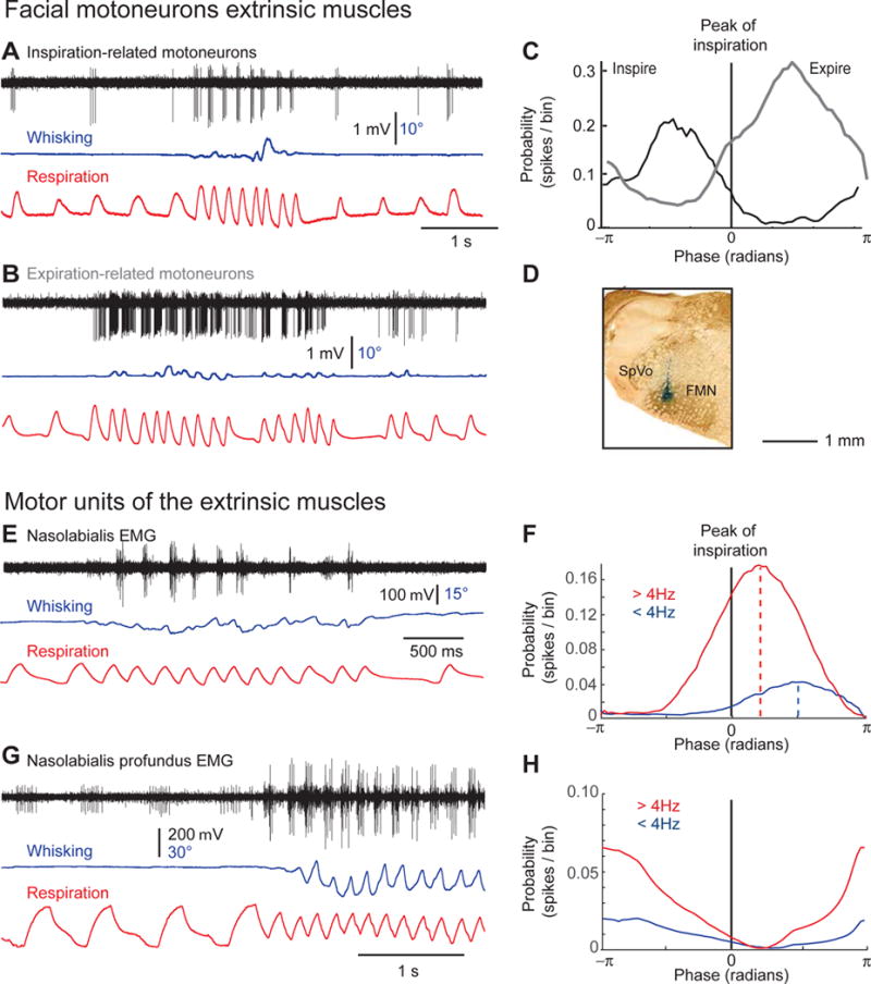
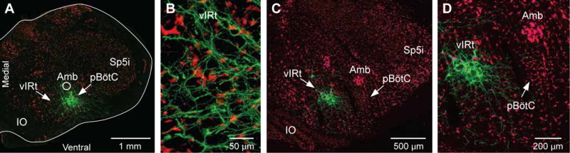
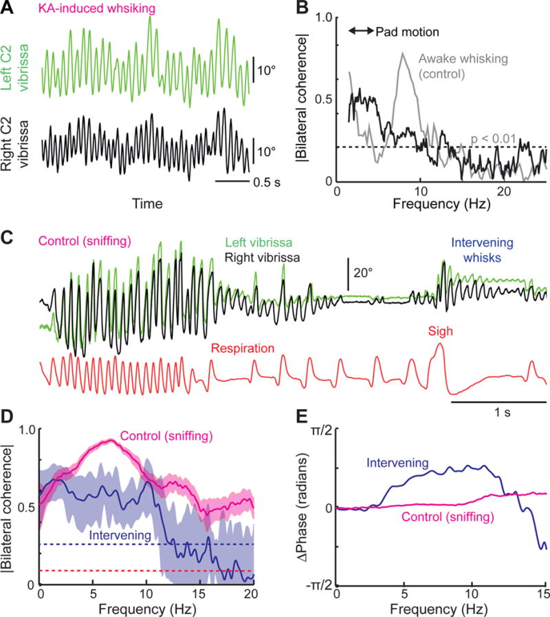
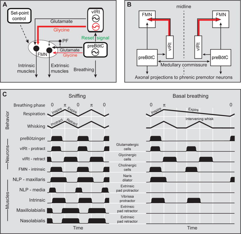
Sniffing and whisking typify the exploratory behavior of rodents. These actions involve separate oscillators in the medulla, located respectively in the pre-Bötzinger complex (preBötC) and the vibrissa-related region of the intermediate reticular formation (vIRt). We examine how these oscillators synergize to control sniffing and whisking. We find that the vIRt contains glycinergic/GABAergic cells that rhythmically inhibit vibrissa facial motoneurons. As a basis for the entrainment of whisking by breathing, but not vice versa, we provide evidence for unidirectional connections from the preBötC to the vIRt. The preBötC further contributes to the control of the mystacial pad. Lastly, we show that bilateral synchrony of whisking relies on the respiratory rhythm, consistent with commissural connections between preBötC cells. These data yield a putative circuit in which the preBötC acts as a master clock for the synchronization of vibrissa, pad, and snout movements, as well as for the bilateral synchronization of whisking.


Figures

Figure 1
Figure 1. Muscle representation in the facial motor nucleus
(A) After insertion of a small pellet of Gelfoam saturated with DiI in the rat’s snout, labeled motoneurons are found at the lateral edge of the facial nucleus. a,c,e, denote corresponding vibrissa rows. (B) Injection of Fluorogold in muscle maxillolabialis produces retrograde labeling in the dorsal sector of the facial nucleus. γ and δ denote corresponding vibrissae. (C) Injection of Fluorogold in muscle deflector nasi labels motoneurons in the dorsolateral sector of the facial nucleus. (D) ΔG-rabies injection in the snout labels motoneurons at the lateral edge of the facial nucleus, and premotor neurons in the preBötC. Amb, ambiguous nucleus. (E) ΔG-rabies injection into the mystacial pad labels motoneurons in the ventral lateral sector of the facial nucleus, and premotor neurons in vIRt. (F) Myotopic map of muscle representation on a Nissl-stained frontal section of the facial nucleus. The cartoon representation was adapted from ref 16. DN, muscle deflector nasi; ML, muscle maxillolabialis; NL, muscle nasolabialis; NLP, muscles nasolabialis profundus.
Figure 2
Figure 2. Differential projections of preBötC and vIRt to the facial nucleus
(A–D) Sindbis-GFP injection in preBötC labels commissural fibers (B), and axonal terminals in the lateral most sector of the facial nucleus (C). The framed area in C is enlarged in D. Frontal sections were stained for cytochrome oxidase, and colors were inverted to enhance contrast. (E–G) After Sindbis-GFP injection into vIRt, the lateral most sector of the facial nucleus is devoid of terminal labeling (F), whereas dense labeling is present in the ventral lateral sector of the nucleus (G). Sagittal sections in F and G correspond to section planes indicated by dashed lines in the Nissl-stained frontal section shown in the insert (E). Sections were stained for cytochrome oxidase, and colors were inverted to enhance contrast. (H–I) Fluorogold was injected into the facial nucleus (H), and in situ hybridization was used to reveal the expression of the VGLUT2 and VGAT transcripts in labeled cells (I). Arrows indicate doubly labeled VGLUT2+ neurons. LRT, lateral reticular nucleus.
Figure 3
Figure 3. Oscillatory activity of facial motoneurons during KA-induced whisking
(A) Subthreshold oscillations and spike discharges are phase locked to vibrissa protraction in whisking motoneurons. In all figures vibrissa protraction and inspiration are up. (B) Motoneuron shown in A was labeled with Neurobiotin. (C) Ten superimposed traces of sawtooth-like subthreshold depolarizations associated with vibrissa protraction in another whisking motoneuron. Traces are aligned with respect to the peak of vibrissa protraction. The blue trace is the average of 10 whisks. Note the sharp repolarization just before the onset of vibrissa retraction. (D) The amplitude of subthreshold oscillations is depressed by hyperpolarizing current injection. (E) Chloride injection in the same motoneuron leads to reversal of the intracellular oscillation. (F) Plots of the phase of membrane potential oscillations before (black trace) and after chloride injection (green trace). Phase plots represent the average of 30 whisks, phase zero corresponding to the peak of protraction. Spikes were removed with a median filter (window, 30 ms) before averaging. Dashed lines indicate 95% confidence interval. (G) Membrane potential oscillations in inspiratory motoneurons increase in amplitude during hyperpolarizing current injection. Action potentials are cropped in the lower trace. (H) Membrane potential oscillations in expiratory motoneurons increase in amplitude during hyperpolarizing current injection. Action potentials are cropped in the lower trace. Scale bars in G apply in H.
Figure 4
Figure 4. vIRt cells rhythmically inhibit vibrissa facial motoneurons
(A) During whisking induced by KA injection in the medulla, vIRt cells display antiphasic firing patterns. (B) Polar plot of the coherence between spiking activity and vibrissa motion at the peak frequency of whisking. Red and green dots represent protraction units and retraction units, respectively, which were recorded under ketamine/xylazine anesthesia. Purple circles represent vIRt units that were recorded under urethane anesthesia. Over 200 whisks per cell were used to compute phase angle and coherence. (C) Expression of VGAT and VGluT2 transcripts in individual vIRt cells labeled juxtacellularly with Neurobiotin. (D) Firing of facial motoneurons during whisking in a head-restrained rat. Both cells discharge during vibrissa protraction, including whisks that are not associated with a breath (intervening whisks). The positive unit is recruited as whisk amplitude increases. (E) Labeling of the recording site by an iontophoretic deposit of Chicago sky blue. FMN, facial motor nucleus. (F) Motor units of the intrinsic vibrissa muscles are inhibited during vibrissa retraction. (G–H) Circular phase histograms of intrinsic motor unit discharges during basal respiration, and during sniffing associated with whisking. Histograms were built from 15 sequences containing both basal respiration and sniffing bouts in 3 rats (149 breaths and 120 sniffs).
Figure 5
Figure 5. Conditional activation of facial motoneurons during sniffing in head-restrained rats
(A,B) Discharges of respiratory motoneurons during basal respiration and sniffing. Note that in these two examples sniffing occurs without significant whisking. (C) Phase plots of motoneuronal discharges across a population of five inspiratory motoneurons (solid line; 572 inspirations), and 7 expiratory motoneurons (dashed line; 974 expirations). (D) The recording site of the neuron shown in (B) was labeled with a deposit of Chicago sky blue. (E) EMG recording of nasolabialis motor units during basal respiration and sniffing. Note that muscle nasolabialis preferentially contracts in phase with expiration during sniffing. (F) Phase plots of muscle nasolabialis motor units activity during basal respiration (< 4Hz; 1180 breaths) and sniffing (> 4Hz; 1725 sniffs). Note the stronger modulation during sniffing, and the phase shift of activity between basal respiration and sniffing. (G) EMG recording of nasolabialis profundus motor units during basal respiration and sniffing. Note that the small unit is active during both basal respiration and sniffing, while the large unit is active during sniffing. (H) Phase plots of muscle nasolabialis motor units activity during basal respiration (< 4Hz; 668 breaths) and sniffing (> 4Hz; 756 sniffs). Note the stronger modulation during the inspiratory phase of sniffing.
Figure 6
Figure 6. Unidirectional connections between preBötC and vIRt
(A) Sindbis-GFP injection in preBötC produces anterograde labeling in vIRt. (B–C) In contrast, Sindbis-GFP injection into vIRt (C) does not label terminals in preBötC. (D) Sections were immunostained with an anti-NeuN antibody. Amb, ambiguous nucleus; IO, inferior olive; Sp5i, interpolaris division of the spinal trigeminal nucleus.
Figure 7
Figure 7. The left and right whisking oscillators are independent from one another
(A) Bilateral kainic acid injection produces independent vibrissa movements on the left and right sides of the face. (B) Spectral coherence (black trace) between the movements of each of the vibrissae in (A). The two signals show low coherence in the band of whisking frequencies relative to control data for bilateral active whisking in alert animals (gray trace). (C) Representative traces of whisking and respiration used to compute average bilateral coherence between sniff-related and intervening whisks in three head-restrained rats. (D) Average coherence between left and right intervening whisks is lower than that of sniff-related whisks. Light blue and red areas represent 95% confidence interval. (E) Whisks on the left and right sides of the face display virtually no phase difference during sniff-related whisks (red line), whereas intervening whisks display a clear phase shift (blue line).
Figure 8
Figure 8. Model of the medullary circuitry that generates whisking in coordination with breathing
(A) The core of the whisking CRG consists of glycinergic/GABAergic cells that inhibit motoneurons that innervate the intrinsic vibrissa muscles. The drive for vibrissa protraction arises from a smaller population of glutamatergic vIRt cells, and from other excitatory and modulatory inputs that control the set-point of whisking. The extrinsic muscles are driven by the respiratory CRG, with a potential contribution from parafacial neurons (PF) that receive input from the preBötC. (B) The reset of whisking by breathing is mediated by unidirectional connections from preBötC to vIRT, and commissural fibers that connect the left and right preBötCs ensure bilateral synchronization of whisking. (C) Idealized time-ordered patterns of behavioral, neuronal and muscular activities associated with the different phases of the respiratory and whisking rhythms. Note that the extrinsic pad retractor and protractor muscles may activate during basal respiration when the amplitude of respiration increases.

Comment in

References

    1. Abbott SBG, Stornetta RL, Coates MB, Guyenet PG. Phox2b-expressing neurons of the parafacial region regulate breathing rate, inspiration, and expiration in conscious rats. J Neurosci. 2011;31:16410–16422. - PMC - PubMed
    1. Ashwell KW. The adult mouse facial nerve nucleus: morphology and musculotopic organization. J Anat. 1982;135:531–538. - PMC - PubMed
    1. Bouvier J, Thoby-Brisson M, Renier N, Dubreuil V, Ericson J, Champagnat J, Pierani A, Chédotal A, Fortin G. Hindbrain interneurons and axon guidance signaling critical for breathing. Nat Neurosci. 2010;3:1066–1074. - PubMed
    1. Deschênes M, Haidarliu S, Demers M, Moore J, Kleinfeld D, Ahissar E. Muscles involved in naris dilation and nose motion in rat. Anat Rec. 2015;298:546–553. - PMC - PubMed
    1. Deschênes M, Kleinfeld D. Sniffing and whisking in rodents. Curr Opin Neurobiol. 2012;22:243–250. - PMC - PubMed

Publication types

LinkOut - more resources