. 2016:17:12.
doi: 10.1186/s40510-016-0126-0. Epub 2016 Apr 18.

Maxillary molar distalization with aligners in adult patients: a multicenter retrospective study

Affiliations

Maxillary molar distalization with aligners in adult patients: a multicenter retrospective study

Serena Ravera et al. Prog Orthod. 2016.

Abstract

Background: The aim of the present study was to test the hypothesis that bodily maxillary molar distalization was not achievable in aligner orthodontics.

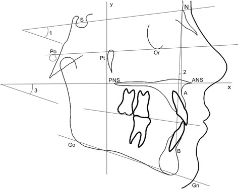
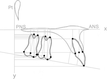
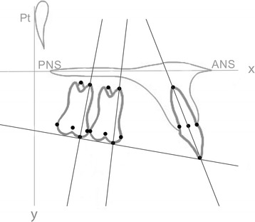
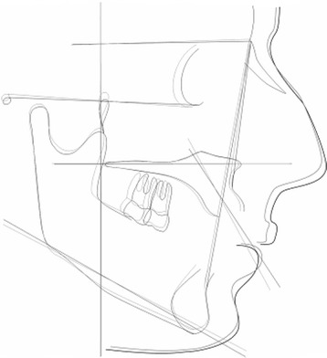
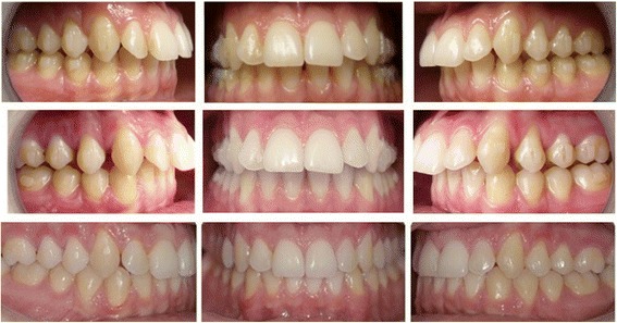
Methods: Forty lateral cephalograms obtained from 20 non-growing subjects (9 male, 11 female; average age 29.73 years) (group S), who underwent bilateral distalization of their maxillary dentition with Invisalign aligners (Align Technology, Inc., San José, CA, USA), were considered for the study. Skeletal class I or class II malocclusion and a bilateral end-to-end class II molar relationship were the main inclusion criteria. Cephalograms were taken at two time points: (T0) pretreatment and (T2) post-treatment. Treatment changes were evaluated between the time points using 39 variables by means of paired t test. The level of significance was set at P < 0.05. Reproducibility of measurements was assessed by the intraclass correlation coefficient (ICC).

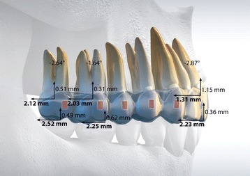
Results: The mean treatment time was 24.3 ± 4.2 months. At the post-treatment point, the first molar moved distally 2.25 mm without significant tipping (P = 0.27) and vertical movements (P = 0.43). The second molar distalization was 2.52 mm without significant tipping (P = 0.056) and vertical movements (P = 0.25). No significant movements were detected on the lower arch. SN^GoGn and SPP^GoGn angles showed no significant differences between pre- and post-treatment cephalograms (P = 0.22 and P = 0.85, respectively).

Conclusions: Aligner therapy in association with composite attachments and class II elastics can distalize maxillary first molars by 2.25 mm without significant tipping and vertical movements of the crown. No changes to the facial height were revealed.

Keywords: Adult patients; Aligners; Class II; Molar distalization.


Figures

Fig. 1
Fig. 1
Schematic illustration of the skeletal variables considered in the study. 1 SN^GoGn (°); 2 SN^A (°), SN^B(°), AN^B (°); 3 SpP^GoGn (°)
Fig. 2
Fig. 2
Schematic Illustration of angular measurements (°) of maxillary second molar (1), maxillary first molar (2), and central incisor (3). The angle between the tooth long axis (passing trough the mesiobuccal cusp and mesiobuccal root’s apex for the first and the second molar; passing trough the incisal edge and root’s apex for the central incisor) and x axis (palatal plane) expressed the inclination of the tooth
Fig. 3
Fig. 3
Schematic illustration of linear measurements (mm) considered in the study. Horizontal measurements were expressed by the distance between the following points and the y axis (a perpendicular line to the palatal plane passing through the Ricketts’ Pt point); second molar horizontal distance from the center of the crown, from the mesiobuccal cusp, from the mesiobuccal root’s apex, from the palatal root’s apex; first molar horizontal distance from the center of the crown, from the mesiobuccal cusp, from the mesiobuccal root’s apex, from the palatal root’s apex; central incisor distance; central incisor edge distance, central incisor radicular apex distance. Vertical distances were expressed by the distance between the same points and the x axis (palatal plane) and between the occlusal plane (except for incisor edge point and mesiobuccal cusp point, both tangential the occlusal plane)
Fig. 4
Fig. 4
Schematic illustration of clinically relevant results for the study
Fig. 5
Fig. 5
Superimposition of one of the patients treated in this study
Fig. 6
Fig. 6
Pre- and post-photos of one of the patients treated in this study

References

    1. Bolla E, Muratore F, Carano A, Bowman SJ. Evaluation of maxillary molar distalization with the distal jet: a comparison with other contemporary methods. Angle Orthod. 2002;72:481–494. - PubMed
    1. Grec RH, Janson G, Branco NC, Moura-Grec PG, Patel MP, Castanha Henriques JF. Intraoral distalizer effects with conventional and skeletal anchorage: a meta-analysis. Am J Orthod Dentofacial Orthop. 2013;143:602–615. doi: 10.1016/j.ajodo.2012.11.024. - DOI - PubMed
    1. Fuziy A, Rodrigues de Almeida R, Janson G, Angelieri F, Pinzan A. Sagittal, vertical and transverse changes consequent to maxillary molar distalization with the pendulum appliance. Am J Orthod Dentofacial Orthop. 2006;130(4):502–10. doi: 10.1016/j.ajodo.2004.12.031. - DOI - PubMed
    1. Fontana M, Cozzani M, Caprioglio A. Non-compliance maxillary molar distalizing appliances: an overview of the last decade. Prog Orthod. 2012;13:173–184. doi: 10.1016/j.pio.2011.10.002. - DOI - PubMed
    1. Caprioglio A, Cozzani M, Fontana M. Comparative evaluation of molar distalization therapy with erupted second molar: segmented versus quad pendulum appliance. Prog Orthod. 2014;15:49. doi: 10.1186/s40510-014-0049-6. - DOI - PMC - PubMed