. 2016 Oct 13;35(41):5412-5421.
doi: 10.1038/onc.2016.79. Epub 2016 Apr 4.

RSK2 signals through stathmin to promote microtubule dynamics and tumor metastasis

Affiliations

RSK2 signals through stathmin to promote microtubule dynamics and tumor metastasis

G N Alesi et al. Oncogene. .

Abstract

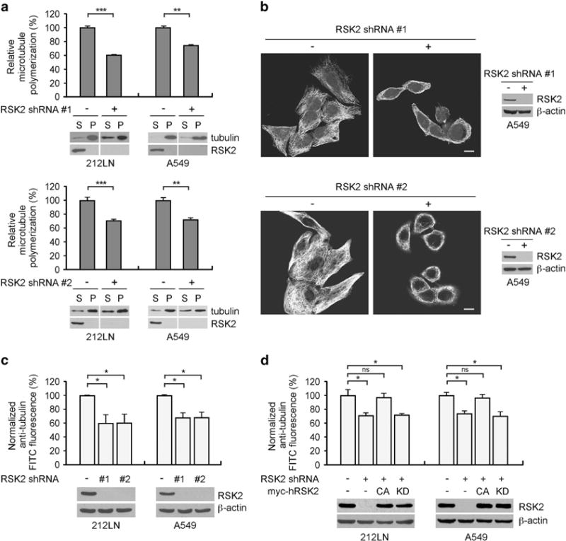
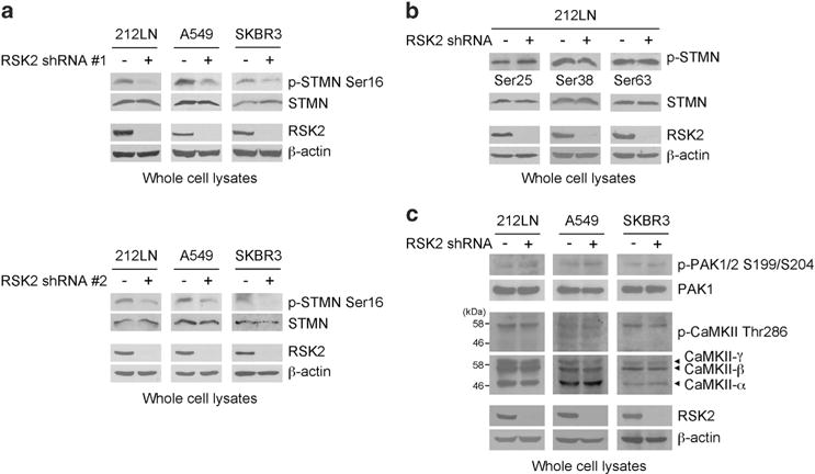
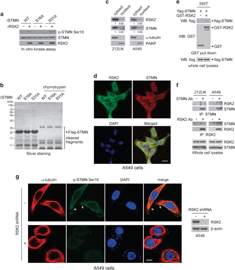
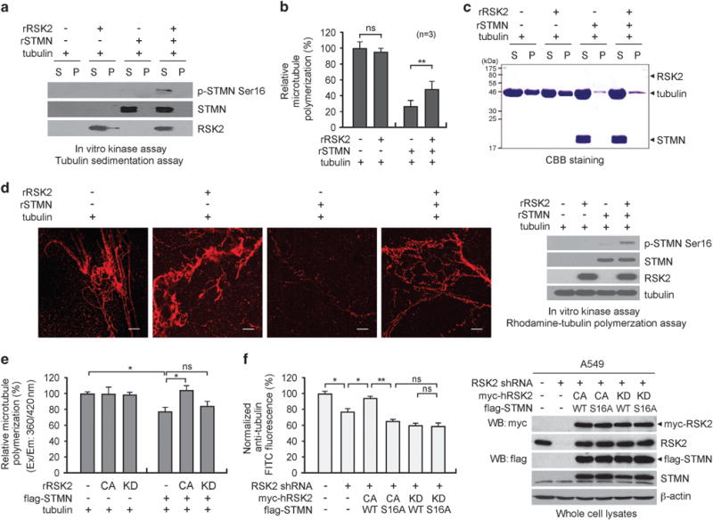
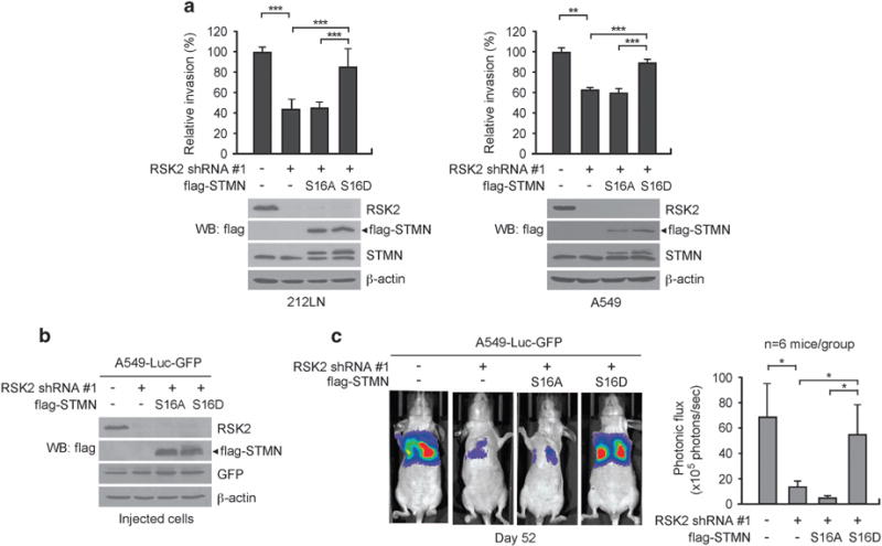
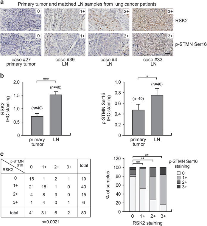
Metastasis is responsible for >90% of cancer-related deaths. Complex signaling in cancer cells orchestrates the progression from a primary to a metastatic cancer. However, the mechanisms of these cellular changes remain elusive. We previously demonstrated that p90 ribosomal S6 kinase 2 (RSK2) promotes tumor metastasis. Here we investigated the role of RSK2 in the regulation of microtubule dynamics and its potential implication in cancer cell invasion and tumor metastasis. Stable knockdown of RSK2 disrupted microtubule stability and decreased phosphorylation of stathmin, a microtubule-destabilizing protein, at serine 16 in metastatic human cancer cells. We found that RSK2 directly binds and phosphorylates stathmin at the leading edge of cancer cells. Phosphorylation of stathmin by RSK2 reduced stathmin-mediated microtubule depolymerization. Moreover, overexpression of phospho-mimetic mutant stathmin S16D significantly rescued the decreased invasive and metastatic potential mediated by RSK2 knockdown in vitro and in vivo. Furthermore, stathmin phosphorylation positively correlated with RSK2 expression and metastatic cancer progression in primary patient tumor samples. Our finding demonstrates that RSK2 directly phosphorylates stathmin and regulates microtubule polymerization to provide a pro-invasive and pro-metastatic advantage to cancer cells. Therefore, the RSK2-stathmin pathway represents a promising therapeutic target and a prognostic marker for metastatic human cancers.


Conflict of interest statement

The authors declare no conflict of interest.

Figures

Figure 1
Figure 1
Targeted downregulation of RSK2 attenuates microtubule polymerization in metastatic human cancer cells. (a) Tubulin sedimentation assay shows that RNAi-mediated stable knockdown of RSK2 results in significantly decreased microtubule polymerization in HNSCC 212LN cells (left) and lung cancer A549 cells (right). Lower: Tubulin immunoblots show soluble and polymerized tubulin in the supernatant (S) and pellet (P), respectively. Upper: Relative tubulin polymerization was determined by density analysis. The amount of polymerized tubulin in cells with RSK2 knockdown was normalized to the tubulin polymerization of control cells with an empty vector. (b) Tubulin immunofluorescence staining shows that stable knockdown of RSK2 disrupts tubulin polymerization in 212LN cells. Scale bar represents 10 μm. Western blotting shows RSK2 knockdown. (c) Polymerized tubulin was quantified by FACS-based whole-cell analysis of microtubules in RSK2 knockdown cells. FITC fluorescence intensity was normalized to a value of 100 for the control cells harboring an empty vector. (d) FACS-based whole-cell microtubule analysis using RSK2 knockdown cells with overexpression of shRNA-resistant CA (Y707A) or KD (Y707A/K100A) human RSK2. Stable RSK2 knockdown cells were transiently transfected with Y707A or Y707A/K100A RSK2 cDNA prior to microtubule analysis. Data represent mean ± s.d. from three technical replicates. Results of one representative experiment from at least two independent experiments are shown. Statistical significance was determined using two-tailed Student’s t-test (NS: not significant; *0.01<P<0.05; **0.001<P<0.01; ***P<0.001).
Figure 2
Figure 2
RSK2 promotes stathmin phosphorylation in diverse metastatic cancer cells. (a and b) Effect of RSK2 stable knockdown on stathmin (STMN) phosphorylation at N-terminal serine residues, S16 (a), S25, S38 and S68 (b) in metastatic human cancer cells, 212LN, A549 and SKBR3. RSK2 knockdown using two different shRNA clones attenuates phosphorylation at stathmin S16 (a). (c) RSK2 knockdown effect on the activity of CaMKII and PAK1, known upstream kinases of stathmin. Activities of PAK1 and CaMKII were assessed by autophosphorylation at S199/S204 and T286, respectively, in diverse metastatic human cancer cells with RSK2 stable knockdown and control vector cells. SKBR3 cells were stimulated with 0.5 M sorbitol for phospho-PAK1 detection. Results of one representative experiment from at least two independent experiments are shown.
Figure 3
Figure 3
RSK2 directly phosphorylates and interacts with stathmin in cells. (a) RSK2 directly phosphorylates stathmin (STMN) at S16. Purified recombinant stathmin (rSTMN) variants, WT, S16A and S31A, were incubated with recombinant active RSK2. Phosphorylation at serine 16 of stathmin was detected by western blotting using specific antibody against phospho-stathmin at S16. (b) Partial protease digestion demonstrates that the global structure of stathmin is not changed by point mutations. In all, 0.5 units of chymotrypsin were incubated with recombinant stathmin variants at 30 °C for 30 min and the digestion patterns were compared. (c) Western blots show the cytosolic/nuclear localization of RSK2 and stathmin in cancer cells. α-Tubulin and PARP (poly ADP-ribose polymerase) were used as control markers for cytosol and nucleus, respectively. (d) Immunofluorescence assay of RSK2 and stathmin in A549 cells. Scale bar represents 20 μm. (e) RSK2 interacts with stathmin. GST or GST-fused RSK2 were enriched by GST pull-down assay from 293T cells transfected with flag-tagged stathmin. Stathmin in the complex of bead-bound GST-RSK2 was detected by western blotting. (f) Co-immunoprecipitation of endogenous RSK2 and stathmin in 212LN and A549 cells. (g) Immunofluorescence assay shows the localization of phosphorylated form of stathmin and tubulin in A549 cells with or without RSK2 shRNA. Scale bar represents 10 μm. Results of one representative experiment from at least two independent experiments are shown.
Figure 4
Figure 4
RSK2-dependent phosphorylation of stathmin promotes microtubule polymerization. Purified stathmin (STMN) was phosphorylated via an in vitro RSK2 kinase assay and incubated with tubulin. (a) Stathmin phosphorylation in supernatant and pellet fractions is shown by western blotting analysis. (b and c) Polymerized tubulin was analyzed by Coomassie blue staining. S: supernatant, P: pellet. Relative tubulin polymerization in the absence or presence of RSK2 and/or stathmin was quantified by analyzing three independent repeats of microtubule polymerization assays (b). Representative image is shown panel (c). (d) Rhodamine-labeled tubulin was polymerized with stathmin alone or with stathmin preincubated with RSK2 and analyzed by fluorescence microscopy. Scale bars represent 30 μm. (e) Tubulin polymerization in the presence of recombinant stathmin, active RSK2 (CA) or inactive RSK2 (KD) in the indicated combination. Polymerized tubulin was quantified using the Fluorimetry-based Tubulin Polymerization Assay Kit. (f) FACS-based whole cell microtubule analysis of RSK2 knockdown cells overexpressing shRNA-resistant CA or KD RSK2 along with WT or S16A stathmin. Stable RSK2 knockdown cells were transiently transfected with distinct RSK2 and stathmin variants prior to whole-cell microtubule analysis. Results of one representative experiment from at least two independent experiments are shown. Data represent mean ± s.d. Statistical significance was determined using two-tailed Student’s t-test (NS: not significant; *0.01<P<0.05; **0.001<P<0.01).
Figure 5
Figure 5
RSK2 promotes cancer cell invasion and tumor metastasis, in part, through phosphorylation of stathmin. (a) Matrigel invasion assay using RSK2 knockdown cells with stathmin (STMN) variants. Stable expression of phospho-mimetic mutant S16D stathmin but not phospho-deficient mutant S16A stathmin restored the cancer cell invasion attenuated by RSK2 knockdown in 212LN and A549 cells. (b) RSK2, flag-tagged stathmin S16A and S16D expression was detected by immunoblotting in A549-luc-GFP cells used for tail-vein injection. (c) Left: BLI imaging of representative mice injected with A549-luc-GFP cells with RSK2 knockdown and S16A or S16D stathmin expression at day 52 after injection. Right: Average photonic flux of each group at weeks 6–8 is shown. (a) Data represent mean ± s.d. from nine (left) and four (right) technical replicates. Results of one representative experiment from at least two independent experiments are shown. (c) Data represent mean ± s.e.m. from six mice for each group. Statistical significance was determined using two-tailed Student’s t-test for panel (a) and one-tailed Student’s t-test for panel (c) (*0.01<P<0.05; **0.001<P<0.01; ***P<0.001).
Figure 6
Figure 6
The levels of RSK2 and phospho-S16 stathmin correlate with metastatic cancer progression in primary human tumor tissue samples from lung cancer patients. The levels of RSK2 and stathmin (STMN) phosphorylation in 40 cases of human lung cancer with matched lymph node (LN) metastasis were determined by IHC staining using lung cancer tissue microarray. (a) Representative tumor specimens with staining intensity of 0 (negative), 1+ (weak), 2+ (moderate) and 3+ (strong) of RSK2 and phospho-stathmin S16 are shown. Scale bar represents 50 μm. (b) Levels of RSK2 expression (left) and stathmin phosphorylation (right) in primary tumors and matched tumors from lymph nodes. The staining intensity was scored from 0 to 3+. Data represent mean ± s.e.m. from n = 40/group. (c) The correlation between RSK2 and phospho-stathmin S16 was determined. Bar graph representation is shown on the right. P-values were determined by two-tailed paired Student’s t-test for panel (b) and chi-square test for panel (c) (*0.01<P<0.05; **0.001<P<0.01; ***P<0.001).

References

    1. Fidler IJ. The pathogenesis of cancer metastasis: the ‘seed and soil’ hypothesis revisited. Nat Rev Cancer. 2003;3:453–458. - PubMed
    1. Nguyen DX, Massague J. Genetic determinants of cancer metastasis. Nat Rev Genet. 2007;8:341–352. - PubMed
    1. Gupta GP, Massague J. Cancer metastasis: building a framework. Cell. 2006;127:679–695. - PubMed
    1. Sahai E. Mechanisms of cancer cell invasion. Curr Opin Genet Dev. 2005;15:87–96. - PubMed
    1. Li Y, Wang JP, Santen RJ, Kim TH, Park H, Fan P, et al. Estrogen stimulation of cell migration involves multiple signaling pathway interactions. Endocrinology. 2010;151:5146–5156. - PMC - PubMed

Publication types

Substances