Review
. 2016 Mar 23:10:39-49.
doi: 10.4137/JEN.S33122. eCollection 2016.

Molecular Mechanisms of Neurodegeneration in Spinal Muscular Atrophy

Affiliations
Review

Molecular Mechanisms of Neurodegeneration in Spinal Muscular Atrophy

Saif Ahmad et al. J Exp Neurosci. .

Abstract

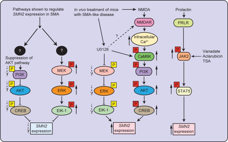
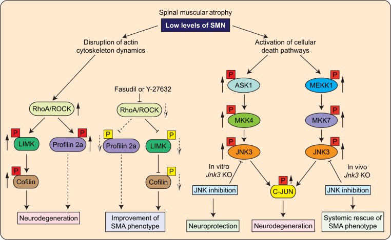
Spinal muscular atrophy (SMA) is an autosomal recessive motor neuron disease with a high incidence and is the most common genetic cause of infant mortality. SMA is primarily characterized by degeneration of the spinal motor neurons that leads to skeletal muscle atrophy followed by symmetric limb paralysis, respiratory failure, and death. In humans, mutation of the Survival Motor Neuron 1 (SMN1) gene shifts the load of expression of SMN protein to the SMN2 gene that produces low levels of full-length SMN protein because of alternative splicing, which are sufficient for embryonic development and survival but result in SMA. The molecular mechanisms of the (a) regulation of SMN gene expression and (b) degeneration of motor neurons caused by low levels of SMN are unclear. However, some progress has been made in recent years that have provided new insights into understanding of the cellular and molecular basis of SMA pathogenesis. In this review, we have briefly summarized recent advances toward understanding of the molecular mechanisms of regulation of SMN levels and signaling mechanisms that mediate neurodegeneration in SMA.

Keywords: JNK; MND; ROCK; SMA; SMN; ZPR1.


Figures

Figure 1
Figure 1
Mechanisms of regulation of SMN2 gene expression in SMA. SMA is caused by low levels of SMN protein translated from full-length transcripts (5%–10%) generated from the SMN2 gene. Increase in the transcription of SMN2 gene generates higher levels of full-length SMN protein. Signaling pathways identified in SMA that may regulate expression of the SMN2 gene are presented. The MEK/ERK/ELK-1 pathway is activated in SMA and negatively regulates SMN2 expression. Inhibition of ERK pathway in vivo in SMA mice using MEK inhibitor (U0126) results in the upregulation of SMN2 expression by the activation of the PI3K/AKT/CREB pathway. Treatment with NMDA also results in the activation of the AKT/CREB pathway that results in the upregulation of SMN2 expression. The activation of JAK2/STAT5 pathway in vivo by treatment with peptide hormone PRL results in an increase in SMN levels in SMA mice. Solid up arrows (red box) show increase in phosphorylation and dotted down arrows (yellow box) show decrease in phosphorylation.
Figure 2
Figure 2
The molecular mechanisms that mediate neurodegeneration in SMA. SMA is characterized by degeneration of spinal motor neurons caused by low levels of SMN. SMN deficiency results in the activation of intracellular signaling pathways that mediate the degeneration of neurons in SMA. Rho/ROCK pathway is activated in mice with intermediate SMA and mediates neurodegeneration by disruption of cytoskeleton stability. Inhibition of Rho/ROCK pathway with inhibitors Y-27632 and Fasudil results in the improvement of NMJ pathology and SMA phenotype. The JNK signaling pathway is activated in the spinal cords of SMA patients and SMAΔ7 mice. Two JNK signaling modules, ASK1/MKK4/JNK3 and MEKK1/MKK7/JNK3, mediate in vivo phosphorylation of c-Jun that causes the degeneration and apoptosis of neurons. Genetic inhibition of the JNK pathway by Jnk3 knockout results in the neuroprotection and systemic amelioration of SMA in mice. Black boxes with question marks represent upstream targets, which mediate the effects of changes stemming from the low levels of SMN that remain to be identified. Solid up arrows (red box) show increase in phosphorylation and dotted down arrows (yellow box) show decrease in phosphorylation. Dotted line connectors represent possibilities that need to be confirmed with further studies.

References

    1. D’Amico A, Mercuri E, Tiziano FD, Bertini E. Spinal muscular atrophy. Orphanet J Rare Dis. 2011;6:71. - PMC - PubMed
    1. Lefebvre S, Burglen L, Reboullet S, et al. Identification and characterization of a spinal muscular atrophy-determining gene. Cell. 1995;80:155–165. - PubMed
    1. Lorson CL, Hahnen E, Androphy EJ, Wirth B. A single nucleotide in the SMN gene regulates splicing and is responsible for spinal muscular atrophy. Proc Natl Acad Sci U S A. 1999;96:6307–6311. - PMC - PubMed
    1. Monani UR, Lorson CL, Parsons DW, et al. A single nucleotide difference that alters splicing patterns distinguishes the SMA gene SMN1 from the copy gene SMN2. Hum Mol Genet. 1999;8:1177–1183. - PubMed
    1. Cusco I, Barcelo MJ, Rojas-Garcia R, et al. SMN2 copy number predicts acute or chronic spinal muscular atrophy but does not account for intrafamilial variability in siblings. J Neurol. 2006;253:21–25. - PubMed

LinkOut - more resources