. 2016 Apr 1:4:6.
doi: 10.1186/s40170-016-0146-8. eCollection 2016.

Inhibition of fatty acid desaturation is detrimental to cancer cell survival in metabolically compromised environments

Affiliations

Inhibition of fatty acid desaturation is detrimental to cancer cell survival in metabolically compromised environments

Barrie Peck et al. Cancer Metab. .

Abstract

Background: Enhanced macromolecule biosynthesis is integral to growth and proliferation of cancer cells. Lipid biosynthesis has been predicted to be an essential process in cancer cells. However, it is unclear which enzymes within this pathway offer the best selectivity for cancer cells and could be suitable therapeutic targets.

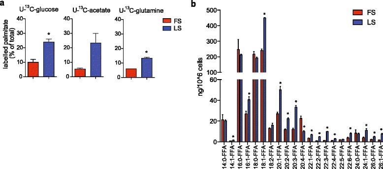
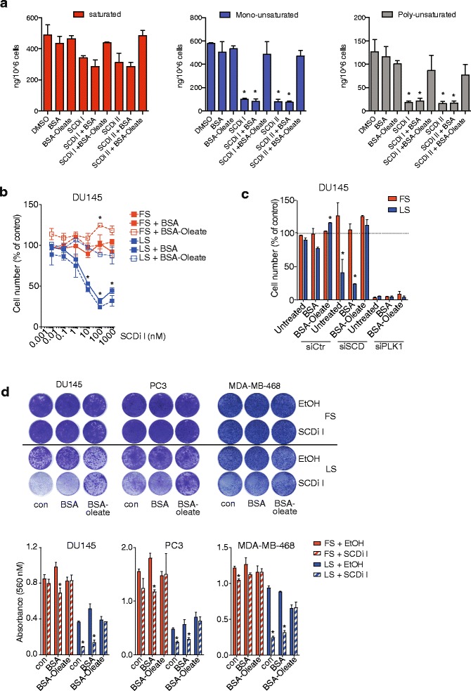
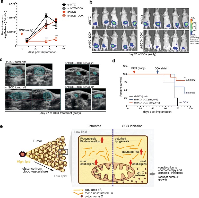
Results: Using functional genomics, we identified stearoyl-CoA desaturase (SCD), an enzyme that controls synthesis of unsaturated fatty acids, as essential in breast and prostate cancer cells. SCD inhibition altered cellular lipid composition and impeded cell viability in the absence of exogenous lipids. SCD inhibition also altered cardiolipin composition, leading to the release of cytochrome C and induction of apoptosis. Furthermore, SCD was required for the generation of poly-unsaturated lipids in cancer cells grown in spheroid cultures, which resemble those found in tumour tissue. We also found that SCD mRNA and protein expression is elevated in human breast cancers and predicts poor survival in high-grade tumours. Finally, silencing of SCD in prostate orthografts efficiently blocked tumour growth and significantly increased animal survival.

Conclusions: Our data implicate lipid desaturation as an essential process for cancer cell survival and suggest that targeting SCD could efficiently limit tumour expansion, especially under the metabolically compromised conditions of the tumour microenvironment.

Keywords: Breast cancer; Lipid desaturation; Lipidomics; Prostate cancer; SCD.


Figures

Fig. 1
Fig. 1
SCD ablation is detrimental to breast and prostate cancer cell lines cultured in low serum. a Strictly standardised mean difference (SSMD) calculated from cell numbers obtained in a functional genomic screen targeting lipid metabolism genes. Genes with the strongest differential (Diff.) between full serum (10 % FCS, FS) and low serum (1 % FCS, LS) are shown. b Screen results for SCD RNAi across the panel of breast and prostate cell lines. SSMD values below −2 indicate statistical significance. RWPE1 cells were cultured in KSFM. c Loss of cell number following silencing of SCD or PLK1 in DU145, MDA-MB-468, PC3 and T47D cells cultured in full serum (FS) or low serum (LS) for 72 h. Cell numbers are relative to non-targeting controls (RISC). Statistical comparisons were performed using Student t test (*p ≤ 0.05). d Breast and prostate cancer cell lines were treated with different doses of SCD inhibitor for 72 h in medium containing full (FS) or low (LS) serum. Cell numbers are relative to untreated controls. Data represent the mean ± SEM of three independent biological replicates. Statistical comparisons were performed using Student t test (*p ≤ 0.05). e DU145 cells were treated with increasing concentrations of two structurally distinct SCD inhibitors (SCDi I and SCDi II) for 72 h in medium containing full (FS) or low (LS) serum. Cell number is relative to untreated controls. Data represent the mean ± SEM of three independent biological replicates. Statistical comparisons were performed using Student t test (*p ≤ 0.05)
Fig. 2
Fig. 2
SCD expression is increased in breast and prostate cancers and associated with poor survival. a Differential expression of SCD between breast and prostate cancers and normal tissue found in different public datasets (Oncomine). b Immunohistochemical analysis of SCD in breast cancer and normal adjacent tissue (NAT) samples from invasive ductal carcinoma (IDC) and invasive lobular carcinoma (ILC). SCD staining intensity was classified as absent, low or high. Representative images of NAT and IDC samples indicating staining intensities. c Correlation of SCD expression in IDC patient samples with tumour grade (Curtis dataset, Oncomine). d Kaplan Meier plots for relapse-free survival (RFS) stratified by SCD expression in breast cancers of different grades. e Kaplan Meier plots for RFS stratified by SCD expression in patients with basal-like breast cancers as determined by expression of the PAM50 signature
Fig. 3
Fig. 3
Low serum conditions induce de novo lipogenesis and alter the composition of cellular free fatty acids. a Incorporation of 13C-carbon from glucose, glutamine or acetate in full (FS) or low (LS) serum in DU145 cells. b Quantitative lipid profiling of FFA content of DU145 cells grown in full (FS) or low (LS) serum for 48 h using LC-MS/MS. Data represent the mean ± SEM of three biologically independent replicates. Statistical comparisons were performed using Student t test (*p ≤ 0.05)
Fig. 4
Fig. 4
SCD maintains fatty acid desaturation to support viability of cancer cells. a DU145 cells were treated with SCD inhibitors (100 nM) or solvent (DMSO) for 48 h in medium with low serum or supplemented with BSA or BSA-coupled oleic acid (BSA-oleate). Quantitative lipid profiling of free fatty acids (FFA) content was performed by LC-MS/MS. Concentrations of saturated, mono-unsaturated and poly-unsaturated FFA are displayed. Data represent the mean ± SEM of three biologically independent replicates. Statistical comparisons were performed using Student t test (*p ≤ 0.05). b DU145 cells were treated with increasing concentrations of SCD inhibitor for 72 h in medium containing full (FS) or low serum (LS) or medium supplemented with BSA or BSA-oleate. Cell numbers are normalised to solvent treated controls. Statistical comparisons were performed using Student t test (*p ≤ 0.05). c DU145 cells were transfected with siRNA targeting SCD (siSCD), PLK1 (siPLK1) or non-targeting controls (siCtrl). Cells were cultured for 72 h in medium containing full (FS) or low serum (LS) or medium supplemented with BSA or BSA-oleate. Cell numbers are displayed relative to untreated siCtrl transfected cells. Data represent the mean ± SEM of two biologically independent experiments with two replicates each. Statistical comparisons were performed using Student t test (*p ≤ 0.05). d DU145, PC3 and MDA-MB-468 cells were treated with SCD inhibitor (SCDi I) or solvent (DMSO) under full (FS) or low (LS) serum conditions for 10 days. Cells were stained using crystal violet and quantified. Data represent mean ± SEM of two biologically independent experiments. Statistical comparisons were performed using Student t test (*p ≤ 0.05)
Fig. 5
Fig. 5
SCD inhibition alters cellular cardiolipin composition leading to cytochrome C release and sensitisation towards apoptosis. a DU145 cells were treated with different concentrations of SCD inhibitor (SCDi II) and proliferation was determined by BrdU labelling. b Cells treated as in a were used to determine apoptosis using Annexin V staining. Data represent mean ± SEM of three independent biological replicates. Statistical comparisons were performed using Student t test (*p ≤ 0.05). c Quantitative lipid profiling of cardiolipin (CL) content of DU145 cells grown for 48 h in medium containing full (FS) or low (LS) serum using LC-MS/MS. Concentrations of mono-unsaturated (≤4 double bonds) and poly-unsaturated (>4 double bonds) CL species are displayed. Data represent the mean ± SEM of three independent biological replicates. Statistical comparisons were performed using Student t test (***p ≤ 0.001). d DU145 cells were treated with SCD inhibitors (100 nM) or solvent (DMSO) for 48 h in medium containing low serum or medium supplemented with BSA or BSA-coupled oleic acid (BSA-Oleate). Quantitative lipid profiling of CL content was determined using LC-MS/MS. Concentrations of mono- and poly-unsaturated CL species are displayed. Data represent the mean ± SEM of three independent biological replicates. Statistical comparisons were performed using Student t tests (*p ≤ 0.05). e DU145 cells were treated with SCD inhibitor (SCDi I) or solvent (DMSO) for 48 h in medium containing low serum or medium supplemented with BSA or BSA-oleate. Cells were lysed by digitonin, and the presence of cytoplasmic cytochrome C was determined. UV treatment was used as positive control. Vinculin is shown as loading control. Levels of phosphorylated Akt (S473) and total Akt were detected in total lysates. f DU145 cells were treated as in e but 3 μM of Akt inhibitor was added prior to addition of BSA-oleate. Cytoplasmic cytochrome C was detected in digitonin lysates. g DU145 cells were treated with SCD inhibitor (SCDi I) or solvent (DMSO) in medium containing full (FS) or low (LS) serum or medium supplemented with BSA or BSA-oleate. The presence of full-length and cleaved (cl.) PARP was determined. Actin is shown as loading control. h DU145 cells were transfected with siRNA targeting SCD (siSCD) or non-targeting controls (siCtrl) and cultured as in e. The presence of full-length and cleaved (cl.) PARP was determined. Actin is shown as loading control. i DU145 or MDA-MB-468 cells were treated with SCD inhibitor (100 nM) for 48 h, and oxygen consumption rate (OCR) before and after addition of oligomycin, FCCP and rotenone was determined using a Seahorse Bioanalyzer. jm DU145 cells were treated with the indicated doses of SCD inhibitor (SCDi II) either alone or in combination with different doses of metformin (Metf, j), rotenone (RN, k), paclitaxel (PTX, l) or staurosporin (STP, m) for 72 h in medium containing low serum. Cell viability was determined by crystal violet staining. Data represent the mean ± SEM of three independent biological replicates. Statistical comparisons were performed using Student t tests (*p ≤ 0.05, **p ≤ 0.01 compared to SCDi alone; #p ≤ 0.05, ##p ≤ 0.01 compared to no SCDi)
Fig. 6
Fig. 6
Spheroid culture reveals metabolic dependency of cancer cells on lipid desaturation. a BT474 cells were cultured in normoxia (20 % O2) or hypoxia (0.1 % O2) for 48 h before lipids were extracted and used to determine the relative amount of diacylglycerol (DG) and phosphatidylcholine (PC) species by LC-MS/MS. Archival tissue from six individual orthotopic xenograft tumours of BT474 cells [14] was also analysed. Statistical comparisons were performed using Student t tests (*p ≤ 0.05). b Lipids were extracted from T47D cells grown in adherent cultures (2D) or as tumour spheroids. Relative distribution of unsaturated and mono- or poly-unsaturated DAG species are shown. c Selected mono-unsaturated lipid species from T47D cells grown in 2D or as tumour spheroids. d, e DU145 cells expressing inducible shRNAs targeting SCD (shSCD #2) or scrambled control (shNTC) were grown as spheroids in the presence of solvent (EtOH) or 0.5 μg/ml doxycycline (DOX). Spheroids taken at day 9 were subjected to quantitative lipid profiling and relative amounts of free fatty acids (FFA, d) and diacylglycerol species (DAG, e) were determined. Data represent the mean ± SEM of three independent biological replicates. Statistical comparisons were performed using Student t tests (*p ≤ 0.05). f Spheroid size was determined at the indicated times. Spheroids taken at day 9 were fixed and subjected to immunohistochemical staining of SCD and H&E staining. g Spheroids of MDA-MB-468 cells were treated with 100 nM of SCD inhibitors for 4 days and spheroid size was determined. Statistical comparisons were performed using Student t test (*p ≤ 0.05)
Fig. 7
Fig. 7
SCD is essential for orthotopic tumour growth of prostate cancer cells. a DU145 cells expressing inducible shRNAs targeting SCD (shSCD #2) or scrambled control (shNTC) were injected orthotopically into the frontal lobe of the prostate of immunocompromised mice (nu/nu). Gene silencing was induced 10 days post-implantation by treatment with doxycycline (DOX). Tumour growth was followed using intravital bioluminescence imaging of luciferase-positive cancer cells. Data represent mean bioluminescence ± SEM of eight mice per treatment group. Statistical comparisons were performed using Student t-tests (***p ≤ 0.0001). b Images of bioluminescence in mice at day 25 after initiation of doxycycline treatment. c Representative images of tumours detected by 3D ultrasound imaging 27 days after initiation of doxycycline treatment. d Survival curves of mice orthotopically implanted with prostate cancer cells and treated with doxycycline from day 10 onwards (early, red line) or day 47 onwards (late, blue line) compared to controls. Statistical comparisons were performed using the log-rank (Mantel-Cox) test (**p ≤ 0.001). e Schematic representation of the vulnerability of cancer cells towards inhibition of FA desaturation under the metabolically compromised conditions of the tumour microenvironment. Our data suggest that tumour cells are exposed to conditions of reduced availability of exogenous lipids, making them vulnerable towards inhibition of FA desaturation. Inhibition of SCD causes relative accumulation of saturated FAs and disturbs CL compositions resulting in release of cytochrome C, reduced mitochondrial activity, enhanced sensitivity towards chemotherapeutic drugs and reduced tumour growth

References

    1. Ward PS, Thompson CB. Metabolic reprogramming: a cancer hallmark even warburg did not anticipate. Cancer Cell. 2012;21(3):297–308. doi: 10.1016/j.ccr.2012.02.014. - DOI - PMC - PubMed
    1. Cairns RA, Harris IS, Mak TW. Regulation of cancer cell metabolism. Nat Rev. 2011;11(2):85–95. doi: 10.1038/nrc2981. - DOI - PubMed
    1. Lunt SY, Vander Heiden MG. Aerobic glycolysis: meeting the metabolic requirements of cell proliferation. Annu Rev Cell Dev Biol. 2011;27:441–64. doi: 10.1146/annurev-cellbio-092910-154237. - DOI - PubMed
    1. Denko NC. Hypoxia, HIF1 and glucose metabolism in the solid tumour. Nat Rev. 2008;8(9):705–13. doi: 10.1038/nrc2468. - DOI - PubMed
    1. Schulze A, Harris AL. How cancer metabolism is tuned for proliferation and vulnerable to disruption. Nature. 2012;491(7424):364–73. doi: 10.1038/nature11706. - DOI - PubMed