. 2016 Feb 2;3(3):200-15.
doi: 10.1002/acn3.292. eCollection 2016 Mar.

Ambroxol chaperone therapy for neuronopathic Gaucher disease: A pilot study

Affiliations

Ambroxol chaperone therapy for neuronopathic Gaucher disease: A pilot study

Aya Narita et al. Ann Clin Transl Neurol. .

Abstract

Objective: Gaucher disease (GD) is a lysosomal storage disease characterized by a deficiency of glucocerebrosidase. Although enzyme-replacement and substrate-reduction therapies are available, their efficacies in treating the neurological manifestations of GD are negligible. Pharmacological chaperone therapy is hypothesized to offer a new strategy for treating the neurological manifestations of this disease. Specifically, ambroxol, a commonly used expectorant, has been proposed as a candidate pharmacological chaperone. The purpose of this study was to evaluate the safety, tolerability, and neurological efficacy of ambroxol in patients with neuronopathic GD.

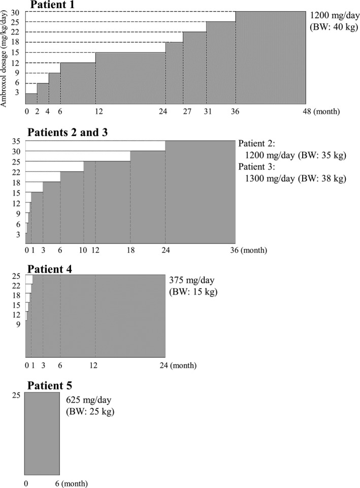
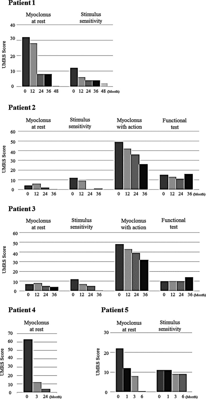
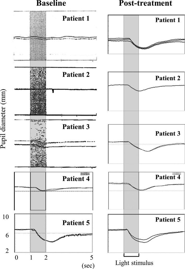
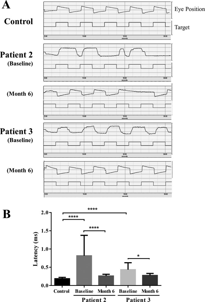
Methods: This open-label pilot study included five patients who received high-dose oral ambroxol in combination with enzyme replacement therapy. Safety was assessed by adverse event query, physical examination, electrocardiography, laboratory studies, and drug concentration. Biochemical efficacy was assessed through evidence of glucocerebrosidase activity in the lymphocytes and glucosylsphingosine levels in the cerebrospinal fluid. Neurological efficacy was evaluated using the Unified Myoclonus Rating Scale, Gross Motor Function Measure, Functional Independence Measure, seizure frequency, pupillary light reflex, horizontal saccadic latency, and electrophysiologic studies.

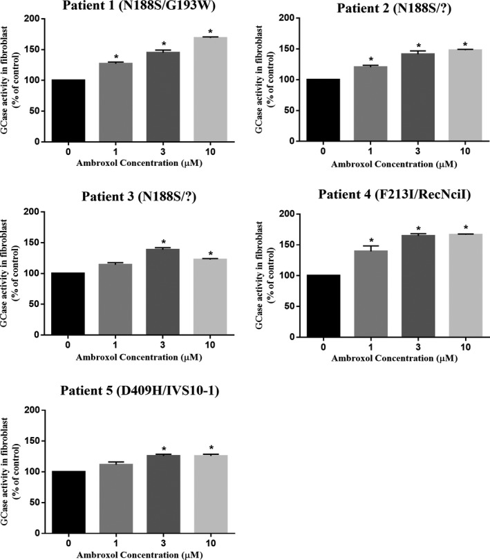
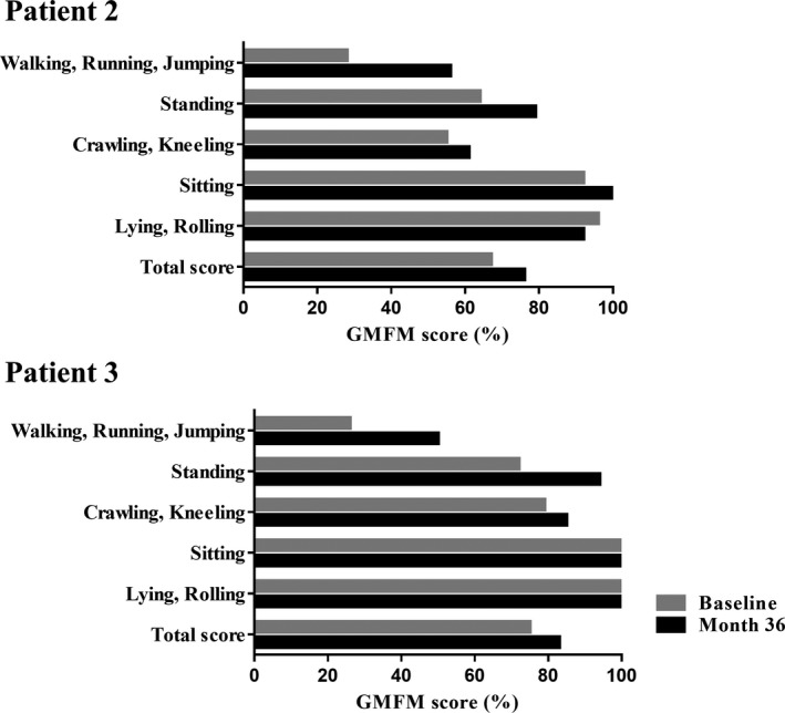
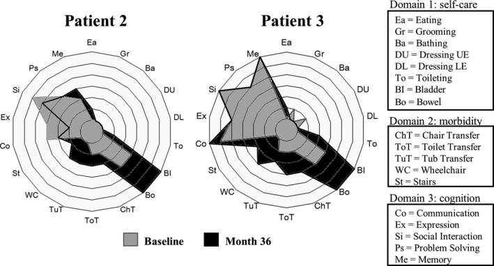
Results: High-dose oral ambroxol had good safety and tolerability, significantly increased lymphocyte glucocerebrosidase activity, permeated the blood-brain barrier, and decreased glucosylsphingosine levels in the cerebrospinal fluid. Myoclonus, seizures, and pupillary light reflex dysfunction markedly improved in all patients. Relief from myoclonus led to impressive recovery of gross motor function in two patients, allowing them to walk again.

Interpretation: Pharmacological chaperone therapy with high-dose oral ambroxol shows promise in treating neuronopathic GD, necessitating further clinical trials.


Figures

Figure 1
Figure 1
Overview of the dosing regimen for each patient. Ambroxol administration was initiated with a starting dose of 3 mg/kg/day divided into three equal doses in the first three patients (patients 1, 2, and 3), because safety information in humans about long‐term high‐dose ambroxol administration was limited. The dose was subsequently increased in increments of 3 mg/kg to reach the target doses (25 mg/kg/day or a maximum dose of 1300 mg/day) over several months to years. For the remaining two patients, ambroxol was initiated at 9 mg/kg/day in patient 4 or 25 mg/kg/day in patient 5. ERT and concomitant medications were continued during the study.
Figure 2
Figure 2
Chaperone effect of ambroxol on mutant GCase activities in GD fibroblasts. Primary skin fibroblasts derived from each patient were incubated with the indicated concentrations of ambroxol for 4 days. In all panels, the data were expressed as the relative increase in GCase activity in the presence of ambroxol compared with that in untreated cells. The results represent the mean ± SEM of three independent experiments. Statistically significant differences between the treated and untreated fibroblasts were elicited, using the Mann–Whitney U‐test. *P < 0.05.
Figure 3
Figure 3
Effects of ambroxol treatment on myoclonus. The Unified Myoclonus Rating Scale (UMRS) was used to evaluate the response of myoclonus to ambroxol therapy in patients with nGD. We evaluated myoclonus at rest in all patients and myoclonus with action, functional tests, and stimulus sensitivity in testable patients. High scores on UMRS reflect a severe condition.
Figure 4
Figure 4
Effects of ambroxol treatment on gross motor function. The graph shows changes in the Gross Motor Function Measure (GMFM) scores after ambroxol treatment for patients 2 and 3, who could be sufficiently examined. The GMFM score of 100% means that the patient's gross motor functions are equivalent to those of a typical 5‐year‐old individual.
Figure 5
Figure 5
Effects of ambroxol treatment on functional status. The polar graph shows changes in the Functional Independence Measure (FIM) ratings after ambroxol treatment for each individual item. The background rings represent scores of 1–7 with an inside‐out order. All items were graded on a 1‐ to 7‐point scale (score 1: total assistance, score 7: complete independence) and the expanding rings indicate the FIM rating.
Figure 6
Figure 6
Effects of ambroxol on PLR to monochromatic light stimulation in nGD patients. Assessments were performed at the following times posttreatment: patient 1, month 48; patients 2 and 3, month 36; patient 4, month 24; and patient 5, month 6.
Figure 7
Figure 7
Effects of ambroxol on horizontal saccadic eye movements. Data were acquired from two testable patients (patients 2 and 3) and six healthy controls (median age = 22 years; range = 22–34 years). (A) Representation of slowed and stepped saccadic eye movements presenting with large latencies in patients at baseline. (B) Comparison of horizontal saccadic latencies in patients with nGD and normal controls.The results present the mean ± SEM. Statistically significant differences between the control and baseline were elicited, using Mann‐Whitney U‐test. Statistically significant differences between the baseline and month 6 were elicited, using Wilcoxon signed‐rank test. *P < 0.05, ****P < 0.0001.

References

    1. Orvisky E, Park JK, LaMarca ME, et al. Glucosylsphingosine accumulation in tissues from patients with Gaucher disease: correlation with phenotype and genotype. Mol Genet Metab 2002;76:262–270. - PubMed
    1. Schueler UH, Kolter T, Kaneski CR, et al. Toxicity of glucosylsphingosine (glucopsychosine) to cultured neuronal cells: a model system for assessing neuronal damage in Gaucher disease type 2 and 3. Neurobiol Dis 2003;14:595–601. - PubMed
    1. Nilsson O, Svennerholm L. Accumulation of glucosylceramide and glucosylsphingosine (psychosine) in cerebrum and cerebellum in infantile and juvenile Gaucher disease. J Neurochem 1982;39:709–718. - PubMed
    1. Grabowski GA, Petsko GA, Kolodny EH. The Online Metabolic and Molecular Basis of Inherited Disease (OMMBID) Chapter 146, 2014.
    1. Grabowski GA. Phenotype, diagnosis, and treatment of Gaucher's disease. Lancet 2008;372:1263–12671. - PubMed