. 2016 Apr 28;532(7600):527-30.
doi: 10.1038/nature17391. Epub 2016 Apr 4.

Crystal structure of the human σ1 receptor

Affiliations

Crystal structure of the human σ1 receptor

Hayden R Schmidt et al. Nature. .

Abstract

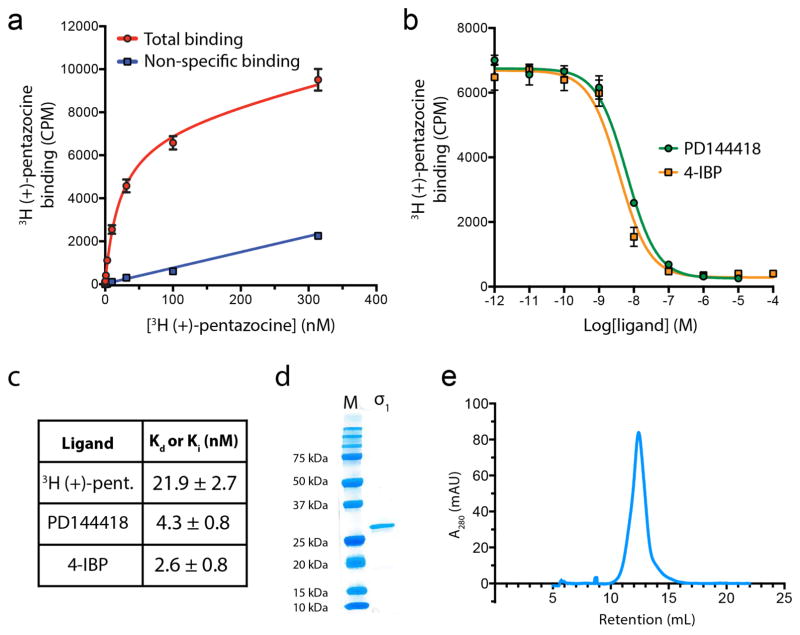
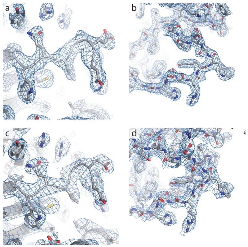
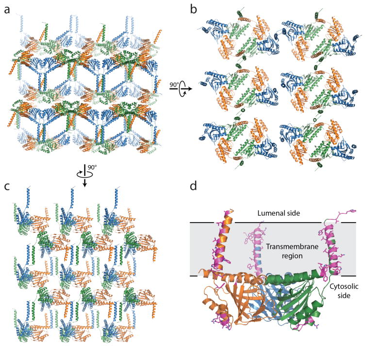
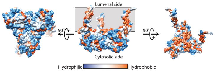
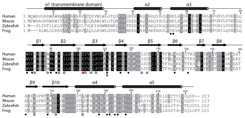
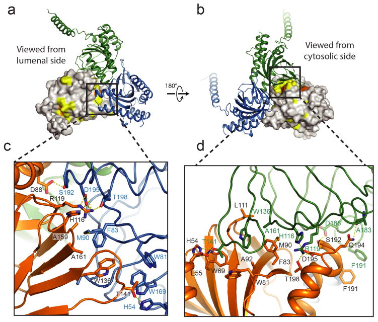
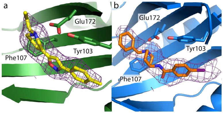
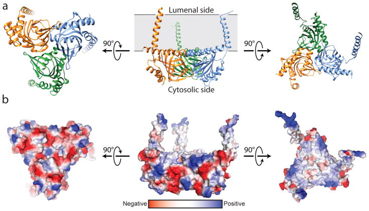
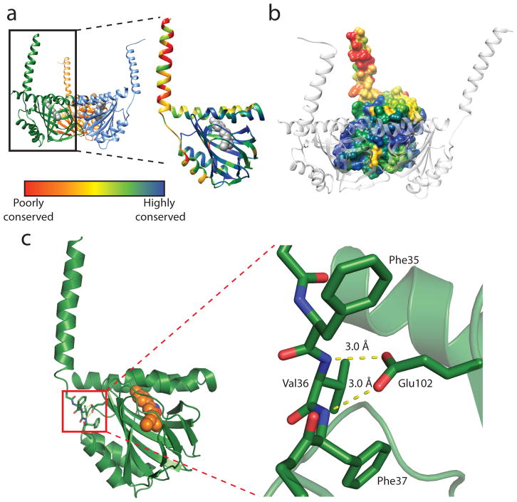
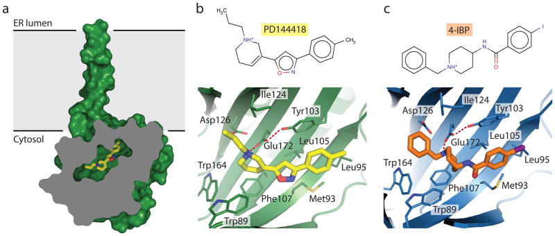
The human σ1 receptor is an enigmatic endoplasmic-reticulum-resident transmembrane protein implicated in a variety of disorders including depression, drug addiction, and neuropathic pain. Recently, an additional connection to amyotrophic lateral sclerosis has emerged from studies of human genetics and mouse models. Unlike many transmembrane receptors that belong to large, extensively studied families such as G-protein-coupled receptors or ligand-gated ion channels, the σ1 receptor is an evolutionary isolate with no discernible similarity to any other human protein. Despite its increasingly clear importance in human physiology and disease, the molecular architecture of the σ1 receptor and its regulation by drug-like compounds remain poorly defined. Here we report crystal structures of the human σ1 receptor in complex with two chemically divergent ligands, PD144418 and 4-IBP. The structures reveal a trimeric architecture with a single transmembrane domain in each protomer. The carboxy-terminal domain of the receptor shows an extensive flat, hydrophobic membrane-proximal surface, suggesting an intimate association with the cytosolic surface of the endoplasmic reticulum membrane in cells. This domain includes a cupin-like β-barrel with the ligand-binding site buried at its centre. This large, hydrophobic ligand-binding cavity shows remarkable plasticity in ligand recognition, binding the two ligands in similar positions despite dissimilar chemical structures. Taken together, these results reveal the overall architecture, oligomerization state, and molecular basis for ligand recognition by this important but poorly understood protein.


Conflict of interest statement

The authors declare no competing financial interests

Figures

Extended Data Figure 1
Extended Data Figure 1. Assessment of σ1 functional properties and biochemical quality
a, Saturation binding curve to measure Kd for 3H (+)-pentazocine, with points shown as mean +/− SEM. b, Competition binding measurement of affinities for the two co-crystallized ligands with points shown as mean +/− SEM. c, Summary of binding affinities with 95% confidence intervals for Kd/Ki values. d, Analysis of receptor purity by SDS-PAGE. e, Analytical size exclusion of purified σ1 receptor in LMNG/CHS detergent buffer on a Superdex 200 column.
Extended Data Figure 2
Extended Data Figure 2. Representative electron density
a, Composite omit 2Fo−Fc electron density contoured at 1.0 σ for σ1 receptor bound to PD144418, showing a loop from Val73 to Glu78 as well as surrounding residues. b, The same map over a loop from His116 to Ser125. c, The equivalent map to that in panel (a), calculated for σ1 receptor bound to 4-IBP. d, The equivalent map to that in panel (b), calculated for σ1 receptor bound to 4-IBP.
Extended Data Figure 3
Extended Data Figure 3. Lattice contacts
a, Lattice packing of the σ1 receptor viewed parallel to the membrane plane. b and c show a view normal to the membrane and another parallel view, respectively. d, A single σ1 trimer is shown, with lattice contact residues highlighted in magenta sticks. Lattice contacts are formed primarily through interactions of the relatively poorly conserved transmembrane helices.
Extended Data Figure 4
Extended Data Figure 4. Hydrophobicity analysis
a, The structure of σ1 receptor shows a hydrophilic (blue) surface on the cytosolic face (left), while transmembrane domains and the membrane-facing surface of the receptor trimer are hydrophobic (orange; right panels). Hydrophobicity analysis was conducted using UCSF Chimera.
Extended Data Figure 5
Extended Data Figure 5. Sequence conservation
The results of an alignment of 277 sigma receptor sequences from a vertebrates with Homo sapiens, Mus musculus, Danio rerio, and Xenopus laevis displayed. Residues with 98%, 80%, and 60% similarity are shown in black, grey, and light grey respectively. Secondary structure elements are shown above the alignment based on the human σ1 receptor crystal structure. Open black circles mark residues within 4 Å of the ligand binding site, solid black circles below the alignment denote residues located in the trimerization interface, and a red circle marks the site of the ALS-associated mutation E102Q.
Extended Data Figure 6
Extended Data Figure 6. Trimerization interface
a, b, Two views of the trimerization interface are shown, colored by sequence conservation. Residues highlighted in yellow are more than 80% conserved among a selection of 300 σ1 receptor homologs, and residues in orange surface are more than 98% conserved. c, d, Closeup views of the interface, showing the extensive hydrophobic and polar contacts at the oligomerization interface.
Extended Data Figure 7
Extended Data Figure 7. Omit maps of PD144418 and 4-IBP
a, An Fo−Fc omit map contoured at 1 σ showing the electron density (purple) of PD144418 (yellow). b, An equivalent map showing the electron density (purple) of 4-IBP (orange).
Extended Data Figure 8
Extended Data Figure 8. Oligomerization state
a, Analysis of receptor oligomerization by size exclusion chromatography with multi-angle light scattering (SEC-MALS) in the presence of the classical antagonist NE-100 or b, the classical agonist SKF-10,047. The peak is 38% detergent and 62% protein by mass. The total mass of each component varies throughout the peak, indicating a mix of oligomeric species. c, Analysis of oligomerization state by blue native PAGE (left) and a higher resolution detergent-supplemented tris-glycine native PAGE gel (right), showing a similar polydisperse profile. Discrete oligomers are marked with red dots, corresponding to possible trimers, hexamers, and higher-order species. d, In a mixed micelle of lauryl maltose neopentyl glycol and cholesterol hemisuccinate modest differences in SEC profile are observed between agonist- and antagonist-treated receptor.
Figure 1
Figure 1. Overall structure of the σ1 receptor
a, Viewed perpendicular to the membrane plane, the σ1 receptor shows a triangular structure comprised of three tightly associated protomers, each with a single transmembrane domain at a corner of the oligomeric triangle. From the side, the receptor reveals a flat membrane-associated surface. The location of the membrane plane is shown in grey, based on PPM server prediction. b, Coloring by electrostatic potential reveals a polar cytosolic surface (left side), and a non-polar membrane-interacting surface flanked by positive charges, suggesting it is partially buried in the membrane.
Figure 2
Figure 2. Structure of the σ1 protomer
a, The receptor shows a cupin-like β-barrel fold flanked by four α-helices with the ligand (grey) bound at the center of the cupin domain. The receptor is colored by sequence conservation, revealing a high degree of conservation in the ligand-binding domain, and relatively lower conservation of the transmembrane helices, which may simply act to tether the receptor to the membrane. b, The intermolecular interface among protomers of the receptor trimer is likewise highly conserved. c, Glu102 forms a pair of hydrogen bonds (yellow dashed lines) with backbone amide nitrogen atoms, providing a structural explanation for receptor destabilization due to the ALS-associated mutation E102Q.
Figure 3
Figure 3. Ligand recognition
a, Cross-section view of the receptor bound to PD144418, showing the deeply buried antagonist and occlusion of the binding pocket from solvent. The ligand is shown in yellow sticks. b, View of PD144418 binding pose, showing charge-charge interaction with Glu172 (red dotted line) and extensive hydrophobic contacts with other binding pocket residues. A hydrogen bond between Glu172 and Tyr103 is also shown as a red dotted line. c, Corresponding structure of the 4-IBP binding pose.

References

    1. Cobos EJ, Entrena JM, Nieto FR, Cendán CM, Del Pozo E. Pharmacology and Therapeutic Potential of Sigma(1) Receptor Ligands. Curr Neuropharmacol. 2008;6:344–366. - PMC - PubMed
    1. Mavlyutov TA, Guo LW, Epstein ML, Ruoho AE. Role of the Sigma-1 receptor in Amyotrophic Lateral Sclerosis (ALS) J Pharmacol Sci. 2015;127:10–16. - PMC - PubMed
    1. Brownstein MJ. A brief history of opiates, opioid peptides, and opioid receptors. Proc Natl Acad Sci USA. 1993;90:5391–5393. - PMC - PubMed
    1. Largent BL, Wikstrom H, Gundlach AL, Snyder SH. Structural determinants of sigma receptor affinity. Mol Pharmacol. 1987;32:772–784. - PubMed
    1. Hanner M, et al. Purification, molecular cloning, and expression of the mammalian sigma1-binding site. Proc Natl Acad Sci USA. 1996;93:8072–8077. - PMC - PubMed

Publication types

MeSH terms