. 2016 Jun;12(6):411-8.
doi: 10.1038/nchembio.2060. Epub 2016 Apr 4.

Decoding Polo-like kinase 1 signaling along the kinetochore-centromere axis

Affiliations

Decoding Polo-like kinase 1 signaling along the kinetochore-centromere axis

Robert F Lera et al. Nat Chem Biol. 2016 Jun.

Abstract

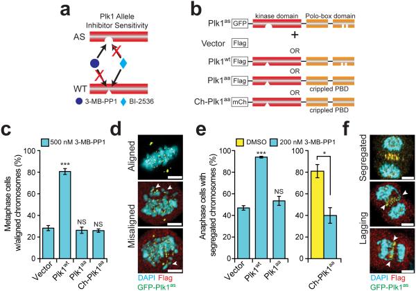
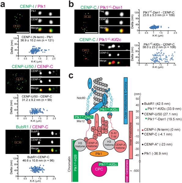
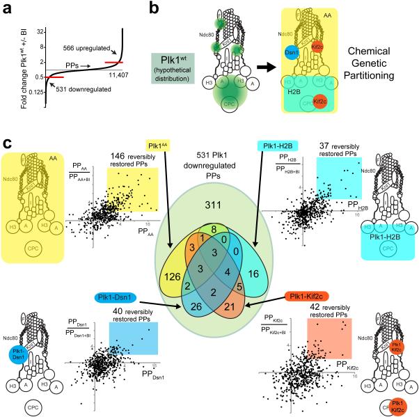
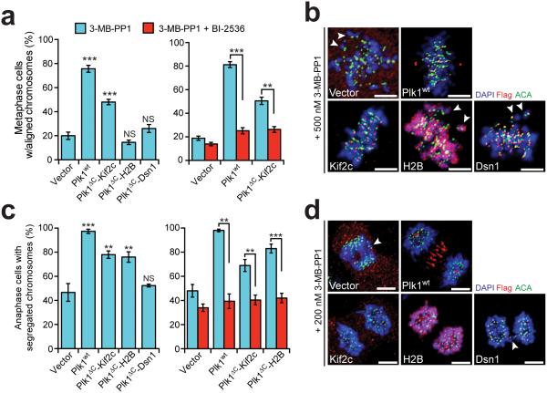
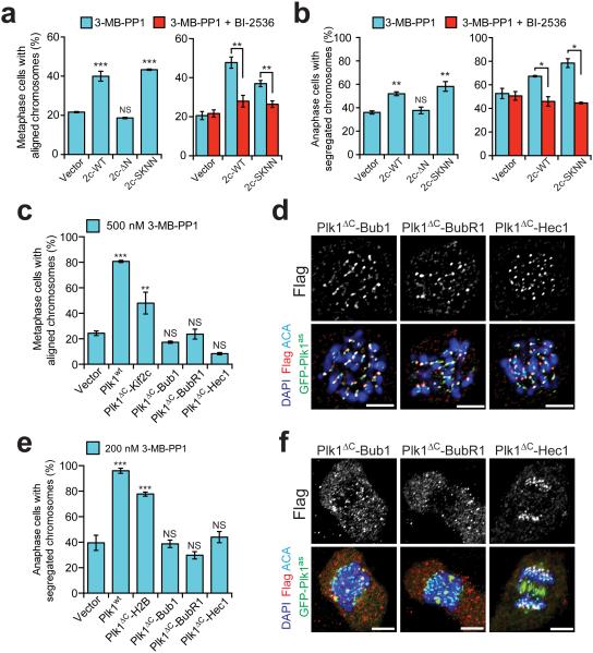
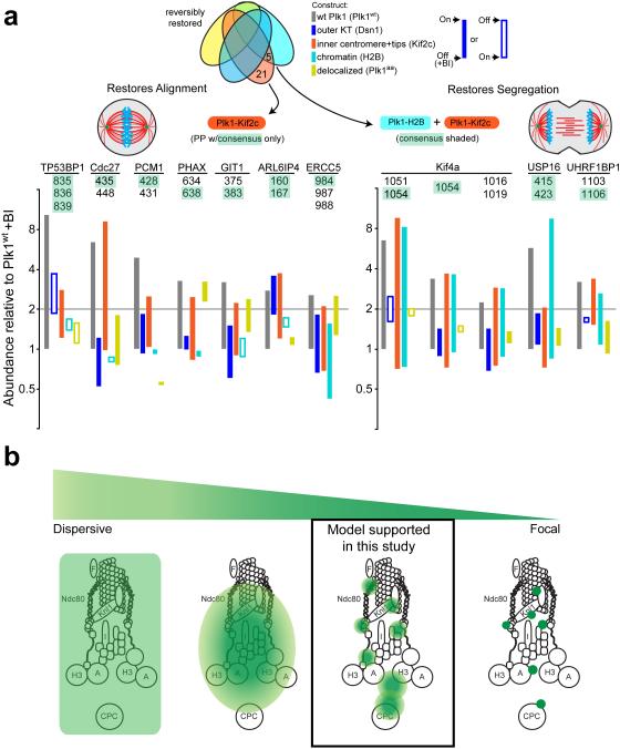
Protein kinase signaling along the kinetochore-centromere axis is crucial to assure mitotic fidelity, yet the details of its spatial coordination are obscure. Here, we examined how pools of human Polo-like kinase 1 (Plk1) within this axis control signaling events to elicit mitotic functions. To do this, we restricted active Plk1 to discrete subcompartments within the kinetochore-centromere axis using chemical genetics and decoded functional and phosphoproteomic signatures of each. We observe distinct phosphoproteomic and functional roles, suggesting that Plk1 exists and functions in discrete pools along this axis. Deep within the centromere, Plk1 operates to assure proper chromosome alignment and segregation. Thus, Plk1 at the kinetochore is a conglomerate of an observable bulk pool coupled with additional functional pools below the threshold of microscopic detection or resolution. Although complex, this multiplicity of locales provides an opportunity to decouple functional and phosphoproteomic signatures for a comprehensive understanding of Plk1's kinetochore functions.


Figures

Figure 1
Figure 1. Plk1 signaling at the kinetochore requires binding via its PBD
(a) Schematic indicating orthogonal control of Plk1 alleles. (b) Schematic of complementation strategy. RPE1 cells expressing GFP-Plk1as (Plk1as) were stably infected with an empty Flag vector (Vector), Flag-tagged wild-type Plk1 (Plk1wt), or Plk1 with a wild-type kinase domain, but crippled Polo-box domain that delocalizes the protein (Plk1aa and Ch-Plk1aa). (c-f) Delocalized Plk1 fails to restore metaphase chromosome alignment or anaphase chromosome segregation when GFP-Plk1as is inhibited with 3-MB-PP1. (c) Graph shows average percentage (± SEM) of pre-anaphase mitotic cells at metaphase with fully aligned chromosomes for each cell line challenged with 500 nM 3-MB-PP1 (n=150 cells/experiment; 3 independent experiments). (d) Representative maximal intensity projection micrographs from (c). Arrowheads indicate misaligned chromosomes. (e) Graph shows average percentage (± SEM) of anaphase cells with fully segregated chromosomes for each cell line challenged with DMSO (yellow bar) or 200 nM 3-MB-PP1 (cyan bars) (n=50 cells/experiment; 4 independent experiments). (f) Representative maximal intensity projection micrographs from (e). Arrowheads indicate lagging chromosomes. *P<.05, ***P<0.0001 by one-way ANOVA or unpaired t-test; NS, not significant (panels c,e). Scale bars, 5 μm (panels d,f).
Figure 2
Figure 2. High-resolution microscopy identifies discrete localization of endogenous Plk1 and kinetochore-tethered Plk1 constructs along the kinetochore-centromere axis
(a) High-resolution single plane images and Delta analysis of endogenous Plk1, CENP-U/50 (also known as PBIP), and BubR1 used for kinetochore mapping in RPE1 cells. K-K indicates distances between the two centroids for CENP-I, CENP-U/50 or BubR1. (b) High-resolution single plane images and Delta analysis of kinetochore-tethered Plk1 constructs. K-K indicates distances between the two centroids for the tethered constructs. (c) Map of kinetochore with summary of Delta measurements from panels a-b set relative to the N-terminus of CENP-I (red). Endogenous proteins indicated in black and Plk1 fusion constructs in green. Positive values are outward toward the spindle microtubules and negative values are inward toward the inner centromere. *CENP-A position was reported previously. Scale bars, 5 μm (panels a-b).
Figure 3
Figure 3. 10-plex TMT phosphoproteomic analysis of Plk1 partitioned by locale within the KT axis
(a) Distribution of phosphopeptides (PPs) regulated by Plk1 activity. Out of a total of 11,407 phosphopeptides encountered, 566 PPs were upregulated (> 2-fold increase +BI/−BI) and 531 downregulated (> 2-fold decrease +/−BI) with exposure to the Plk1wt inhibitor, BI-2536. (b) Hypothetical distribution of Plk1wt (green) within the KT axis and partitioning function by locale within the KT axis (Dsn1-outer KT, Kif2c-outer KT/inner centromere, H2B-chromatin) or by a delocalized control, (Ch-Plk1AA) (c) Identification of phosphopeptides (PPs) that are restored with each tethered Plk1 construct. Each dot on plot represents one of 531 physiologically Plk1-regulated PPs (downregulated >2x with Plk1wt+BI). The x-axis displays its abundance relative to control (Plk1wt+BI) with values >2 indicating that phosphorylation is restored by the construct in question. The y-axis indicates the ratio PP/PP+BI, with values >2 indicating that the restored PP is depleted with chemical inactivation of the regionally-restricted construct. Color-coded regions indicate the PPs that are reversibly restored with each regional construct. The Venn diagram at center illustrates the number of PPs that overlap by the distinct color-coded constructs.
Figure 4
Figure 4. Restricting Plk1 activity along the kinetochore-centromere axis produces distinct phosphoproteomic and functional signatures
Cells expressing GFP-Plk1as and kinetochore-tethered Plk1 constructs were challenged with 3-MB-PP1 (inhibits Plk1as) ± BI-2536 (inhibits Plk1wt/ Plk1ΔC constructs) and assayed for ability of constructs to rescue chromosome alignment during metaphase or segregation during anaphase. (a) Graphs show average percentage (± SEM) of pre-anaphase mitotic cells at metaphase with fully aligned chromosomes for each cell line (n≥100 cells/experiment; 3 independent experiments). (b) Representative maximal intensity projection micrographs from (a). Arrowheads indicate misaligned chromosomes. (c) Graph shows average percentage (± SEM) of anaphase cells with fully segregated chromosomes for each cell line (n≥40 cells/experiment; at least 3 independent experiments). (d) Representative maximal intensity projection micrographs from (c). Arrowheads indicate lagging chromosomes. **P<0.005, ***P<0.0001 by one-way ANOVA or unpaired t-test; NS, not significant (panels a,c). Scale bars, 5 μm (panels b,d).
Figure 5
Figure 5. Outer kinetochore tethering of Plk1 fails to restore chromosome alignment or segregation
(a-b) Cells expressing GFP-Plk1as and Kif2c localization mutants were challenged with 3-MB-PP1 ± BI-2536 and assayed for ability of constructs to rescue chromosome alignment during metaphase or segregation during anaphase. (a) Graphs show average percentage (± SEM) of pre-anaphase mitotic cells at metaphase with fully aligned chromosomes for each cell line (n=150 cells/experiment; 3 independent experiments). (b) Graph shows average percentage (± SEM) of anaphase cells with fully segregated chromosomes for each cell line (n=50 cells/experiment; 4 independent experiments). (c-f) Cells expressing GFP-Plk1as and outer kinetochore-tethered Plk1 constructs were challenged with 3-MB-PP1 and assayed for ability of constructs to rescue chromosome alignment during metaphase or segregation during anaphase. (c) As in panel a (n=150 cells/experiment; 3 independent experiments). (d) Representative single-plane micrographs indicating kinetochore localization of Plk1 tethers in cells failing to restore chromosome alignment. (e) As in panel b (n≥30 cells/experiment; 4 independent experiments). (f) Representative maximal intensity micrographs indicating localization of Plk1 tethers in cells failing to restore chromosome segregation. *P< 0.05, **P<0.005, ***P<0.0001 by one-way ANOVA or unpaired t-test; NS, not significant (panels a,b,c,e). Scale bars, 5 μm (panel d,f).
Figure 6
Figure 6. Functional/proteomic signatures and a model for Plk1 activity in the kinetochore
(a) Phosphopeptides that are restored by constructs concordant with phenotypic rescue of chromosome alignment (left) and accurate chromosome segregation (right). Phosphopeptides (PPs) containing minimal Plk1 consensus are illustrated for each phenotype with bars illustrating difference between +/− BI-2536 for each construct. Left, phosphopeptides that are restored by Plk1wt and Kif2c only. Right, phosphopeptides that are restored with both H2B and Kif2c. (b) A model of Plk1 operation within the kinetochore could range from a highly dispersive operation where one binding partner provides access to all substrates (extreme left), versus focal operation, where Plk1 binds each substrate directly (extreme right). Our data support an intermediate model where Plk1 at each subcompartment can access elicit ~40 phosphorylation events, with minimal overlap between inner centromere, chromatin, and the outer kinetochore.

References

    1. Godek KM, Kabeche L, Compton DA. Regulation of kinetochore-microtubule attachments through homeostatic control during mitosis. Nat Rev Mol Cell Biol. 2015;16:57–64. - PMC - PubMed
    1. London N, Biggins S. Signalling dynamics in the spindle checkpoint response. Nat. Rev. Mol. Cell Biol. 2014;15:736–748. - PMC - PubMed
    1. Foley EA, Kapoor TM. Microtubule attachment and spindle assembly checkpoint signalling at the kinetochore. Nat. Rev. Mol. Cell Biol. 2013;14:25–37. - PMC - PubMed
    1. Liu D, Vader G, Vromans MJM, Lampson MA, Lens SMA. Sensing chromosome bi-orientation by spatial separation of aurora B kinase from kinetochore substrates. Science. 2009;323:1350–1353. - PMC - PubMed
    1. Welburn JPI, et al. Aurora B Phosphorylates Spatially Distinct Targets to Differentially Regulate the Kinetochore-Microtubule Interface. Mol. Cell. 2010;38:383–392. - PMC - PubMed

MeSH terms

Substances

LinkOut - more resources