. 2016 May;19(5):742-748.
doi: 10.1038/nn.4285. Epub 2016 Apr 4.

A GABAergic nigrotectal pathway for coordination of drinking behavior

Affiliations

A GABAergic nigrotectal pathway for coordination of drinking behavior

Mark A Rossi et al. Nat Neurosci. 2016 May.

Abstract

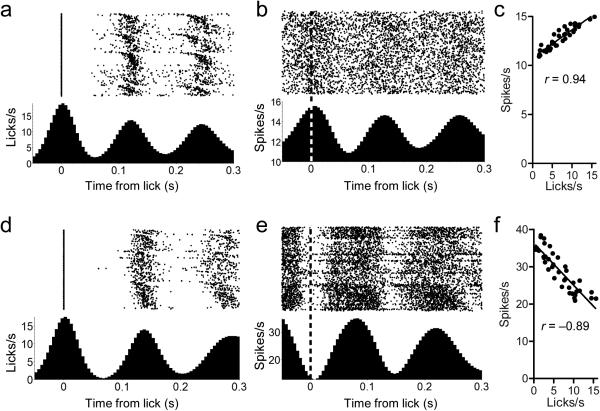
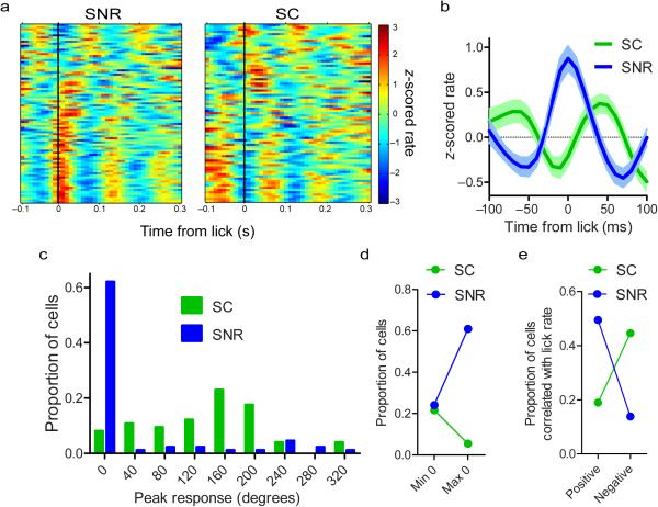
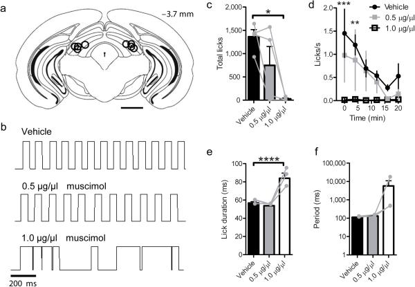
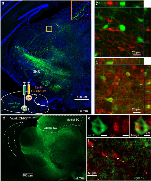
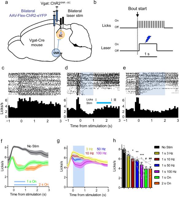
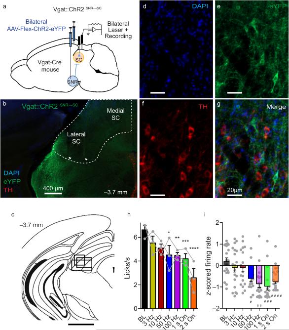
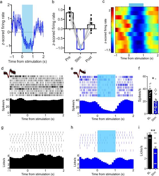
The contribution of basal ganglia outputs to consummatory behavior remains poorly understood. We recorded from the substantia nigra pars reticulata (SNR), the major basal ganglia output nucleus, during self-initiated drinking in mice. The firing rates of many lateral SNR neurons were time-locked to individual licks. These neurons send GABAergic projections to the deep layers of the orofacial region of the lateral tectum (superior colliculus, SC). Many tectal neurons were also time-locked to licking, but their activity was usually in antiphase with that of SNR neurons, suggesting inhibitory nigrotectal projections. We used optogenetics to selectively activate the GABAergic nigrotectal afferents in the deep layers of the SC. Photo-stimulation of the nigrotectal projections transiently inhibited the activity of the lick-related tectal neurons, disrupted their licking-related oscillatory pattern and suppressed self-initiated drinking. These results demonstrate that GABAergic nigrotectal projections have a crucial role in coordinating drinking behavior.


Figures

Figure 1
Figure 1. SNR and SC activity is time-locked to the lick cycle
(a) Rate histogram of licks occurring within a bout of licking in a mouse during SNR recording. Licks are aligned to the start of spout contact. Licking proceeds in rhythmic oscillations, shown as peaks and valleys in lick rate. (b) Representative lick-related oscillatory activity of SNR neuron. The oscillations in the neural activity are tightly coupled with the oscillations of the lick cycle. Dashed line indicates the time of spout contact. (c) Firing rate of the neuron in (b) is positively correlated with instantaneous lick rate, y axis value shown in (a) (p = 1.91.53 × 10−19). (d) Rate histogram of licks occurring within a bout of licking of a mouse during SC recording. (e) Representative lick-related oscillatory activity of SC neuron. The oscillations in the neural activity are tightly coupled with the oscillations of the lick cycle but are antiphase to the SNR oscillations. Dashed line indicates the time of spout contact. (f) Firing rate of the neuron in (e) is negatively correlated with instantaneous lick rate—y axis value shown in (d) (p = 8.38 × 10⁻¹⁵).
Figure 2
Figure 2. SNR and SC exhibit antiphase oscillations during licking
(a) Population spike density functions showing oscillatory activity of SNR (n = 87 neurons from 6 mice) and SC (n = 74 neurons from 4 mice) neurons. Each row corresponds to the activity of one neuron. (b) Population average and s.e.m. of neural activity aligned to the start of each lick. (c) Distribution of the point within the lick cycle (in degrees; 0 is the start of contact) of the peak neural response of SNR and SC neurons. (d) Proportion of neurons whose maximum or minimum response rate at the 0 phase of the lick cycle (χ2 = 13.31, p = 1.90 × 10⁻⁵). (e) Proportion of neurons whose firing rate is positively or negatively correlated (p < 0.05) with the instantaneous lick rate during a bout of licking (χ2 = 24.08, p = 9.20 × 10⁻⁷).
Figure 3
Figure 3. Muscimol injection into the lateral SC suppresses voluntary licking
(a) Summary of bilateral cannula placements within lateral SC (n = 4 mice). Scale bar is 1 mm. (b) Lick traces from a single mouse following injection of vehicle or muscimol. Upward deflections indicate contact with the lick spout. (c) The number of licks was dose-dependently reduced by muscimol infusion (p = 0.019 Vehicle vs 1.0 μg/μl muscimol). (d) Lick rate over time (two-way repeated measures ANOVA [Time × Dose]: Main effect of time (F (7,48) = 3.75, p = 0.007), main effect of Dose (F (2,48) = 12.95, p = 0.00015), no Interaction (F (14, 48) = 1.16, p = 0.33). Bonferroni post hoc test Vehicle vs 1.0 μg/μl muscimol at 0 (*** p = 0.0003) and 4 min (** p = 0.0021). (e-f) For mice that licked at all doses of muscimol (n = 3), lick duration was increased (**** p = 0.008), and the period was not significantly affected (p = 0.41). Values are mean and s.e.m. Individual data points are overlayed in gray.
Figure 4
Figure 4. SNR GABA neurons project to lateral SC
(a) Lenti-FuGB2-Cre was injected into lateral SC yielding retrogradely-transported Cre. AAV-FLEX-GFP was injected in SNR, resulting in selective expression of GFP in nigrotectal neurons. Bottom inset shows nigrotectal axons in the lateral SC. (b) No overlap is observed between TH-expressing and GFP-expressing neurons. (c) GFP colocalizes with Vgat-expressing neurons. Experiment was conducted in a single mouse. (d-f) Nigrotectal projections were targeted with ChR2. AAV-Flex-ChR2-eYFP was injected into the SNR of Vgat-Cre mice. Optic fibers targeted the nigrotectal axons within the lateral SC. (d) Example of Vgat-ChR2-eYFP expression. (e) eYFP overlap with Vgat. (f) eYFP does not overlap with TH. Experiment was successfully repeated on 7 mice.
Figure 5
Figure 5. Vgat::ChR2SNR→SC activation disrupts consummatory behavior
(a) Schematic representation of optogenetic stimulation strategy. Cre-dependent ChR2 was bilaterally injected into the SNR of Vgat-Cre mice (n = 7). Optic fibers targeted nigrotectal axon terminals in the lateral SC. (b) Depiction of lick-triggered optogenetic stimulation. A computer program triggered the laser after the start of a licking bout. (c-e) Peri-stimulation lick plots from one mouse. When no stimulation is present (c), the lick rate remains elevated following the initiation of a lick bout. When 1 s (d) or 2 s (e) constant illumination (~10 mW) is delivered during a bout, licking is temporarily interrupted and rebounds following the termination of stimulation. The inset in (d) is a single trial example of when licking was completely abolished during stimulation. (f-g) Group (n = 7 mice) average (±s.e.m) peri-stimulus lick rate with constant (f) or pulsed (g) illumination. (h) Lick rate during stimulation decreased as a function of stimulation frequency. * p = 0.04, ** p = 0.018, *** p = 0.003, # p = 8.88 × 10⁻⁴, ## p = 9.00 × 10⁻⁴ compared to No Stim condition. Bars are mean + s.e.m.
Figure 6
Figure 6. Vgat::ChR2SNR→SC stimulation inhibits SC neurons and suppresses consummatory behavior
(a) Schematic representation of optrode recording strategy. Cre-dependent ChR2 was bilaterally injected into the SNR of Vgat-Cre mice (n = 3). An optic fiber was placed in the SC of one hemisphere and an optrode in the other. (b) Representative placement of optrode within the lateral SC. Electrode locations are indicated by arrows. The SC is outlined in white. (c) The spread of electrode placements is indicated by a single rectangle corresponding to each mouse. Scale bar is 1 mm. (d-g) eYFP-expressing neurons do not overlap with TH-expressing neurons. For panels b and d-g, the experiment was successfully repeated on three mice. (h) Vgat::ChR2SNR→SC stimulation reduced lick rate. (i) SC population activity is suppressed by Vgat::ChR2SNR→SC activation (n = 135 units recorded from 3 mice). * p = 0.007, ** p = 0.006, ***p = 0.002, **** p = 7.8 × 10⁻⁵, # p = 6.0 × 10⁻⁵, ## p = 5.65 × 10⁻³, ### p = 1.02 × 10⁻⁴, #### p = 1.86 × 10⁻⁴ compared to BL. Error bars are s.e.m.
Figure 7
Figure 7. When Vgat::ChR2SNR→SC stimulation inhibits SC neurons, consummatory behavior is suppressed
(a-c) 65% (11/17 neurons from 3 mice) of neurons were inhibited during 1 s constant illumination. (a) Average response of all inhibited neurons. (b) Inhibition was transient, and spike rates rebounded immediately after stimulation. (c) Spike density function showing activity of inhibited neurons during stimulation. (d-e) Single unit activity recorded during licking. (d) The neuron is tonically active during licking on baseline trials in which the laser was off. (e) Constant illumination during licking suppresses firing. Waveforms recorded during baseline trials and when the laser was on are shown above the rasters (red lines are median waveforms). Waveform shape is unchanged by laser. (f) This neuron is inhibited by Vgat::ChR2SNR→SC stimulation (t-test, 10 baseline trials and 26 stimulation trials: t(34) = 3.85, * p = 0.0005). (g) Licking behavior during the trials in a. (h) When the laser is on, the lick rate is suppressed. (i) Lick rate is significantly reduced by Vgat::ChR2 SNR→SC stimulation (t-test with Welch's correction for unequal variance, 10 baseline trials and 26 stimulation trials: t(31) = 6.27, ** p = 5.31 × 10⁻⁷). Values are mean ±s.e.m.

References

    1. Fowler SC, Mortell C. Low doses of haloperidol interfere with rat tongue extensions during licking: a quantitative analysis. Behav Neurosci. 1992;106:386–395. - PubMed
    1. Nakamura S, Muramatsu S, Yoshida M. Role of the basal ganglia in manifestation of rhythmical jaw movement in rats. Brain research. 1990;535:335–338. - PubMed
    1. Travers JB, Dinardo LA, Karimnamazi H. Motor and premotor mechanisms of licking. Neurosci Biobehav Rev. 1997;21:631–647. - PubMed
    1. Alexander GE, Crutcher MD. Functional architecture of basal ganglia circuits: neural substrates of parallel processing. Trends Neurosci. 1990;13:266–271. - PubMed
    1. Yin HH, Ostlund SB, Balleine BW. Reward-guided learning beyond dopamine in the nucleus accumbens: the integrative functions of cortico-basal ganglia networks. Eur J Neurosci. 2008;28:1437–1448. - PMC - PubMed

Publication types