. 2016 Apr 1;21(4):443.
doi: 10.3390/molecules21040443.

Silymarin Prevents Restraint Stress-Induced Acute Liver Injury by Ameliorating Oxidative Stress and Reducing Inflammatory Response

Affiliations

Silymarin Prevents Restraint Stress-Induced Acute Liver Injury by Ameliorating Oxidative Stress and Reducing Inflammatory Response

Sou Hyun Kim et al. Molecules. .

Abstract

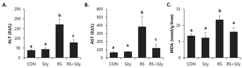
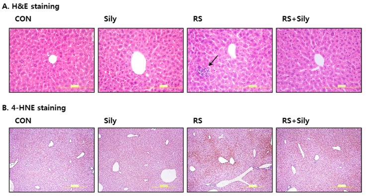
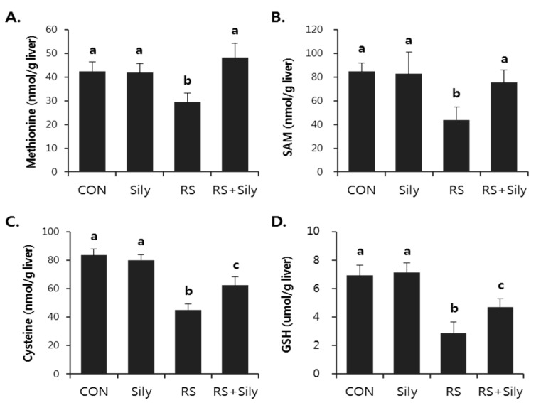
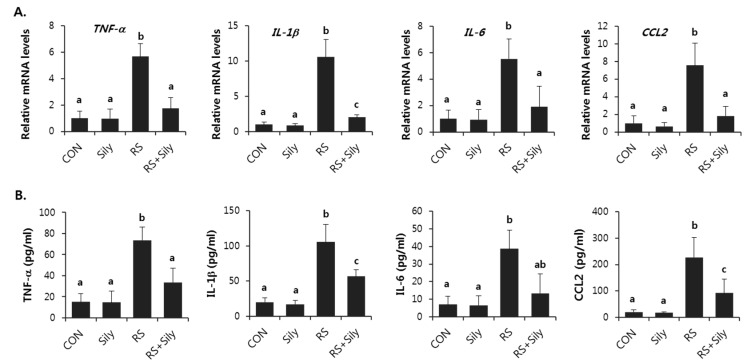
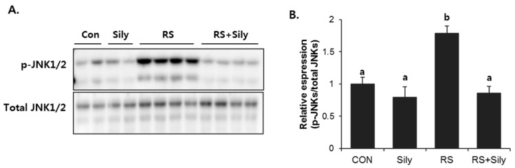
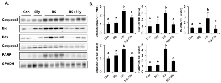
Silymarin is a flavonoid extracted from the milk thistle Silybum marianum. It has been reported to prevent liver injuries induced by various chemicals or toxins. Our recent study suggested that silymarin induces hepatic synthesis of glutathione by increasing cysteine availability, which may consequently contribute to increased antioxidant capacity of the liver. In the present study, we investigated the effects of silymarin on acute liver injury induced by restraint stress. Silymarin (100 mg/kg) was orally administered to BALB/c mice every 12 h (3 times in total). After the last dose, mice were subjected to restraint stress for 6 h, and serum levels of aspartate and alanine aminotransferases, and hepatic levels of lipid peroxidation were determined. Hepatic levels of sulfur-containing metabolites such as methionine, S-adenosylmethionine, cysteine, and glutathione were also measured. The level of pro-inflammatory mediators in both liver and serum was determined. To study the mechanism of the effects of silymarin, we assessed Jun N-terminal kinase (JNK) activation and apoptotic signaling. Restraint stress induced severe oxidative stress and increased mRNA levels of pro-inflammatory mediators; both effects of restraint stress were significantly inhibited by silymarin. Moreover, administration of silymarin significantly prevented acute liver injury induced by restraint stress by blocking JNK activation and subsequently apoptotic signaling. In conclusion, these results suggest that the inhibition of restraint stress-induced liver injury by silymarin is due at least in part to its anti-oxidant activity and its ability to suppress the inflammatory response.

Keywords: acute liver injury; inflammation; oxidative stress; restraint stress; silymarin.


Conflict of interest statement

The authors declare that they have no financial or nonfinancial competing interests.

Figures

Figure 1
Figure 1
Silymarin inhibited restraint-induced acute liver injury. Serum activity of ALT (A) and AST (B); and hepatic level of MDA (C) were analyzed to examine liver injury. Each value is the mean ± SD for 4 mice. Values with different letters (a, b, c) are significantly different from one another (one-way ANOVA followed by Newman-Keuls multiple range test, p < 0.05). CON; control mice, Sily; only silymarin-treated mice, RS; restraint stressed mice, RS + Sily; restraint-stressed mice with silymarin treatment.
Figure 2
Figure 2
Effect of silymarin on histological changes of the liver in mice subjected to restraint stress. (A) Hematoxylin and eosin (H & E) staining of representative liver sections shown at the same magnification (400×). Only mice subjected to restraint showed infilterated cells in the liver parenchyma. Black arrow represents inflammatory cell infiltration; (B) Representative pictures of 4-HNE protein adducts with brown color. 4-HNE positivity was observed mostly in hepatocytes around the central vein and it was most clear in the liver of restraint stressed mice. (CON; control mice, Sily; only silymarin-treated mice, RS; restraint stressed mice, RS + Sily; restraint-stressed mice with silymarin treatment).
Figure 3
Figure 3
Silymarin ameliorated restraint-induced GSH depletion via down-regulation of sulfur-containing amino acid metabolism. Hepatic level of methionine (A); SAM (B); cysteine (C); and GSH (D) were determined. Each value is the mean ± SD for four mice. Values with different letters (a, b, c) are significantly different from one another (one-way ANOVA followed by Newman-Keuls multiple range test, p < 0.05). CON; control mice, Sily; only silymarin-treated mice, RS; restraint stressed mice, RS + Sily; restraint-stressed mice with silymarin treatment.
Figure 4
Figure 4
Silymarin prevented increase in mRNA (A) and serum levels (B) of pro-inflammatory mediators induced by restraint stress. Each value is the mean ± SD for four mice. Values with different letters (a, b, c) are significantly different from one another (one-way ANOVA followed by Newman-Keuls multiple range test, p < 0.05). CON; control mice, Sily; only silymarin-treated mice, RS; restraint stressed mice, RS + Sily; restraint-stressed mice with silymarin treatment.
Figure 5
Figure 5
Silymarin treatment prevented restraint-induced JNKs phosphorylation. (A) Immunoblot analysis of phopho-JNK1/2 and total JNK1/2. (B) Quantitative analysis of blots. Each value is the mean ± SD for four mice. Values with different letters (a, b) are significantly different from one another (one-way ANOVA followed by Newman-Keuls multiple range test, p < 0.05). CON; control mice, Sily; only silymarin-treated mice, RS; restraint stressed mice, RS + Sily; restraint-stressed mice with silymarin treatment.
Figure 6
Figure 6
Silymarin treatment reduced restraint-induced apoptotic cascade. (A) Immunoblot analysis of caspase8, Bid, Bax, caspase3, PARP, and GAPDH. (B) Quantitative analysis of blots. Each value is the mean ± SD for four mice. Values with different letters (a, b) are significantly different from one another (one-way ANOVA followed by Newman-Keuls multiple range test, p < 0.05). CON; control mice, Sily; only silymarin-treated mice, RS; restraint stressed mice, RS + Sily; restraint-stressed mice with silymarin treatment.

References

    1. Moore C.J., Cunningham S.A. Social position, psychological stress, and obesity: A systematic review. J. Acad. Nutr. Diet. 2012;112:518–526. doi: 10.1016/j.jand.2011.12.001. - DOI - PubMed
    1. Rosenberger C., Elsenbruch S., Scholle A., de Greiff A., Schedlowski M., Forsting M., Gizewski E.R. Effects of psychological stress on the cerebral processing of visceral stimuli in healthy women. Neurogastroenterol. Motil. 2009;21:e740–e745. doi: 10.1111/j.1365-2982.2009.01295.x. - DOI - PubMed
    1. Stojanovich L., Marisavljevich D. Stress as a trigger of autoimmune disease. Autoimmun. Rev. 2008;7:209–213. doi: 10.1016/j.autrev.2007.11.007. - DOI - PubMed
    1. Cao H.J., Tan R.R., He R.R., Tang L.P., Wang X.L., Yao N., Duan W.J., Hu Y.A., Yao X.S., Kurihara H. Sarcandra glabra extract reduces the susceptibility and severity of influenza in restraint-stressed mice. Evid. Based Complement. Altern. Med. 2012;2012 doi: 10.1155/2012/236539. - DOI - PMC - PubMed
    1. He R.R., Wang M., Wang C.Z., Chen B.T., Lu C.N., Yao X.S., Chen J.X., Kurihara H. Protective effect of apple polyphenols against stress-provoked influenza viral infection in restraint mice. J. Agric. Food Chem. 2011;59:3730–3737. doi: 10.1021/jf104982y. - DOI - PubMed

Publication types