

Morphologic Analysis of the Temporomandibular Joint Between Patients With Facial Asymmetry and Asymptomatic Subjects by 2D and 3D Evaluation: A Preliminary Study
- PMID: 27043669
- PMCID: PMC4998530
- DOI: 10.1097/MD.0000000000003052
Morphologic Analysis of the Temporomandibular Joint Between Patients With Facial Asymmetry and Asymptomatic Subjects by 2D and 3D Evaluation: A Preliminary Study
Abstract
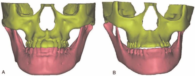
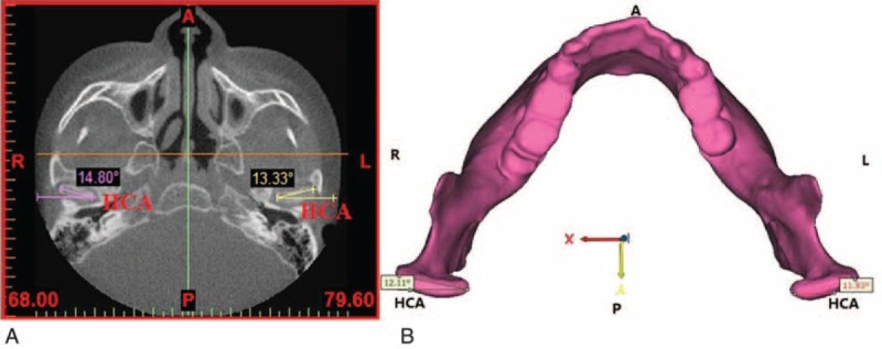
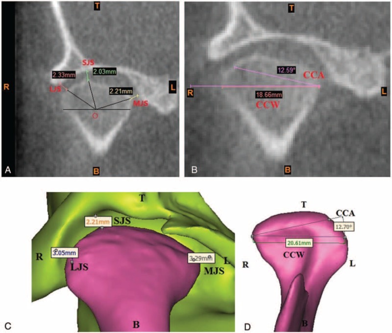
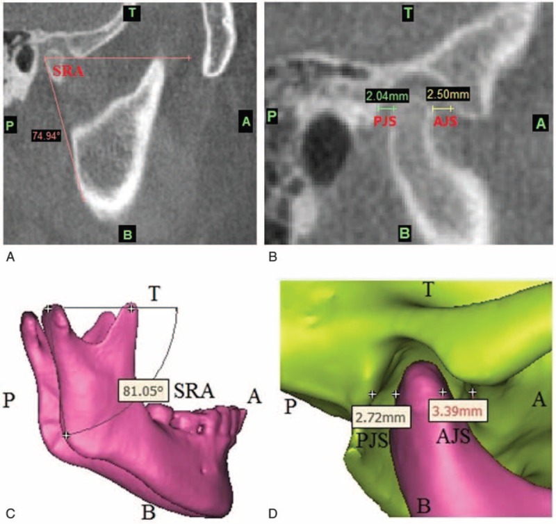
Signs and symptoms of temporomandibular joint (TMJ) dysfunction are commonly found in patients with facial asymmetry. Previous studies on the TMJ position have been limited to 2-dimensional (2D) radiographs, computed tomography (CT), or cone-beam computed tomography (CBCT). The purpose of this study was to compare the differences of TMJ position by using 2D CBCT and 3D model measurement methods. In addition, the differences of TMJ positions between patients with facial asymmetry and asymptomatic subjects were investigated. We prospectively recruited 5 patients (cases, mean age, 24.8 ± 2.9 years) diagnosed with facial asymmetry and 5 asymptomatic subjects (controls, mean age, 26 ± 1.2 years). The TMJ spaces, condylar and ramus angles were assessed by using 2D and 3D methods. The 3D models of mandible, maxilla, and teeth were reconstructed with the 3D image software. The variables in each group were assessed by t-test and the level of significance was 0.05. There was a significant difference in the horizontal condylar angle (HCA), coronal condylar angle (CCA), sagittal ramus angle (SRA), medial joint space (MJS), lateral joint space (LJS), superior joint space (SJS), and anterior joint space (AJS) measured in the 2D CBCT and in the 3D models (P < 0.05). The case group had significantly smaller SJS compared to the controls on both nondeviation side (P = 0.009) and deviation side (P = 0.004). In the case group, the nondeviation SRA was significantly larger than the deviation side (P = 0.009). There was no significant difference in the coronal condylar width (CCW) in either group. In addition, the anterior disc displacement (ADD) was more likely to occur on the deviated side in the case group. In conclusion, the 3D measurement method is more accurate and effective for clinicians to investigate the morphology of TMJ than the 2D method.
Conflict of interest statement
The authors have no conflicts of interest to disclose.
Figures
Similar articles
-
Morphological study of the changes after sagittal split ramus osteotomy in patients with facial asymmetry: measurements of 3-dimensional modelling.Br J Oral Maxillofac Surg. 2018 Dec;56(10):925-930. doi: 10.1016/j.bjoms.2018.10.005. Epub 2018 Oct 31. Br J Oral Maxillofac Surg. 2018. PMID: 30391085
-
Comparison of Morphologic Parameters of Temporomandibular Joint for Asymptomatic Subjects Using the Two-Dimensional and Three-Dimensional Measuring Methods.J Healthc Eng. 2017;2017:5680708. doi: 10.1155/2017/5680708. Epub 2017 May 2. J Healthc Eng. 2017. PMID: 29065621 Free PMC article.
-
Changes in temporomandibular joint and ramus after sagittal split ramus osteotomy in mandibular prognathism patients with and without asymmetry.J Craniomaxillofac Surg. 2012 Dec;40(8):821-7. doi: 10.1016/j.jcms.2012.03.003. Epub 2012 Apr 14. J Craniomaxillofac Surg. 2012. PMID: 22507292
-
Application of cone beam computed tomography for assessment of the temporomandibular joints.Aust Dent J. 2012 Mar;57 Suppl 1:109-18. doi: 10.1111/j.1834-7819.2011.01663.x. Aust Dent J. 2012. PMID: 22376103 Review.
-
Temporomandibular Joint Anatomy Assessed by CBCT Images.Biomed Res Int. 2017;2017:2916953. doi: 10.1155/2017/2916953. Epub 2017 Feb 2. Biomed Res Int. 2017. PMID: 28261607 Free PMC article. Review.
Cited by
-
Morphological Analysis of the Temporomandibular Joint in Patients with Malocclusion Using 2D and 3D Measuring Methods.Iran J Public Health. 2020 Sep;49(9):1796-1798. doi: 10.18502/ijph.v49i9.4101. Iran J Public Health. 2020. PMID: 33643956 Free PMC article. No abstract available.
-
Correlation between temporomandibular joints and craniocervical posture in patients with bilateral anterial disc displacement.BMC Oral Health. 2024 Feb 1;24(1):159. doi: 10.1186/s12903-024-03892-9. BMC Oral Health. 2024. PMID: 38297238 Free PMC article.
-
Disc Displacement of the Temporomandibular Joint and Facial Asymmetry in Children and Adolescents: A Systematic Review and Meta-Analysis.Children (Basel). 2022 Aug 27;9(9):1297. doi: 10.3390/children9091297. Children (Basel). 2022. PMID: 36138607 Free PMC article. Review.
-
Effects of osseous structure based on three-dimensional reconstructive imaging evaluation in the assessment of temporomandibular joint disc position.Clin Oral Investig. 2023 Apr;27(4):1449-1463. doi: 10.1007/s00784-023-04936-0. Epub 2023 Mar 6. Clin Oral Investig. 2023. PMID: 36877406
-
Correlations Between Mandibular Asymmetries and Temporomandibular Disorders: A Systematic Review.J Int Soc Prev Community Dent. 2021 Jul 3;11(5):481-489. doi: 10.4103/jispcd.JISPCD_130_21. eCollection 2021 Sep-Oct. J Int Soc Prev Community Dent. 2021. PMID: 34760791 Free PMC article. Review.
References
-
- Bishara SE, Burkey PS, Kharouf JG. Dental and facial asymmetries: a review. Angle Orthod 1994; 64:89–98. - PubMed
-
- Bergersen EO. Enlargement and distortion in cephalometric radiography: compensation tables for linear measurements. Angle Orthod 1980; 50:230–244. - PubMed
-
- Wang H, Wang B, Ji C, et al. The prevalence of malocclusion and mandible deviation in Beijing. Beijing J Stomatology 2002; 10:70–72.
-
- Hua Z, Song J, Yanqiong L. Clinical study of orthognathic surgery for the treatment of dentofacial deformities. J Stomatology 2003; 23:219–223.
-
- Ueki K, Nakagawa K, Marukawa K, et al. The relationship between temporomandibular joint disc morphology and stress angulation in skeletal Class III patients. Eur J Orthod 2005; 27:501–506. - PubMed
Publication types
MeSH terms
LinkOut - more resources
Full Text Sources
Other Literature Sources
