. 2016 Apr 19;113(16):4494-9.
doi: 10.1073/pnas.1601720113. Epub 2016 Apr 4.

Proteus mirabilis fimbriae- and urease-dependent clusters assemble in an extracellular niche to initiate bladder stone formation

Affiliations

Proteus mirabilis fimbriae- and urease-dependent clusters assemble in an extracellular niche to initiate bladder stone formation

Jessica N Schaffer et al. Proc Natl Acad Sci U S A. .

Abstract

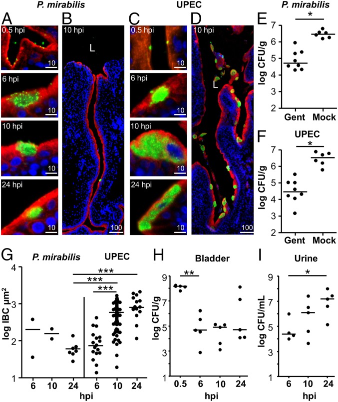
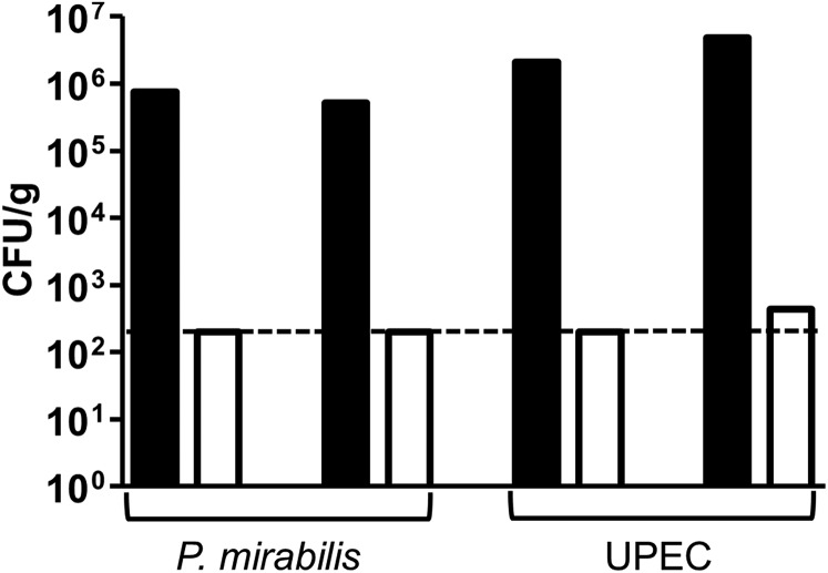
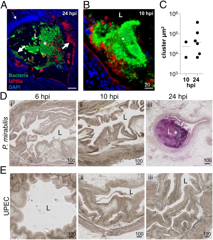
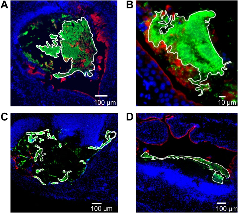
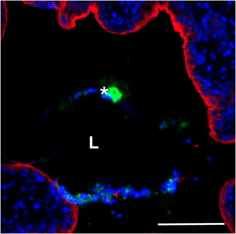
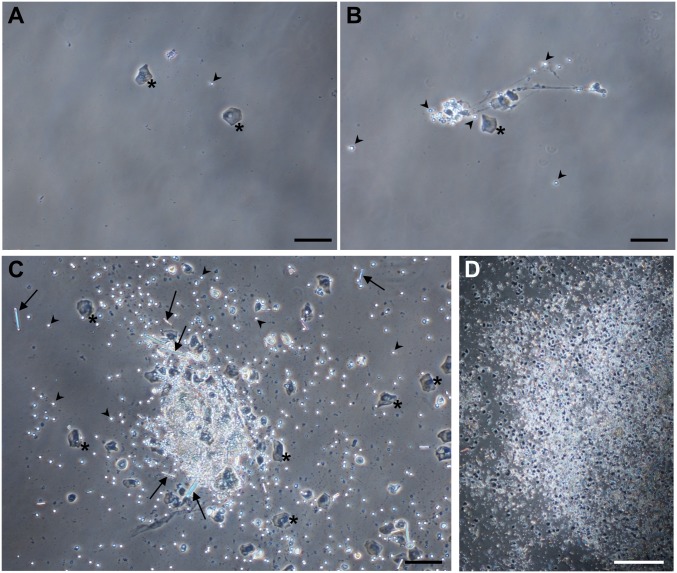
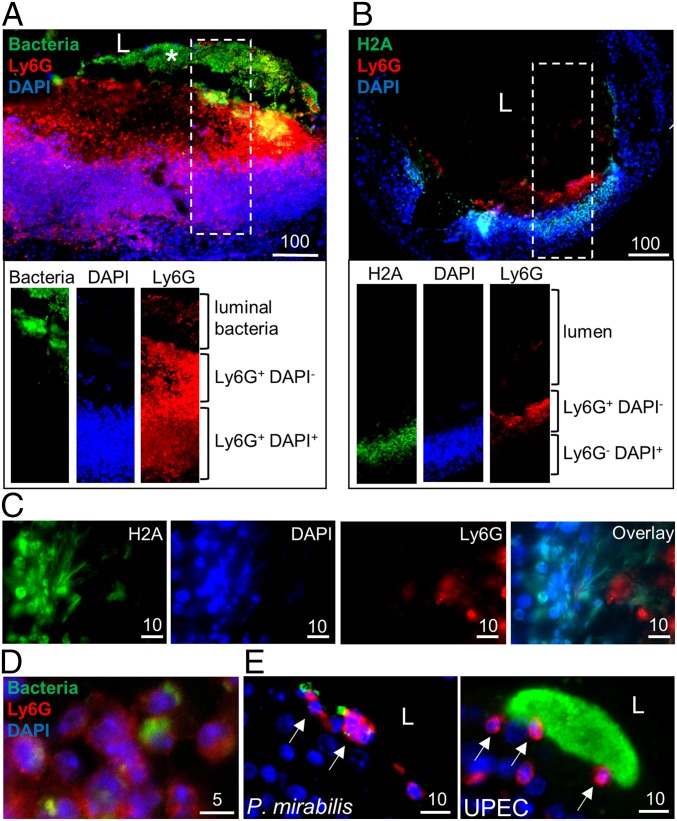
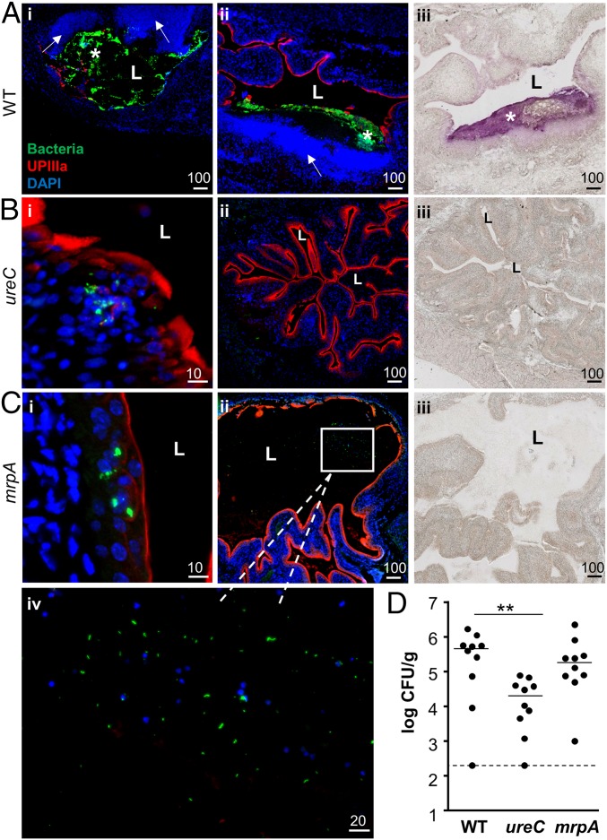
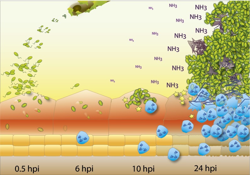
The catheter-associated uropathogenProteus mirabilisfrequently causes urinary stones, but little has been known about the initial stages of bladder colonization and stone formation. We found thatP. mirabilisrapidly invades the bladder urothelium, but generally fails to establish an intracellular niche. Instead, it forms extracellular clusters in the bladder lumen, which form foci of mineral deposition consistent with development of urinary stones. These clusters elicit a robust neutrophil response, and we present evidence of neutrophil extracellular trap generation during experimental urinary tract infection. We identified two virulence factors required for cluster development: urease, which is required for urolithiasis, and mannose-resistantProteus-like fimbriae. The extracellular cluster formation byP. mirabilisstands in direct contrast to uropathogenicEscherichia coli, which readily formed intracellular bacterial communities but not luminal clusters or urinary stones. We propose that extracellular clusters are a key mechanism ofP. mirabilissurvival and virulence in the bladder.

Keywords: Proteus mirabilis; bladder stones; fimbriae; urease; urinary tract infection.


Conflict of interest statement

The authors declare no conflict of interest.

Figures

Fig. 1.
Fig. 1.
P. mirabilis invades the urothelium, but infrequently forms IBCs. (AD) Representative images of P. mirabilis (A and B) and UPEC (C and D) attachment and invasion. Bacteria (green), UPIIIa (red), and DNA (blue) show localization of the bacteria relative to the apical surface of the urothelium. (Scale bars, 10 µm.) (B and D) A regional view of the bladder section containing the 10 hpi IBC shown in A and C, respectively. (Scale bars, 100 µm.) L, bladder lumen. (E and F) Quantification of P. mirabilis (E) and UPEC (F) bladder invasion at 0.5 hpi following either ex vivo gentamicin treatment (Gent) or mock treatment (Mock) (n = 6–8). *P < 0.05. (G) Size of P. mirabilis and UPEC IBCs. Every observed IBC was measured from P. mirabilis-infected sections, as well as from UPEC-infected sections at 6 and 24 hpi. However, due to the high number of IBCs in UPEC-infected mice at 10 hpi, sizes were determined from two sections, each from a different mouse. See Table 1 for details on the IBC frequency relative to the number of sections and mice. (H and I) Quantification of the cfu detected during P. mirabilis infection of the bladder (H) and urine (I). Each data point represents an individual animal.
Fig. S1.
Fig. S1.
Gentamicin treatment kills accessible bacteria. To test the efficacy of the gentamicin treatment protocol, one-half of a murine bladder was homogenized in 1.2 mL DMEM. A total of 500 µL of the homogenized bladder was either mock-treated or treated with 100 µg/mL gentamicin at 37 °C for 1 h with gentle shaking. The homogenates were washed three times with PBS, serially diluted, and plated (n = 2 per bacterial species). Black bars, mock treatment. White bars, gentamicin treatment. The 200 cfu/g limit of detection is indicated with a dashed line.
Fig. 2.
Fig. 2.
P. mirabilis extracellular clusters are precursors to stone formation. (A and B) A representative image of a P. mirabilis cluster at 24 hpi (A). (Scale bar, 100 µm.) (B) A P. mirabilis cluster at 10 hpi with limited urothelial destruction and normal DAPI staining. (Scale bar, 20 µm.) In both A and B, staining of bacteria (green), UPIIIa (red), and DNA (blue) show accumulation of bacterial clusters at the bacteria–bladder interface. An asterisk indicates an extracellular cluster; L, bladder lumen. The thin arrow indicates a region with increased DAPI signal, whereas thick arrows indicate areas of extensive urothelial damage. (C) Area of every extracellular cluster detected in P. mirabilis-infected murine bladders (n = 7 mice each at 10 and 24 hpi). (D and E) Detection of mineral deposition using Alizarin Red staining of (D) P. mirabilis- and (E) UPEC-infected bladder sections at (i) 6, (ii) 10, and (iii) 24 hpi. (Scale bars, 100 µm.) L, bladder lumen; an asterisk indicates extracellular cluster (purple staining).
Fig. S2.
Fig. S2.
Determination of cluster size. For each cluster observed, the paraffin section with the largest bacterial cross-section was selected for sizing. FIJI software was used to outline the contiguous edge of the cluster and calculate the enclosed area. Here, the outlines were emphasized using Photoshop. (A and B) Clusters shown in Fig. 2. (C and D) Clusters shown in Fig. 4.
Fig. S3.
Fig. S3.
P. mirabilis minor cluster at 24 hpi. The staining of bacteria (green), UPIIIa (red), and DNA (blue) show a small cluster, unattached to the urothelium, with limited nonurothelial DAPI signal. The asterisk indicates an extracellular cluster; L, bladder lumen. (Scale bar, 100 µm.)
Fig. S4.
Fig. S4.
Visualization of clusters in voided urine. Mice (n = 7) were infected with wild-type P. mirabilis. At 24 hpi, urine was collected and Giemsa-stained for microscopic viewing. (A) Typical urine image with single epithelial cells, leukocytes, and planktonic bacteria but no clusters. (B) A mass of leukocytes and epithelial cells connected by stringy material is suggestive of NET formation; this image is from the same mouse as in A. (C and D) Urine from one mouse was visibly thicker and contained multiple clusters consisting of epithelial cells, leukocytes, bacteria, and rod-shaped crystalline material. Asterisks indicate an epithelial cell; arrowhead, leukocyte; arrow, crystalline material; bacteria are among the pinpoint background dots. (Scale bars in AC, 100 μm; in D, 500 μm.)
Fig. 3.
Fig. 3.
P. mirabilis induces neutrophil recruitment and NET formation. (A and B) Visualization of neutrophil recruitment at extracellular clusters at 24 hpi. Individual channels for the region enclosed in the dashed rectangle are shown below at the same magnification. (Scale bars, 100 µm.) (C) Identification of NET formation in regions of neutrophil recruitment at 24 hpi. Individual channels representing nuclear stains (H2A and DAPI) and membrane stains (Ly6G) show the overlap of DAPI and H2A distant from Ly6G staining. (Scale bars, 10 µm.) (D) Neutrophil phagocytosis of P. mirabilis at 24 hpi. Arrows indicate neutrophils which have phagocytosed bacteria. (E) Individual neutrophil recruitment in sections of murine bladders infected with P. mirabilis without clusters at 24 hpi and UPEC-infected sections at 10 hpi. Bacteria (green), Ly6G (red), and DNA (blue) show neutrophils adjacent to bacteria. Arrows indicate intact neutrophils.
Fig. 4.
Fig. 4.
P. mirabilis mrpA and ureC mutants are defective in cluster formation. (AC) Representative sections of (A) P. mirabilis wild-type, (B) ureC, and (C) mrpA-infected murine bladders at 24 hpi. The wild-type–infected bladders show two regional views of clusters, whereas the mutant-infected bladders show a close-up of the urothelial surface (B, i; C, i] and a regional view (B, ii; C, ii). A region with a loose aggregate of mrpA mutant bacteria is boxed in C, ii, and magnified in C, iv. Bacteria are in green, UPIIIa in red, and DAPI in blue. (Scale bars, in micrometers, are as marked.) (AC, iii) Alizarin Red staining of a section proximate to the regional view shown by immunofluorescent staining. Only wild-type P. mirabilis-infected bladders contain significant mineral deposition. L, bladder lumen; an asterisk indicates an extracellular cluster. Arrows indicate regions with increased DAPI signal. (D) Quantification of P. mirabilis wild type and mutant bladder colonization at 24 hpi. Dashed line indicates the limit of detection (200 cfu/g). n = 10 mice per strain tested.
Fig. S5.
Fig. S5.
The ureC mutant does not have an in vitro growth defect. The ureC mutant has identical growth kinetics in LB medium compared with the wild-type parent strain HI4320, consistent with its initial report (53). The same was true when these strains were cultured in minimal medium, even if the sole nitrogen source, ammonium sulfate, was halved. Results are from three independent experiments. Although the ureC mutant is readily cultured in pooled human urine (28), the parent strain rapidly generates alkaline conditions (pH > 9.0) with consequent growth inhibition and mineral precipitation.
Fig. S6.
Fig. S6.
Urinary pH during P. mirabilis UTI. Urinary pH was measured in mice before infection and 24 hpi. Connected lines show the change in pH in an individual mouse over time. The median increase in pH was 0.6 and 0.3 for wild type and the mrpA mutant, respectively.
Fig. 5.
Fig. 5.
Model of initial bladder colonization and cluster development by P. mirabilis. P. mirabilis infections are frequently associated with urinary catheters (Top). At early time points after gaining access to the bladder (0.5 hpi), bacteria (green) adhere to and invade urothelial umbrella cells; however, by 6 hpi, intracellular bacteria have largely disappeared (dashed outlining). Instead, fimbriated bacteria begin to form clusters on the apical surface of umbrella cells (10 hpi), which continue to grow (24 hpi), leading to increased local concentrations of toxins (yellow stars) and urease-generated ammonia (NH3) as well as massive neutrophil infiltration (blue cells). The combined effect leads to crystalline mineral deposition (gray) and destruction of the urothelial surface.

Comment in

Similar articles

Cited by

References

    1. Coker C, Poore CA, Li X, Mobley HLT. Pathogenesis of Proteus mirabilis urinary tract infection. Microbes Infect. 2000;2(12):1497–1505. - PubMed
    1. Stickler DJ. Bacterial biofilms in patients with indwelling urinary catheters. Nat Clin Pract Urol. 2008;5(11):598–608. - PubMed
    1. Schaffer JN, Pearson MM. Proteus mirabilis and urinary tract infections. Microbiol Spectr. 2015;3(5) :UTI-0017-2013. - PMC - PubMed
    1. Bahrani FK, et al. Construction of an MR/P fimbrial mutant of Proteus mirabilis: Role in virulence in a mouse model of ascending urinary tract infection. Infect Immun. 1994;62(8):3363–3371. - PMC - PubMed
    1. Jansen AM, Lockatell V, Johnson DE, Mobley HLT. Mannose-resistant Proteus-like fimbriae are produced by most Proteus mirabilis strains infecting the urinary tract, dictate the in vivo localization of bacteria, and contribute to biofilm formation. Infect Immun. 2004;72(12):7294–7305. - PMC - PubMed

Publication types

MeSH terms

LinkOut - more resources