Where is the exact origin of narrow premature ventricular contractions manifesting qR in inferior wall leads?
- PMID: 27044385
- PMCID: PMC4820958
- DOI: 10.1186/s12872-016-0240-4
Where is the exact origin of narrow premature ventricular contractions manifesting qR in inferior wall leads?
Abstract
Background: In recent years, radiofrequency catheter ablation(RFCA) has been established as an effective therapy for idiopathic premature ventricular contractions (PVCs), however, its effect on the narrow PVCs (QRS duration < 130 msec) with qR pattern in inferior leads, may not been fully concluded.
Methods: Characteristics of 12-lead electrocardiogram (ECG) and electrophysiologic recordings were analyzed in 40 patients with symptomatic PVCs manifesting narrow QRS complex with qR pattern in inferior leads. The procedure of RFCA was performed based on pace mapping and activation mapping.
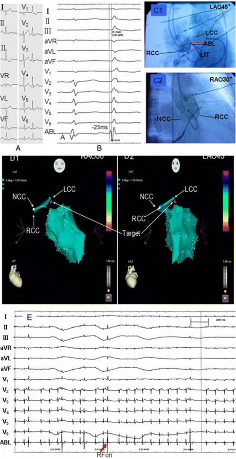
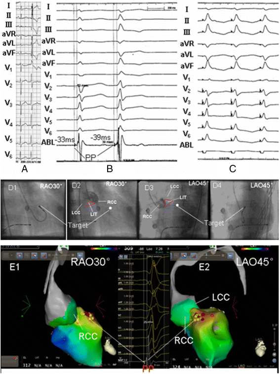
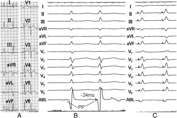
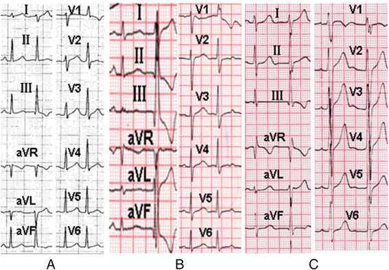
Results: Among the 40 patients with narrow PVCs, complete elimination of PVCs was achieved by RFCA in 35 patients during a median follow-up period of 23 months. Successful ablation was achieved on 19 patients at the sites where earliest Purkinje potentials were recorded in left ventricular anterosuperior septum, thus PVCs arising from left anterior fascicle (LAF) were confirmed, for these PVCs, the QRS morphology were right bundle branch and left posterior fascicle block (RBBB + LPFB) with rightward axis, the average QRS duration 116.07 ± 7.96 ms, R or rsR'in lead V1,with transition zone ahead of lead V1 in precordial leads. Another 16 successful RFCA were achieved by energy delivery at interleaflet triangle(ILT) between right coronary cusp(RCC) and left coronary cusp(LCC) where no Purkinje potentials were recorded, for narrow PVCs arising from the L-RCC ILT, the QRS morphology were similar to the PVCs arising from LAF but much narrower in QRS duration (100.44 ± 3.49 vs. 116.07 ± 7.96 ms, p < 0.05), they were also R or Rs in lead V1 with the transition zone ahead of lead V1. For 5 symptomatic narrow PVCs failed to the procedure of RFCA, their electrocardiographic characteristics showed that the narrowest QRS duration (91.80 ± 6.94 vs. 100.44 ± 3.49, 116.07 ± 7.96 ms, p < 0.05), rs or rS (r/s or r/S≦1) morphology in lead V1 with the precordial transition zone behind lead V3.
Conclusions: Most of idiopathic PVCs of narrow QRS duration (<130 msec) with qR pattern in inferior leads can be cured by the procedure of RFCA. On the basis of our study, we proposed that for narrow PVCs presenting qR pattern in inferior leads, when the ablation procedure failed at proximity of LAF within left anterosuperior septum, mapping and ablation in L-RCC ILT can be tried. The present findings can be helpful for planning catheter ablation for narrow PVCs manifesting qR in inferior leads.
Keywords: L-RCC ILT; Left anterior fascicle; Premature ventricular contractions; Radiofrequency catheter ablation.
Figures


Similar articles
-
Origins location of the outflow tract ventricular arrhythmias exhibiting qrS pattern or QS pattern with a notch on the descending limb in lead V1.BMC Cardiovasc Disord. 2017 May 15;17(1):124. doi: 10.1186/s12872-017-0561-y. BMC Cardiovasc Disord. 2017. PMID: 28506214 Free PMC article.
-
Ablation at Right Coronary Cusp as an Alternative and Favorable Approach to Eliminate Premature Ventricular Complexes Originating From the Proximal Left Anterior Fascicle.Circ Arrhythm Electrophysiol. 2020 May;13(5):e008173. doi: 10.1161/CIRCEP.119.008173. Epub 2020 Apr 17. Circ Arrhythm Electrophysiol. 2020. PMID: 32302210
-
Premature ventricular contractions originating from the left ventricular septum: results of radiofrequency catheter ablation in twenty patients.BMC Cardiovasc Disord. 2011 Jun 2;11:27. doi: 10.1186/1471-2261-11-27. BMC Cardiovasc Disord. 2011. PMID: 21635765 Free PMC article.
-
[Localization of ventricular premature contractions by 12-lead ECG].Herzschrittmacherther Elektrophysiol. 2021 Mar;32(1):21-26. doi: 10.1007/s00399-021-00746-7. Epub 2021 Feb 3. Herzschrittmacherther Elektrophysiol. 2021. PMID: 33533995 Review. German.
-
An organized approach to the localization, mapping, and ablation of outflow tract ventricular arrhythmias.J Cardiovasc Electrophysiol. 2013 Oct;24(10):1189-97. doi: 10.1111/jce.12237. Epub 2013 Sep 9. J Cardiovasc Electrophysiol. 2013. PMID: 24015911 Review.
Cited by
-
Approach selection of radiofrequency catheter ablation for ventricular arrhythmias originating from the left ventricular summit: potential relevance of Pseudo Delta wave, Intrinsicoid deflection time, maximal deflection index.BMC Cardiovasc Disord. 2017 May 30;17(1):140. doi: 10.1186/s12872-017-0575-5. BMC Cardiovasc Disord. 2017. PMID: 28558750 Free PMC article.
-
Palpitations in a young male: a case of dual node physiology unmasked by 'junctional' ectopics.Eur Heart J Case Rep. 2022 Mar 28;6(4):ytac136. doi: 10.1093/ehjcr/ytac136. eCollection 2022 Apr. Eur Heart J Case Rep. 2022. PMID: 35481250 Free PMC article. No abstract available.
References
-
- Kamakura S, Shimizu W, Matsuo K, Taguchi A, Suyama K, Kurita T, Aihara N, Ohe T, Shimomura K. Localization of optimal ablation site of idiopathic ventricular tachycardia from right and left ventricular outflow tract by body surface ECG. Circulation. 1998;98(15):1525–1533. doi: 10.1161/01.CIR.98.15.1525. - DOI - PubMed
-
- Kanagaratnam L, Tomassoni G, Schweikert R, Pavia S, Bash D, Beheiry S, Neibauer M, Saliba W, Chung M, Tchou P, et al. Ventricular tachycardias arising from the aortic sinus of valsalva: an under-recognized variant of left outflow tract ventricular tachycardia. J Am Coll Cardiol. 2001;37(5):1408–1414. doi: 10.1016/S0735-1097(01)01127-5. - DOI - PubMed
-
- Tada H, Tadokoro K, Ito S, Naito S, Hashimoto T, Kaseno K, Miyaji K, Sugiyasu A, Tsuchiya T, Kutsumi Y, et al. Idiopathic ventricular arrhythmias originating from the tricuspid annulus: Prevalence, electrocardiographic characteristics, and results of radiofrequency catheter ablation. Heart Rhythm. 2007;4(1):7–16. doi: 10.1016/j.hrthm.2006.09.025. - DOI - PubMed
Publication types
MeSH terms
LinkOut - more resources
Full Text Sources
Other Literature Sources

