. 2016 Apr 4:7:49.
doi: 10.1186/s13287-016-0297-0.

VCAM-1+ placenta chorionic villi-derived mesenchymal stem cells display potent pro-angiogenic activity

Affiliations

VCAM-1+ placenta chorionic villi-derived mesenchymal stem cells display potent pro-angiogenic activity

Wenjing Du et al. Stem Cell Res Ther. .

Abstract

Introduction: Mesenchymal stem cells (MSCs) represent a heterogeneous cell population that is promising for regenerative medicine. The present study was designed to assess whether VCAM-1 can be used as a marker of MSC subpopulation with superior angiogenic potential.

Methods: MSCs were isolated from placenta chorionic villi (CV). The VCAM-1(+/-) CV-MSCs population were separated by Flow Cytometry and subjected to a comparative analysis for their angiogenic properties including angiogenic genes expression, vasculo-angiogenic abilities on Matrigel in vitro and in vivo, angiogenic paracrine activities, cytokine array, and therapeutic angiogenesis in vascular ischemic diseases.

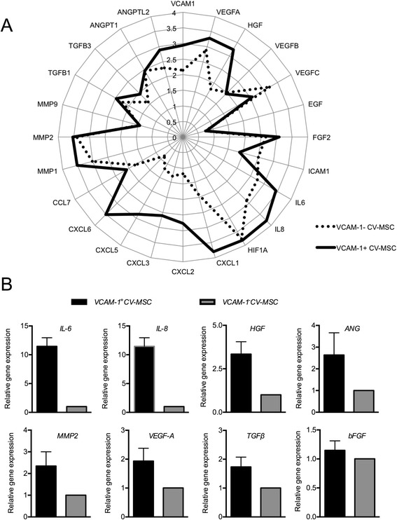
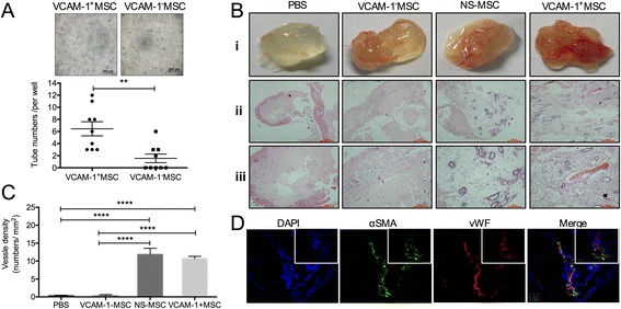
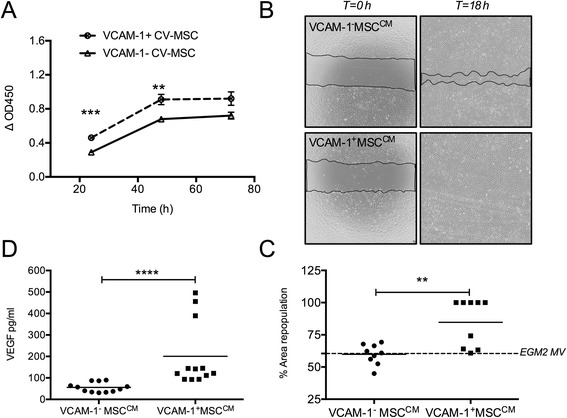
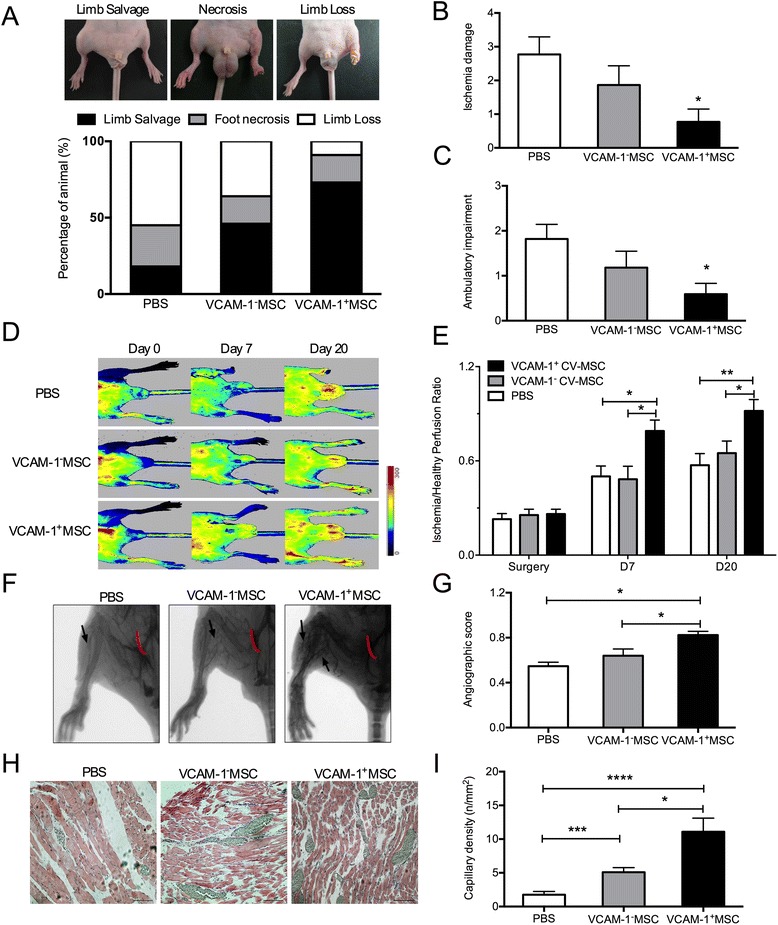
Results: Angiogenic genes, including HGF, ANG, IL8, IL6, VEGF-A, TGFβ, MMP2 and bFGF, were up-regulated in VCAM-1(+)CV-MSCs. Consistently, angiogenic cytokines especially HGF, IL8, angiogenin, angiopoitin-2, μPAR, CXCL1, IL-1β, IL-1α, CSF2, CSF3, MCP-3, CTACK, and OPG were found to be significantly increased in VCAM-1(+) CV-MSCs. Moreover, VCAM-1(+)CV-MSCs showed remarkable vasculo-angiogenic abilities by angiogenesis analysis with Matrigel in vitro and in vivo and the conditioned medium of VCAM-1(+) CV-MSCs exerted markedly pro-proliferative and pro-migratory effects on endothelial cells compared to VCAM-1(-)CV-MSCs. Finally, transplantation of VCAM-1(+)CV-MSCs into the ischemic hind limb of BALB/c nude mice resulted in a significantly functional improvement in comparison with VCAM-1(-)CV-MSCs transplantation.

Conclusions: VCAM-1(+)CV-MSCs possessed a favorable angiogenic paracrine activity and displayed therapeutic efficacy on hindlimb ischemia. Our results suggested that VCAM-1(+)CV-MSCs may represent an important subpopulation of MSC for efficient therapeutic angiogenesis.

Keywords: Angiogenesis; Mesenchymal stem cells; Paracrine; Placenta; Vascular cell adhesion molecule-1 (CD106).


Figures

Fig. 1
Fig. 1
Phenotype of CV-MSCs and flow cell sorting. a Surface markers of CV-MSCs were evaluated by FACS analysis. CV-MSCs positively expressed CD105, CD73, CD166, CD29, CD90, HLA-ABC, CD54, and VCAM-1, and hardly expressed CD14, CD45, CD31, CD144, CD133 and HLA-DR. A representative sample is shown. b VCAM-1+CV-MSCs and VACM-1CV-MSCs were separated by the BD Aria III cell sorting system. c Morphology of VCAM-1+CV-MSCs and VCAM-1CV-MSCs (scale bar = 200 μm). CV chorionic villi, MSC mesenchymal stem cell, SSC side scatter, VCAM-1 vascular cell adhesion molecule 1
Fig. 2
Fig. 2
Angiogenic genes were upregulated in VCAM-1+CV-MSCs. a Gene expression profile of VCAM-1+CV-MSCs and VCAM-1CV-MSCs determined using Affymetrix oligoarray, with the angiogenic genes valued and expressed in log10. b Several raised angiogenic genes in VCAM-1+CV-MSCs were confirmed by real-time PCR, including IL-6, IL-8 [23], HGF, ANG, MMP2, VEGF-A, TGFβ, and bFGF (n = 3–5). ANG angiogenin, ANGPT2 angiopoietin-2, ANGPTL2 angiopoietin-like protein 2, BFGF basic fibroblast growth factor, CCL Chemokine (C-C motif) ligand, CV chorionic villi, CXCL chemokine (C-X-C motif) ligand, EGF epidermal growth factor, HGF hepatocyte growth factor, HIF hypoxia-induced factor, IL interleukin, MMP matrix metalloproteinase, MSC mesenchymal stem cell, TGF transforming growth factor, VCAM-1 vascular cell adhesion molecule 1, VEGF vascular endothelial cell growth factor
Fig. 3
Fig. 3
VCAM-1+CV-MSCs revealed vasculoangiogenic potential by angiogenesis analysis with Matrigel in vitro and vivo. a VCAM-1+CV-MSCs spontaneously formed much more intact tube-structures on Matrigel than VCAM-1CV-MSCs (n = 3, ** p <0.01), indicating that VCAM-1+CV-MSCs possessed vasculogenic potential. Representative images are shown (scale bar = 500 μm). Each sample was performed in triplicate. b Macroscopic and microscopic view of Matrigel plugs. The Matrigel plug was harvested 21 days later; macroscopic vessels were seen in the Matrigel plug in the VCAM-1+CV-MSCs and NS CV-MSCs groups i. H & E staining was performed to reveal the vessel density in Matrigel plug (scale bar: ii = 500 μm; iii = 200 μm). c Vessel densities in the VCAM-1+CV-MSCs and NS CV-MSCs groups were much greater than in the PBS and VCAM-1CV-MSCs groups (n = 3, **** p <0.0001). d New outgrowth in Matrigel plug was immunostained with vWF and α-SMA antibodies to indicate the endothelial cells and smooth muscle cells, respectively. Photographs were taken under × 20 (bottom) and × 60 (upper) magnifications. α-SMA alpha smooth muscle actin, CV chorionic villi, DAPI 4',6-diamidino-2-phenylindole, MSC mesenchymal stem cell, NS nonseparated, PBS phosphate-buffered saline, VCAM-1 vascular cell adhesion molecule 1, vWF von Willebrand factor
Fig. 4
Fig. 4
VCAM-1+CV-MSCCM exerted angiogenic paracrine effects on endothelial cells. a Endothelial cell proliferation assay was used to study the pro-proliferative activity of VCAM-1+CV-MSCs and VCAM-1CV-MSCs. By comparison with VCAM-1CV-MSCCM, VCAM-1+CV-MSCCM significantly promoted endothelial cell proliferation during 48 hours, but this effect was not significant at 72 hours (n = 3, **p <0.01, ***p <0.001). Each sample was done in quadruplicate. b Wound healing assay was performed to study the pro-migratory effect of VCAM-1+/−CV-MSCs. Representative photographs were shown at 0 and 18 hours under × 40 magnification. c Result of area repopulation (%) indicating that VCAM-1+CV-MSCCM efficiently accelerated the endothelial cell wound healing process compared with VCAM-1CV-MSCCM (n = 3, **p <0.01). Each sample was performed in triplicate. d VEGF concentration in VCAM-1+CV-MSCCM and VCAM-1CV-MSCCM measured by ELISA (n = 4, ****p <0.0001). Each sample was tested in triplicate. CM conditioned medium, CV chorionic villi, MSC mesenchymal stem cell, VCAM-1 vascular cell adhesion molecule 1, VEGF vascular endothelial cell growth factor
Fig. 5
Fig. 5
Human cytokine antibody array displayed the angiogenic secretome of VCAM-1+CV-MSCs. a Expression of 120 cytokines in SN of VCAM-1+CV-MSCs and VCAM-1CV-MSCs was determined by human cytokine antibody array (AAH-CYT-G1000). VCAM-1+CV-MSC and VCAM-1CV-MSC SN derived from two healthy donors were used. Each sample was performed in duplicate. The differential angiogenic cytokines between VCAM-1+CV-MSCs and VCAM-1CV-MSCs were similar in two healthy donors. b Cytokine signal >200 was analyzed, and ratio of cytokine signal in VCAM-1+CV-MSCs to VCAM-1CV-MSCs was calculated. This was statistically significant if the cytokine signal ratio was >1.3 or <0.75. Data revealed that VCAM-1+CV-MSCs secreted more abundant angiogenic cytokines than VCAM-1CV-MSCs, including HGF, IL-8, ANG, ANGPT2, μPAR, CXCL1, IL-1β, IL-1α, CSF2, CSF3, MCP-3, CTACK, and OPG. CV chorionic villi, MSC mesenchymal stem cell, VCAM-1 vascular cell adhesion molecule 1. See Abbreviations for cytokine definitions
Fig. 6
Fig. 6
Transplantation of VCAM-1+CV-MSCs significantly enhanced the blood perfusion and the generation of collateral vessels in the ischemic sites. a VCAM-1+/−CV-MSCs or PBS were injected into the ischemic site of nude mice. Percentage distributions of limb salvage, foot necrosis, and limb loss in the three groups are shown and analyzed by the Fisher’s exact test (n = 11, p = 0.102). Ischemia damage and physiological function of ischemic limbs were semiquantified by ischemia scores b and ambulatory impairment scores c (n = 11, *p <0.05). d Blood perfusion in ischemic/healthy limb was detected by the Kodak In-Vivo FX Pro Imaging System on days (D) 0, 7, and 20. Different colors indicate different blood perfusion. Blood flow increased from dark blue to red. e Blood perfusion ratio in ischemic to healthy limbs was used to quantitatively analyze the blood flow restoration in ischemic limbs (n = 11, *p <0.05, **p <0.01). f Angiography was performed to assess the collateral vessel generation in the ischemic site. Red curves indicated the site of the femoral arteries incision; black arrows showed the collateral vessels in the ischemic hind limb. g Angiography score indicated that VCAM-1+CV-MSCs were superior to VCAM-1CV-MSCs in augmenting collateral vessels (n = 3–5, *p <0.05). h H & E staining was performed to study the vessel density in ischemia limbs. Pictures showed that the blood vessels were full of barium sulfate (silver, scale bar = 100 μm). i Vessel density in VCAM-1+CV-MSC or VCAM-1CV-MSCs group was significantly greater than the PBS group (n = 7, ***p <0.001, ****p <0.0001). Furthermore, VCAM-1+CV-MSC transplantation apparently promoted the vessel generation compared with the VCAM-1CV-MSCs group (**p <0.05). CV chorionic villi, MSC mesenchymal stem cell, PBS phosphate-buffered saline, VCAM-1 vascular cell adhesion molecule 1 (Color figure online)

Similar articles

Cited by

References

    1. Ouriel K. Peripheral arterial disease. Lancet. 2001;358(9289):1257–64. doi: 10.1016/S0140-6736(01)06351-6. - DOI - PubMed
    1. Kinnaird T, Stabile E, Burnett MS, Shou M, Lee CW, Barr S, et al. Local delivery of marrow-derived stromal cells augments collateral perfusion through paracrine mechanisms. Circulation. 2004;109(12):1543–9. doi: 10.1161/01.CIR.0000124062.31102.57. - DOI - PubMed
    1. Xu Y, Meng H, Li C, Hao M, Wang Y, Yu Z, et al. Umbilical cord-derived mesenchymal stem cells isolated by a novel explantation technique can differentiate into functional endothelial cells and promote revascularization. Stem Cells Dev. 2010;19(10):1511–22. doi: 10.1089/scd.2009.0321. - DOI - PubMed
    1. Kim SW, Zhang HZ, Kim CE, An HS, Kim JM, Kim MH. Amniotic mesenchymal stem cells have robust angiogenic properties and are effective in treating hindlimb ischaemia. Cardiovasc Res. 2012;93(3):525–34. doi: 10.1093/cvr/cvr328. - DOI - PubMed
    1. Hongyan Tao ZH, Zhong Chao Han, and Zongjin Li. Proangiogenic features of mesenchymal stem cells and their therapeutic applications. Stem Cells Int. 2016;2016:1314709. doi:10.1155/2016/1314709. - PMC - PubMed

Publication types

Substances