. 2016 May 27;291(22):11939-53.
doi: 10.1074/jbc.M115.702746. Epub 2016 Apr 4.

The Carbohydrate-linked Phosphorylcholine of the Parasitic Nematode Product ES-62 Modulates Complement Activation

Affiliations

The Carbohydrate-linked Phosphorylcholine of the Parasitic Nematode Product ES-62 Modulates Complement Activation

Umul Kulthum Ahmed et al. J Biol Chem. .

Abstract

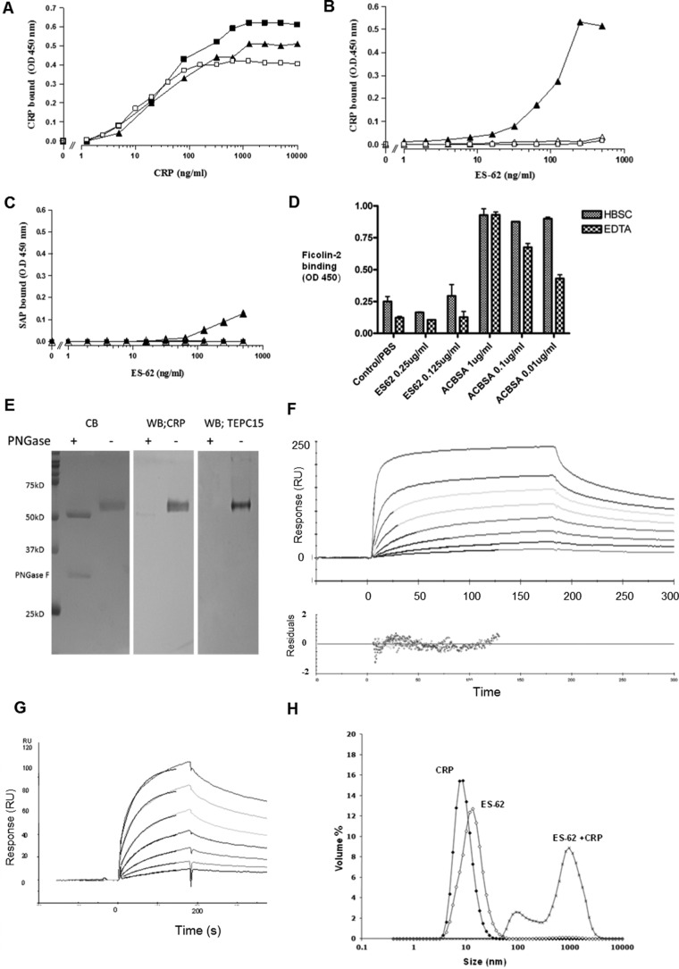
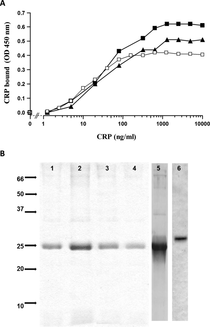
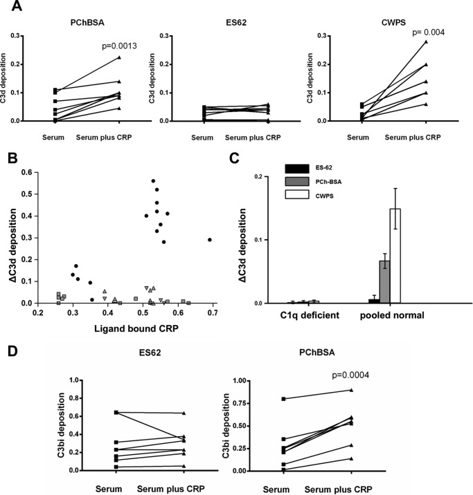
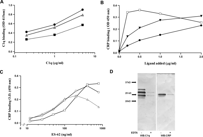
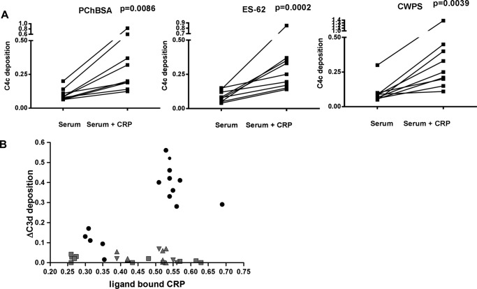
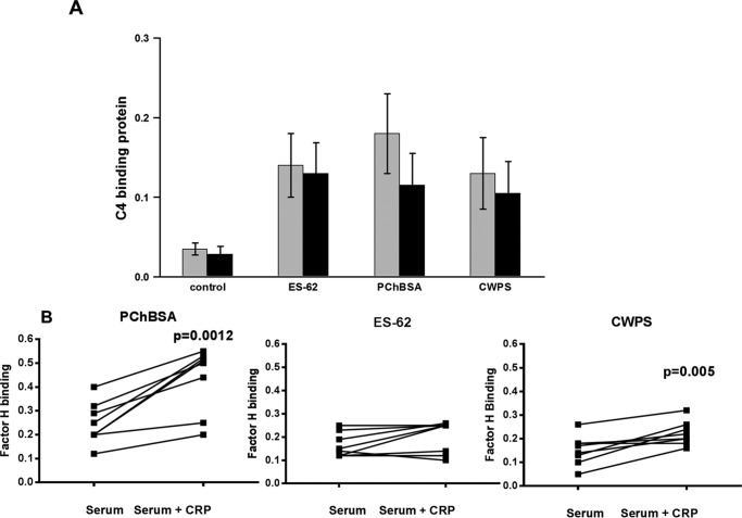
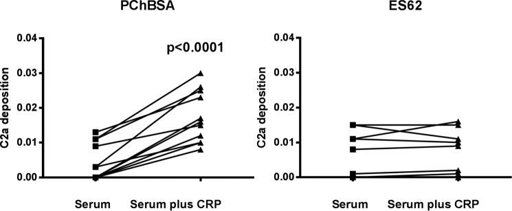
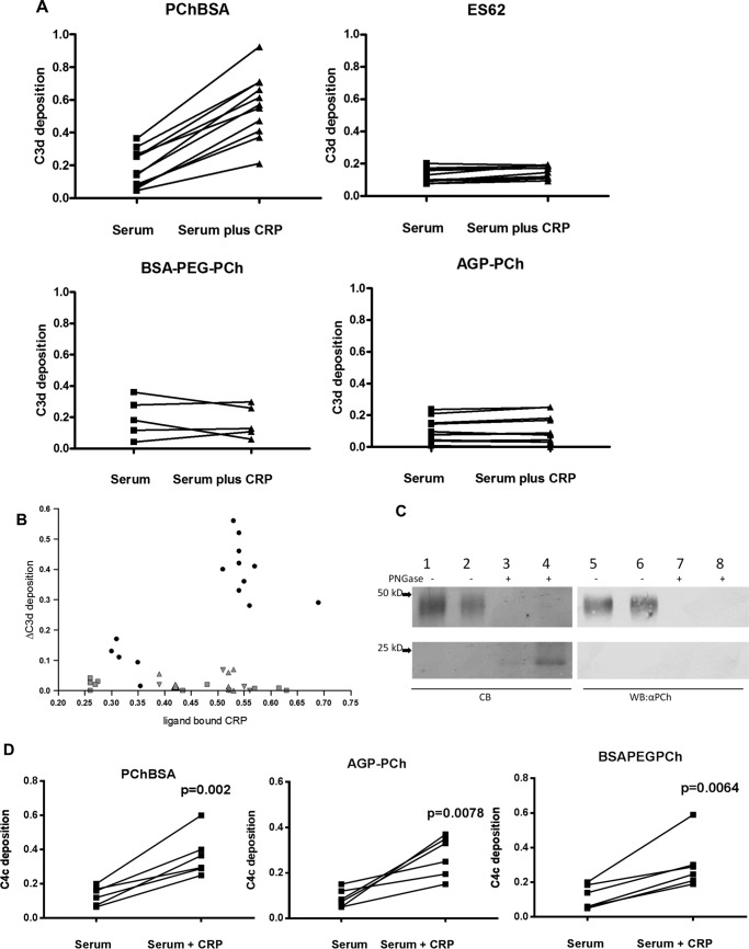
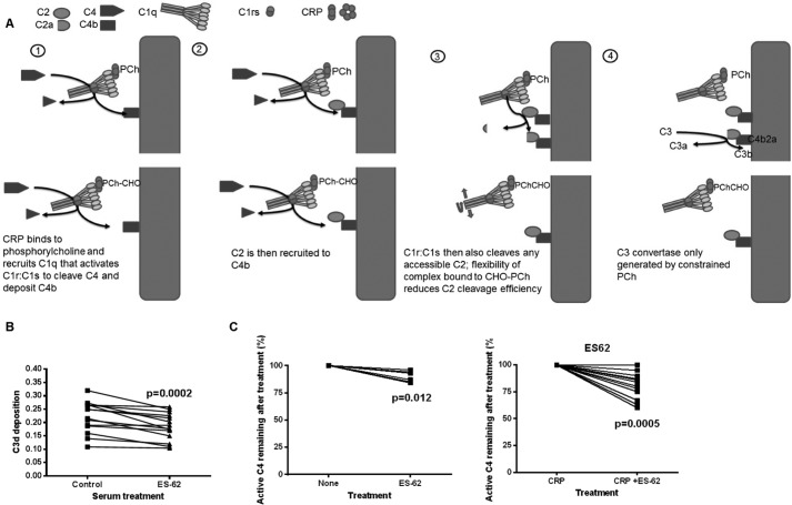
Parasitic nematodes manufacture various carbohydrate-linked phosphorylcholine (PCh)-containing molecules, including ES-62, a protein with an N-linked glycan terminally substituted with PCh. The PCh component is biologically important because it is required for immunomodulatory effects. We showed that most ES-62 was bound to a single protein, C-reactive protein (CRP), in normal human serum, displaying a calcium-dependent, high-avidity interaction and ability to form large complexes. Unexpectedly, CRP binding to ES-62 failed to efficiently activate complement as far as the C3 convertase stage in comparison with PCh-BSA and PCh-containing Streptococcus pneumoniae cell wall polysaccharide. C1q capture assays demonstrated an ES-62-CRP-C1q interaction in serum. The three ligands all activated C1 and generated C4b to similar extents. However, a C2a active site was not generated following ES-62 binding to CRP, demonstrating that C2 cleavage was far less efficient for ES-62-containing complexes. We proposed that failure of C2 cleavage was due to the flexible nature of carbohydrate-bound PCh and that reduced proximity of the C1 complex was the reason that C2 was poorly cleaved. This was confirmed using synthetic analogues that were similar to ES-62 only in respect of having a flexible PCh. Furthermore, ES-62 was shown to deplete early complement components, such as the rate-limiting C4, following CRP interaction and thereby inhibit classical pathway activation. Thus, flexible PCh-glycan represents a novel mechanism for subversion of complement activation. These data illustrate the importance of the rate-limiting C4/C2 stage of complement activation and reveal a new addition to the repertoire of ES-62 immunomodulatory mechanisms with possible therapeutic applications.

Keywords: C-reactive protein; C1Q complex (C1QA); ES-62; acute phase reactant; carbohydrate function; complement; immunomodulation; parasite; pentraxin; phosphorylcholine.


Figures

FIGURE 1.
FIGURE 1.
High-avidity binding of C-reactive protein to ES-62 is calcium-dependent and can be inhibited by PCh. A, dose response of binding of purified CRP to immobilized ES-62 (2.0 μg/ml, ■), PCh-BSA (0.5 μg/ml, ▴), or CWPS (5 μg/ml, □) on microtiter plates. Various concentrations of CRP were offered, and binding of CRP was detected using polyclonal anti-human CRP-HRP. OD, optical density. B, CRP binding from ES-62 is calcium-dependent and can be inhibited by PCh. Various concentrations of ES-62 were coated onto microtiter plates, and normal serum diluted 1 in 50 to give a final CRP concentration of 50 ng/ml was added. Binding of CRP was detected with the anti-native human CRP monoclonal antibody 2C10 and anti-mouse IgG HRP and 1,1,3,3 tetramethylbenzidine substrate (optical density, 450 nm). Serum was diluted in HBS containing 1 mm CaCl2 (▴), HBS with 10 mm EDTA (▵), or HBS with 1 mm CaCl2 and 50 mm phosphorylcholine (□). C, SAP provided in serum diluted 1 in 50 binds weakly to ES-62 (▴) but not PCh-BSA-coated plates (■). SAP was determined using monoclonal anti-SAP and anti-mouse IgG HRP. Controls show binding to ES-62 in the presence of EDTA (▵). D, plates were coated with ES-62 or the positive control acetylated BSA (ACBSA) at various concentrations, serum was added in the presence or absence of calcium, and binding was detected with biotinylated anti-ficolin 2 and streptavidin HRP. Data are mean ± S.E. of triplicates. E, ligand blotting of ES-62 following SDS-PAGE demonstrates binding of C-reactive protein to PCh attached to N-linked glycan. Left panel, ES-62 or ES-62 deglycosylated with PNGase stained directly with Coomassie Blue (CB). Center panel, ES-62 was transferred to PVDF, and CRP binding in TBSC was detected with anti-CRP and anti-mouse-alkaline phosphatase. Right panel, as for the center panel, but PCh was detected with anti-PCh myeloma protein, TEPC15. WB, Western blotting. F, surface plasmon resonance analysis of interaction. ES-62 was immobilized, and CRP was offered at concentrations of 10, 2.5, 1.25, 0.62, 0.3, 0.16, 0.08, and 0.04 μg/ml. Langmuir 1:1 analysis was performed. Residuals from the association analysis are shown below. RU, response unit. G, surface plasmon resonance analysis of ES-62 (12.5, 6.25, 3, 1.6, 0.8, 0.4, and 0.2 μg/ml) binding to biotinylated CRP immobilized on a streptavidin surface. The superimposed lines show modeled fit. H, CRP and ES-62 form large complexes in fluid phase. The size of the complex was determined using light scattering 5 min after mixing for 50 μg/ml CRP and 65 μg/ml ES-62 in HBS in the presence of 1 mm CaCl2.
FIGURE 2.
FIGURE 2.
CRP is the major serum protein binding to ES-62. A, other serum proteins do not inhibit CRP binding to ES-62. ES-62 (0.25 μg/ml) was coated onto microtiter plates, and CRP at indicated concentrations was added alone (□) or in the presence of two individual normal serum samples (■ and ▴). CRP binding was detected as for Fig. 1B. OD, optical density. B, CRP is the major protein pulled out of normal serum by ES-62 (lanes 1 and 2) or AGP-PCh (lanes 3 and 4) coupled to magnetic beads. Bound protein was eluted with EDTA (lanes 1 and 3) or by SDS-PAGE lysis buffer (lanes 2 and 4). SDS-PAGE gels were run and analyzed by Coomassie Blue staining (lanes 1–4). Gels were also immunoblotted, and CRP eluted from ES-62-coated beads was detected by Western blotting (lane 5), as was SAP to a lesser degree (lane 6).
FIGURE 3.
FIGURE 3.
ES-62, in contrast to PChBSA and CWPS, does not lead to C3d or C3bi deposition. A, CRP increases deposition of C3d onto PCh in PChBSA and CWPS but not ES-62. Individual sera from healthy donors with or without added CRP in VBSCaMg were incubated at 37 °C in ligand-coated plates in VBSCaMg, and C3d deposition was determined. The background level of complement activation for ES-62 seen without added CRP was not diminished in sera depleted of PCh binding activity by passage through an anti-PCh-Sepharose column. Statistical analysis was undertaken by paired t test. B, the same experiment was performed, but the increase in C3d deposition mediated by CRP was plotted against the amount of CRP bound to the plate under each condition. Ligands: PCh-BSA (■), CPWS (●), or ES-62 (▴). Small symbols represent data obtained for individual donor serum. Larger symbols represent data for pooled serum. C, complement activation by ES-62 and other PCh ligands is through C1q. Normal serum or C1q-depleted pooled sera were used to determine C3d deposition against ES-62, PCh-BSA, and CWPS. Data are mean ± S.E. of four replicates. D, CRP addition to serum increases complement C3bi deposition to ligand PCh-BSA but not ES-62. Following incubation as in A at 37 °C for 30 min, C3bi bound to the surface was detected with biotinylated anti-C3bi and streptavidin HRP. In A, B, and D, the data are for between seven and nine different donors measured in three different experiments.
FIGURE 4.
FIGURE 4.
Demonstration of the CRP·ES-62·C1q complex in serum. A, C1q binds to CRP/ES-62. ES-62 (▴), PCh-BSA (■), or S. pneumoniae CWPS (●) were immobilized on microtiter plates at concentrations that lead to equivalent CRP binding, determined by polyclonal anti-CRP binding. CRP (0.4 μg/ml) was added, followed by C1q at various concentrations (0, 0.2, 2.0, and 5.0 μg/ml), and following washing, bound C1q was detected with anti-C1q-AP and p-nitrophenyl phosphate substrate (optical density (OD) 415). B, CRP binds to C1q only when ligand is added to serum. Plates were coated with C1q, and ligand (□, CWPS; ▾, PCh-BSA; ●, ES-62) was added to wells, with serum diluted 1 in 5 in VBSCaMg so that the final CRP concentration was 0.4 μg/ml. Complex was measured by CRP binding, determined using polyclonal anti-CRP -HRP. C, maximal CRP: ES-62 complex is captured to C1q at equal molarity of CRP and ES-62. Plates were coated with C1q, and serum was diluted at 1:100 added with CRP at 0.03 μg/ml (▵), 0.06 μg/ml (○), and 0.125 μg/ml (□). Various amounts of ES-62 were added, and CRP bound to the C1q was measured using an anti-CRP monoclonal antibody as in Fig. 1B. D, C1q was recruited to CRP bound to ES-62. Plates were coated with ES-62 and incubated with serum diluted in VBSCaMg or VBSEDTA at 4 °C. The plates were washed, and bound protein was removed from the surface with SDS sample buffer, run on a 12% SDS-PAGE gel, and Western-blotted with anti-C1q and CRP. Lanes 1 and 2, blotting for C1q (to reveal C1qA, C1qB, and C1qC chains); lanes 3 and 4, blotting for CRP. Representative data from two to three donor sera are shown.
FIGURE 5.
FIGURE 5.
ES-62-bound CRP leads to C4 deposition. A, CRP bound to ES-62 leads to C4 product deposition. Plates were coated with ES-62 or other CRP ligand at concentrations that bound similar amounts of CRP. Serum diluted in VBSCaMg from eight to ten normal healthy donors was added with or without CRP at 0.4 μg/ml and incubated at 37 °C for 30 min. Deposited C4c was determined with biotinylated anti-C4c and streptavidin HRP. Statistical analysis was undertaken by paired t test (n = 9). B, bound CRP correlates with increased C4c deposition for all three ligands. Data were obtained as in A, but different serum and CRP concentrations were used, and bound CRP was measured and plotted against the deposited C4c. PChBSA, ■; ES-62, ●; CWPS, ▴.
FIGURE 6.
FIGURE 6.
Lack of a role for complement regulatory factors. A, no significant difference between binding of C4 binding protein to immobilized ES-62-, PChBSA-, or CWPS-coated microtiter plates. Plates were coated with ES-62, PCh-BSA, or CWPS as described previously and incubated with serum with (black bars) or without CRP (gray bars). Following incubation for 30 min with serum diluted in VBSCaMg or VBSMgEGTA buffer with or without CRP (0.4 μg/ml), the plates were washed, and C4bp was detected with biotinylated antiC4bp. Data are presented as mean ± S.E. of five different donor sera. B, factor H recruitment to the plate surface following CRP-mediated complement activation in response to PCh-BSA and CWPS but not ES-62. PCh ligand was coated to the plate to recruit equivalent CRP amounts, and following incubation with serum with or without added 0.4 μg/ml CRP at 37 °C for 30 min, the amount of factor H bound was determined. Two experiments on eight different donors were analyzed by paired t test.
FIGURE 7.
FIGURE 7.
ES-62 does not efficiently generate a C3 convertase. CRP addition to serum leads to active C2a generated in response to immobilized PCh-BSA but poorly in response to immobilized ES-62. Plates were coated with ligand as described previously and incubated at 37 °C for 30 min, and active C2a was detected with the biotinylated monoclonal antibody 175-62. Statistical analysis was undertaken by paired t test on 11 different sera in two different experiments.
FIGURE 8.
FIGURE 8.
Highly mobile PCh ligands bind CRP but do not activate complement. A, CRP increases deposition of C3d onto PCh in PChBSA but not ES-62 or the synthetic PCh ligands AGP-PCh and BSA-PEG4-PCh. Individual sera from healthy donors with or without added CRP in VBSCaMg were incubated at 37 °C in ligand-coated plates in VBSCaMg, and C3d deposition was determined. Statistical analysis was undertaken by paired t test on 5–11 different sera in two different experiments. B, the same experiment was performed, but the increase in C3d deposition mediated by CRP was plotted against the amount of CRP bound to the plate under each condition when ligand concentration was varied. Ligands: PCh-BSA (●), ES-62 (▴), AGP-PCh (▾), and BSA PEG4 PCh (■). C, PCh of synthetic PCh-AGP is attached to N-linked carbohydrate. Shown is SDS-PAGE and Coomassie Blue (CB) staining (lanes 1–4) or immunoblot (lanes 5–8) with anti-PCh (TEPC15) of AGP-PCh and an equivalent amount of PNGase-treated AGP-PCh. Lanes 1, 4, 5, and 8, 5 μg/ml; lanes 2, 3, 6, and 7, 2.5 μg/ml. WB, Western blotting. D, CRP addition to serum increases C4c deposition onto AGP-PCh and BSA PEG4-PCh. Data for six different donor sera were analyzed by paired t test.
FIGURE 9.
FIGURE 9.
ES-62 added to serum reduces classical complement activation and the rate-limiting factor active C4. A, diagram of the effects of different PCh forms on the complement pathway. B, classical complement activation was measured by C3d deposition onto IgM-coated microtiter plates. Serum diluted 1 to 100 in VBSCaMg was either left untreated or treated with ES-62 (0.1 μg/ml) for 30 min at 37 °C prior to addition to the IgM. Statistical analysis was undertaken by Wilcoxon matched pairs test (n = 14 different sera in three different experiments). C, active C4 was measured following addition of ES-62 (0.5 μg/ml) to serum diluted 1 in 100 and incubation for 45 min at 37 °C (n = 6 different donor sera). D, as for B, but CRP was added at 0.4 μg/ml final concentration. Data for 12 different donor sera in two separate experiments were analyzed by Wilcoxon matched pairs test.

References

    1. Maizels R. M., Burke J., and Denham D. A. (1987) Phosphorylcholine bearing antigens in filarial nematode parasites: analysis of somatic extracts, in vitro secretions and infection sera from B. malayi and B. pahangi. Parasite Immunol. 9, 49–66 - PubMed
    1. Hewitson J. P., Harcus Y. M., Curwen R. S., Dowle A. A., Atmadja A. K., Ashton P. D., Wilson A., and Maizels R. M. (2008) The secretome of the filarial parasite, Brugia malayi: proteomic profile of adult excretory-secretory products. Mol. Biochem. Parasitol. 160, 8–21 - PubMed
    1. Lal R. B., and Ottesen E. A. (1989) Phosphocholine epitopes on helminth and protozoal parasites and their presence in the circulation of infected human patients. Trans. R Soc. Trop. Med. Hyg. 83, 652–655 - PubMed
    1. Wuhrer M., Rickhoff S., Dennis R. D., Lochnit G., Soboslay P. T., Baumeister S., and Geyer R. (2000) Phosphocholine-containing, zwitterionic glycosphingolipids of adult Onchocerca volvulus as highly conserved antigenic structures of parasitic nematodes. Biochem. J. 348, 417–423 - PMC - PubMed
    1. Haslam S. M., Houston K. M., Harnett W., Reason A. J., and Morris H. R., and Dell A. (1999) Structural studies of N glycans of filarial parasite: conservation of phosphorylcholine substituted glycans among species and discovery of novel chito-oligomers. J. Biol. Chem. 274, 20953–20960 - PubMed

Publication types

MeSH terms