

Th17 responses and natural IgM antibodies are related to gut microbiota composition in systemic lupus erythematosus patients
- PMID: 27044888
- PMCID: PMC4820712
- DOI: 10.1038/srep24072
Th17 responses and natural IgM antibodies are related to gut microbiota composition in systemic lupus erythematosus patients
Abstract
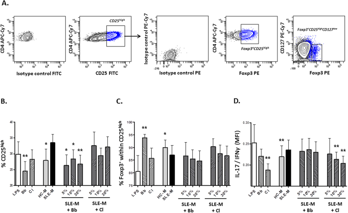
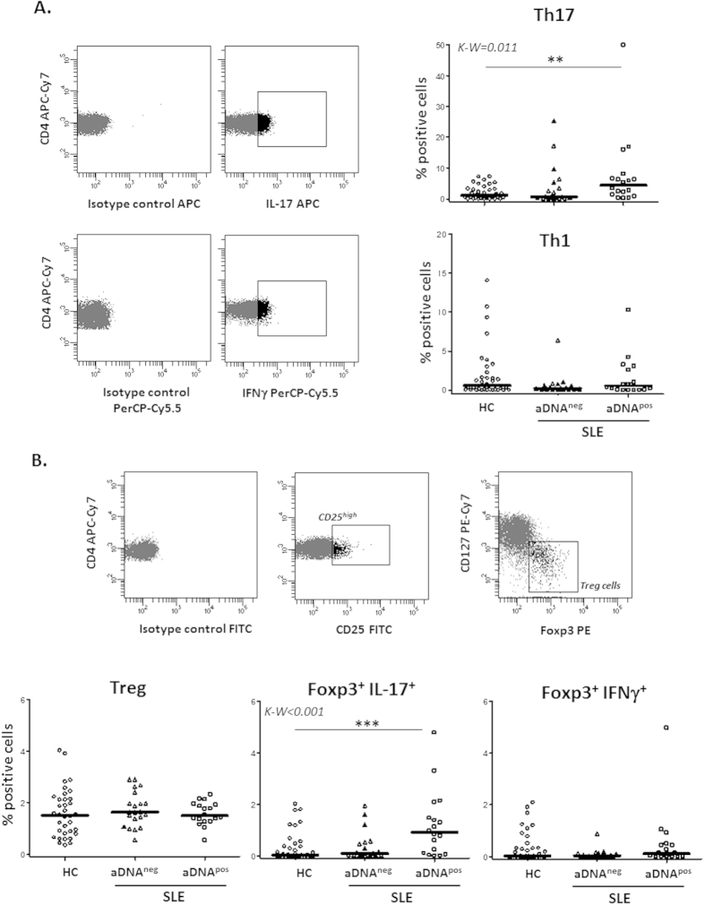
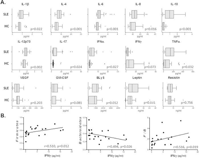
Intestinal dysbiosis, characterized by a reduced Firmicutes/Bacteroidetes ratio, has been reported in systemic lupus erythematosus (SLE) patients. In this study, in vitro cultures revealed that microbiota isolated from SLE patient stool samples (SLE-M) promoted lymphocyte activation and Th17 differentiation from naïve CD4(+) lymphocytes to a greater extent than healthy control-microbiota. Enrichment of SLE-M with Treg-inducing bacteria showed that a mixture of two Clostridia strains significantly reduced the Th17/Th1 balance, whereas Bifidobacterium bifidum supplementation prevented CD4(+) lymphocyte over-activation, thus supporting a possible therapeutic benefit of probiotics containing Treg-inducer strains in order to restore the Treg/Th17/Th1 imbalance present in SLE. In fact, ex vivo analyses of patient samples showed enlarged Th17 and Foxp3(+) IL-17(+) populations, suggesting a possible Treg-Th17 trans-differentiation. Moreover, analyses of fecal microbiota revealed a negative correlation between IL-17(+) populations and Firmicutes in healthy controls, whereas in SLE this phylum correlated directly with serum levels of IFNγ, a Th1 cytokine slightly reduced in patients. Finally, the frequency of Synergistetes, positively correlated with the Firmicutes/Bacteroidetes ratio in healthy controls, tended to be reduced in patients when anti-dsDNA titers were increased and showed a strong negative correlation with IL-6 serum levels and correlated positively with protective natural IgM antibodies against phosphorylcholine.
Figures
Similar articles
-
IgM antibodies against phosphorylcholine promote polarization of T regulatory cells from patients with atherosclerotic plaques, systemic lupus erythematosus and healthy donors.Atherosclerosis. 2018 Jan;268:36-48. doi: 10.1016/j.atherosclerosis.2017.11.010. Epub 2017 Nov 16. Atherosclerosis. 2018. PMID: 29175653
-
Th1/Th2/Th17/Treg cytokine imbalance in systemic lupus erythematosus (SLE) patients: Correlation with disease activity.Cytokine. 2015 Apr;72(2):146-53. doi: 10.1016/j.cyto.2014.12.027. Epub 2015 Jan 31. Cytokine. 2015. PMID: 25647269
-
[Expression and significance of Th17 and Treg cells in peripheral blood of patients with systemic lupus erythematosus].Zhonghua Yi Xue Za Zhi. 2012 Feb 21;92(7):460-3. doi: 10.3760/cma.j.issn.00376-2491-2012.07.008. Zhonghua Yi Xue Za Zhi. 2012. PMID: 22490966 Chinese.
-
Imbalance of Th17 cells, Treg cells and associated cytokines in patients with systemic lupus erythematosus: a meta-analysis.Front Immunol. 2024 Jul 17;15:1425847. doi: 10.3389/fimmu.2024.1425847. eCollection 2024. Front Immunol. 2024. PMID: 39086480 Free PMC article.
-
Recovery of the immune balance between Th17 and regulatory T cells as a treatment for systemic lupus erythematosus.Rheumatology (Oxford). 2011 Aug;50(8):1366-72. doi: 10.1093/rheumatology/ker116. Epub 2011 Apr 12. Rheumatology (Oxford). 2011. PMID: 21489974 Review.
Cited by
-
Gut Microbiota in Lupus: a Butterfly Effect?Curr Rheumatol Rep. 2021 Apr 16;23(4):27. doi: 10.1007/s11926-021-00986-z. Curr Rheumatol Rep. 2021. PMID: 33864162 Review.
-
Abundance and nuclear antigen reactivity of intestinal and fecal Immunoglobulin A in lupus-prone mice at younger ages correlate with the onset of eventual systemic autoimmunity.Sci Rep. 2020 Aug 31;10(1):14258. doi: 10.1038/s41598-020-71272-8. Sci Rep. 2020. PMID: 32868790 Free PMC article.
-
Microbiota and Immune-Mediated Skin Diseases-An Overview.Microorganisms. 2019 Aug 21;7(9):279. doi: 10.3390/microorganisms7090279. Microorganisms. 2019. PMID: 31438634 Free PMC article. Review.
-
Probiotics and Disease: A Comprehensive Summary-Part 7, Immune Disorders.Integr Med (Encinitas). 2017 Oct;16(5):46-57. Integr Med (Encinitas). 2017. PMID: 30936805 Free PMC article. Review.
-
Putting the microbiota to work: Epigenetic effects of early life antibiotic treatment are associated with immune-related pathways and reduced epithelial necrosis following Salmonella Typhimurium challenge in vitro.PLoS One. 2020 Apr 27;15(4):e0231942. doi: 10.1371/journal.pone.0231942. eCollection 2020. PLoS One. 2020. PMID: 32339193 Free PMC article.
References
Publication types
MeSH terms
Substances
LinkOut - more resources
Full Text Sources
Other Literature Sources
Medical
Research Materials
