

Sensitivity of the resting-state haemodynamic response function estimation to autonomic nervous system fluctuations
- PMID: 27044997
- PMCID: PMC4822449
- DOI: 10.1098/rsta.2015.0190
Sensitivity of the resting-state haemodynamic response function estimation to autonomic nervous system fluctuations
Abstract
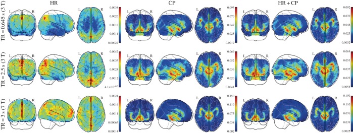
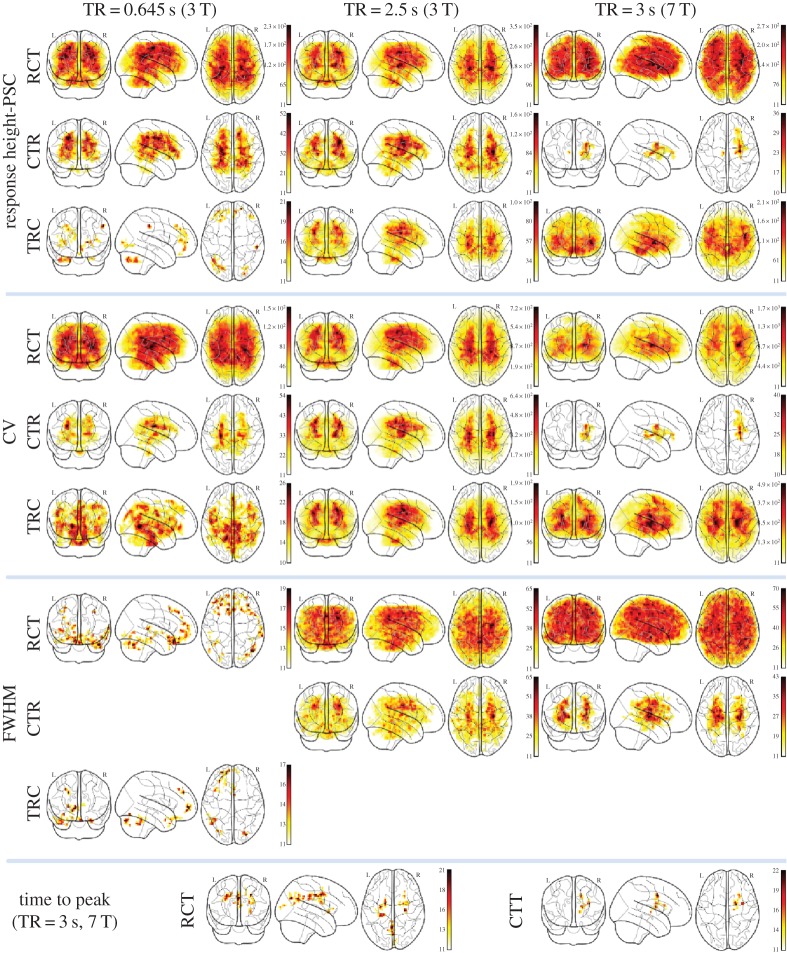
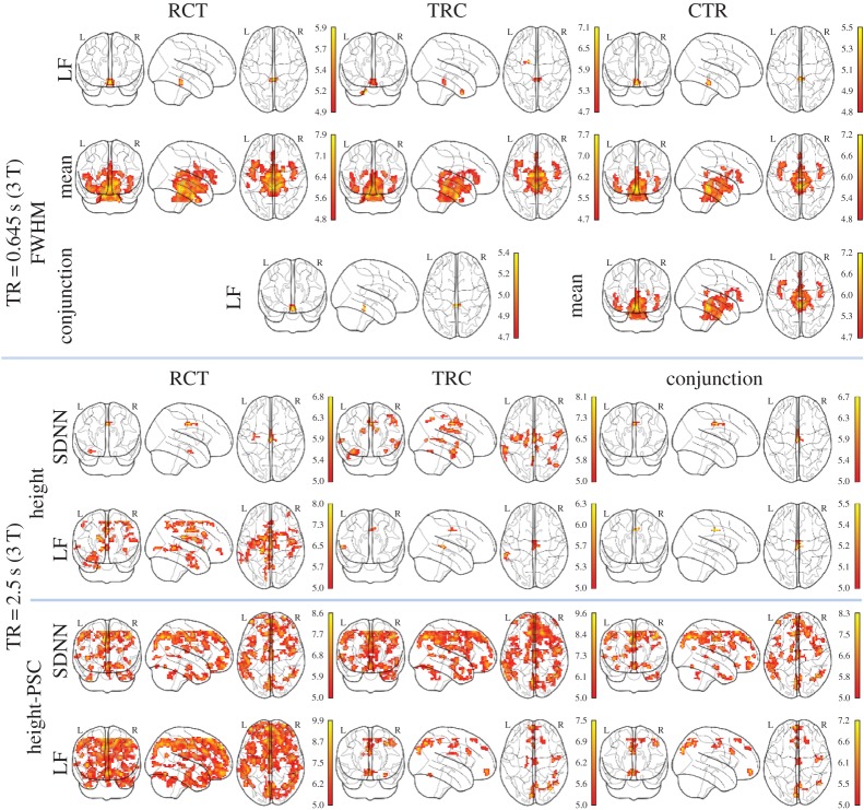
The haemodynamic response function (HRF) is a key component of the blood oxygen level-dependent (BOLD) signal, providing the mapping between neural activity and the signal measured with functional magnetic resonance imaging (fMRI). Most of the time the HRF is associated with task-based fMRI protocols, in which its onset is explicitly included in the design matrix. On the other hand, the HRF also mediates the relationship between spontaneous neural activity and the BOLD signal in resting-state protocols, in which no explicit stimulus is taken into account. It has been shown that resting-state brain dynamics can be characterized by looking at sparse BOLD 'events', which can be retrieved by point process analysis. These events can be then used to retrieve the HRF at rest. Crucially, cardiac activity can also induce changes in the BOLD signal, thus affecting both the number of these events and the estimation of the haemodynamic response. In this study, we compare the resting-state haemodynamic response retrieved by means of a point process analysis, taking the cardiac fluctuations into account. We find that the resting-state HRF estimation is significantly modulated in the brainstem and surrounding cortical areas. From the analysis of two high-quality datasets with different temporal and spatial resolution, and through the investigation of intersubject correlation, we suggest that spontaneous point process response durations are associated with the mean interbeat interval and low-frequency power of heart rate variability in the brainstem.
Keywords: cardiac fluctuations; functional magnetic resonance imaging; haemodynamic response; point process; resting state.
© 2016 The Author(s).
Figures
 –
– . Second column:
  –
– . Third column:
  –
– . MP, motion parameter. (Online version in colour.)
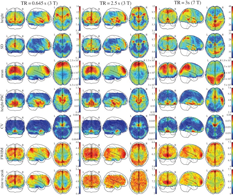
Similar articles
-
The resting-state neurovascular coupling relationship: rapid changes in spontaneous neural activity in the somatosensory cortex are associated with haemodynamic fluctuations that resemble stimulus-evoked haemodynamics.Eur J Neurosci. 2013 Sep;38(6):2902-16. doi: 10.1111/ejn.12295. Epub 2013 Jul 10. Eur J Neurosci. 2013. PMID: 23841797
-
Modeling the Hemodynamic Response Function Using EEG-fMRI Data During Eyes-Open Resting-State Conditions and Motor Task Execution.Brain Topogr. 2022 May;35(3):302-321. doi: 10.1007/s10548-022-00898-w. Epub 2022 Apr 30. Brain Topogr. 2022. PMID: 35488957
-
Multivariate semi-blind deconvolution of fMRI time series.Neuroimage. 2021 Nov 1;241:118418. doi: 10.1016/j.neuroimage.2021.118418. Epub 2021 Jul 22. Neuroimage. 2021. PMID: 34303793
-
The neural basis of the blood-oxygen-level-dependent functional magnetic resonance imaging signal.Philos Trans R Soc Lond B Biol Sci. 2002 Aug 29;357(1424):1003-37. doi: 10.1098/rstb.2002.1114. Philos Trans R Soc Lond B Biol Sci. 2002. PMID: 12217171 Free PMC article. Review.
-
[Functional connectivity analysis of the brain network using resting-state FMRI].Brain Nerve. 2011 Dec;63(12):1307-18. Brain Nerve. 2011. PMID: 22147450 Review. Japanese.
Cited by
-
Time-Resolved Resting-State Functional Magnetic Resonance Imaging Analysis: Current Status, Challenges, and New Directions.Brain Connect. 2017 Oct;7(8):465-481. doi: 10.1089/brain.2017.0543. Brain Connect. 2017. PMID: 28874061 Free PMC article. Review.
-
Unveiling functional-metabolic synergy in the healthy brain: multivariate integration of dynamic [18F]FDG-PET and resting-state fMRI.bioRxiv [Preprint]. 2025 May 27:2025.05.21.655345. doi: 10.1101/2025.05.21.655345. bioRxiv. 2025. PMID: 40501582 Free PMC article. Preprint.
-
Uncovering brain-heart information through advanced signal and image processing.Philos Trans A Math Phys Eng Sci. 2016 May 13;374(2067):20160020. doi: 10.1098/rsta.2016.0020. Philos Trans A Math Phys Eng Sci. 2016. PMID: 27044995 Free PMC article.
-
Dynamic time warping outperforms Pearson correlation in detecting atypical functional connectivity in autism spectrum disorders.Neuroimage. 2020 Dec;223:117383. doi: 10.1016/j.neuroimage.2020.117383. Epub 2020 Sep 17. Neuroimage. 2020. PMID: 32949710 Free PMC article.
-
Sustained versus instantaneous connectivity differentiates cognitive functions of processing speed and episodic memory.Hum Brain Mapp. 2018 Dec;39(12):4949-4961. doi: 10.1002/hbm.24336. Epub 2018 Aug 16. Hum Brain Mapp. 2018. PMID: 30113114 Free PMC article.
References
MeSH terms
LinkOut - more resources
Full Text Sources
Other Literature Sources
