. 2016 Apr 5;10(4):e0004595.
doi: 10.1371/journal.pntd.0004595. eCollection 2016 Apr.

Severe Fever with Thrombocytopenia Syndrome Virus Antigen Detection Using Monoclonal Antibodies to the Nucleocapsid Protein

Affiliations

Severe Fever with Thrombocytopenia Syndrome Virus Antigen Detection Using Monoclonal Antibodies to the Nucleocapsid Protein

Aiko Fukuma et al. PLoS Negl Trop Dis. .

Abstract

Background: Severe fever with thrombocytopenia syndrome (SFTS) is a tick-borne infectious disease with a high case fatality rate, and is caused by the SFTS virus (SFTSV). SFTS is endemic to China, South Korea, and Japan. The viral RNA level in sera of patients with SFTS is known to be strongly associated with outcomes. Virological SFTS diagnosis with high sensitivity and specificity are required in disease endemic areas.

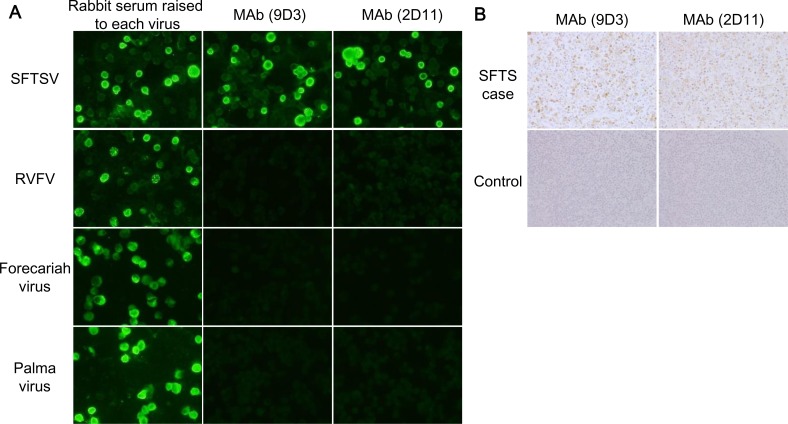
Methodology/principal findings: We generated novel monoclonal antibodies (MAbs) against the SFTSV nucleocapsid (N) protein and developed a sandwich antigen (Ag)-capture enzyme-linked immunosorbent assay (ELISA) for the detection of N protein of SFTSV using MAb and polyclonal antibody as capture and detection antibodies, respectively. The Ag-capture system was capable of detecting at least 350-1220 TCID50/100 μl/well from the culture supernatants of various SFTSV strains. The efficacy of the Ag-capture ELISA in SFTS diagnosis was evaluated using serum samples collected from patients suspected of having SFTS in Japan. All 24 serum samples (100%) containing high copy numbers of viral RNA (>105 copies/ml) showed a positive reaction in the Ag-capture ELISA, whereas 12 out of 15 serum samples (80%) containing low copy numbers of viral RNA (<105 copies/ml) showed a negative reaction in the Ag-capture ELISA. Among these Ag-capture ELISA-negative 12 samples, 9 (75%) were positive for IgG antibodies against SFTSV.

Conclusions: The newly developed Ag-capture ELISA is useful for SFTS diagnosis in acute phase patients with high levels of viremia.


Conflict of interest statement

The authors have declared that no competing interests exist.

Figures

Fig 1
Fig 1. Reactivity of MAbs (9D3 and 2D11) to SFTSV N protein and other TBPVs.
(A) The indirect immunofluorescence staining (IFA) of MAbs. Vero cells infected with SFTSV strain YG1, RVFV strain MP12, FORV, and PALV were stained with MAbs. Rabbit sera obtained from animals immunized with SFTSV or RVFV recombinant N protein, or infected with FORV or PALV, were used as positive controls in the IFA. (B) The immunohistochemical staining of SFTSV antigens with the developed MAbs. The lymph nodes collected from patient with SFTS and patients without SFTS were used for evaluation of utility of the MAbs in SFTS diagnosis with the IHC analysis.
Fig 2
Fig 2. Detection limit and cross reactivity of the Ag-capture ELISA.
(A) The detection limit of SFTSV rN protein by the Ag-capture ELISA using MAb 9D3 or 2D11. (B) The detection of authentic SFTSV by the Ag-capture ELISA. The Ag-capture ELISA were used MAb 9D3 or 2D11 as capture antibodies and anti-rN protein rabbit serum as detecting antibody. The dashed lines indicate the cut-off values (mean + 3×SD derived from OD405 values without antigens) for each ELISA. The detection limits for each MAb are shown in the lower panel.
Fig 3
Fig 3. Correlation between the results of the Ag-capture ELISA and that of the viral RNA copy numbers or OD values of IgG ELISA.
(A) Correlation between the result of the Ag-capture ELISA and viral RNA copy numbers in the serum samples. The Ag-capture ELISA (x axis) and the viral RNA copy number determined by qRT-PCR (y axis) from each sample are plotted as dots. (B) Relationship between the titers of the Ag-capture ELISA and the patients' outcomes. The patients' outcomes (x axis) and the titers determined by the Ag-capture ELISA (y axis) from each sample are plotted as dots. (C) Correlation between the result of the Ag-capture ELISA and OD values of IgG ELISA. The results determined by the Ag-capture ELISA (x axis) and OD405 values determined by IgG ELISA (y axis) from each sample are plotted as dots. The mean of each group is indicated by a horizontal bar. The t-test was used to determine the level of statistical significance. The calculated p values are shown above the groups that were compared.

References

    1. Yu XJ, Liang MF, Zhang SY, Liu Y, Li JD, et al. (2011) Fever with thrombocytopenia associated with a novel bunyavirus in China. N Engl J Med 364: 1523–1532. 10.1056/NEJMoa1010095 - DOI - PMC - PubMed
    1. Xu B, Liu L, Huang X, Ma H, Zhang Y, et al. (2011) Metagenomic analysis of fever, thrombocytopenia and leukopenia syndrome (FTLS) in Henan Province, China: discovery of a new bunyavirus. PLoS Pathog 7: e1002369 10.1371/journal.ppat.1002369 - DOI - PMC - PubMed
    1. Zhang YZ, Zhou DJ, Qin XC, Tian JH, Xiong Y, et al. (2012) The ecology, genetic diversity, and phylogeny of Huaiyangshan virus in China. J Virol 86: 2864–2868. 10.1128/JVI.06192-11 - DOI - PMC - PubMed
    1. Niu G, Li J, Liang M, Jiang X, Jiang M, et al. (2013) Severe fever with thrombocytopenia syndrome virus among domesticated animals, China. Emerg Infect Dis 19: 756–763. 10.3201/eid1905.120245 - DOI - PMC - PubMed
    1. Li Z, Hu J, Bao C, Li P, Qi X, et al. (2014) Seroprevalence of antibodies against SFTS virus infection in farmers and animals, Jiangsu, China. J Clin Virol 60: 185–189. 10.1016/j.jcv.2014.03.020 - DOI - PubMed

Publication types

MeSH terms

LinkOut - more resources