. 2016 Apr 5;6(4):e774.
doi: 10.1038/tp.2016.42.

Regulation of prefrontal cortex myelination by the microbiota

Affiliations

Regulation of prefrontal cortex myelination by the microbiota

A E Hoban et al. Transl Psychiatry. .

Abstract

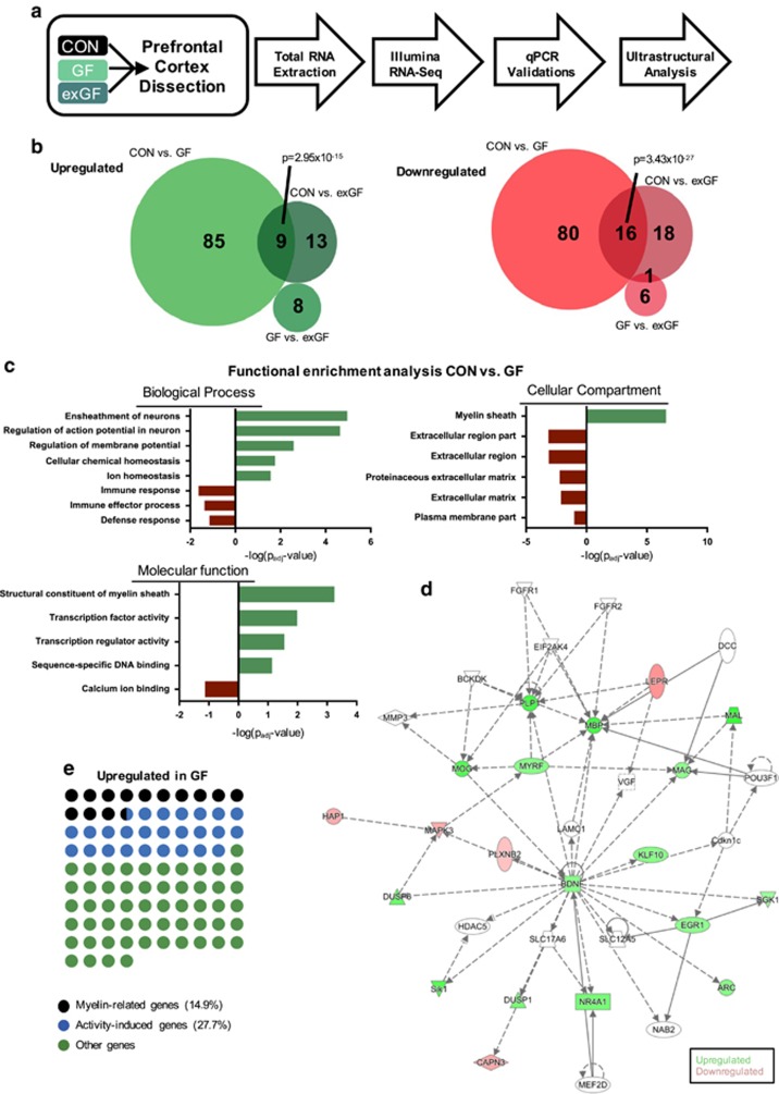
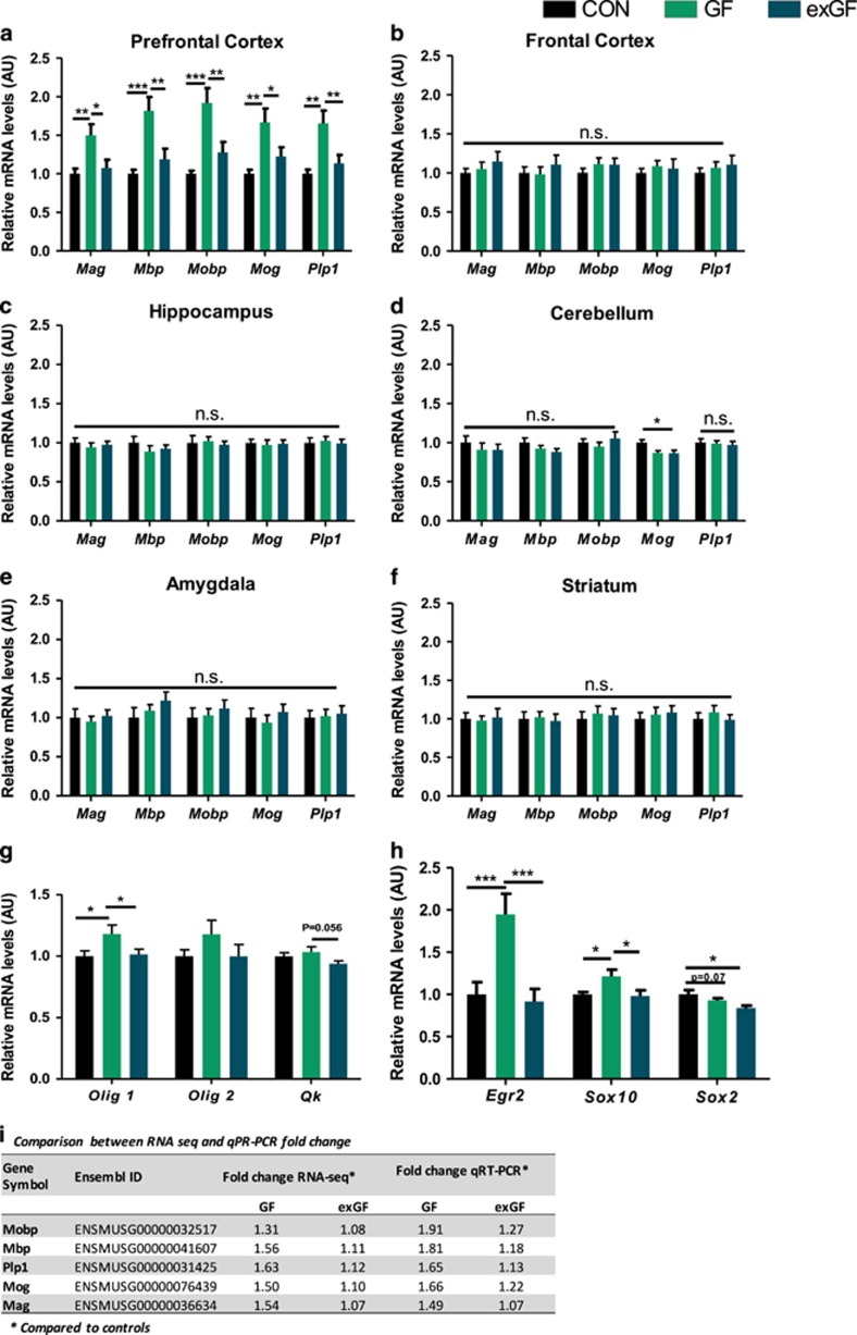
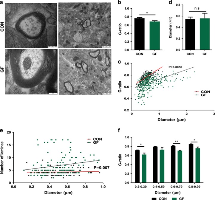
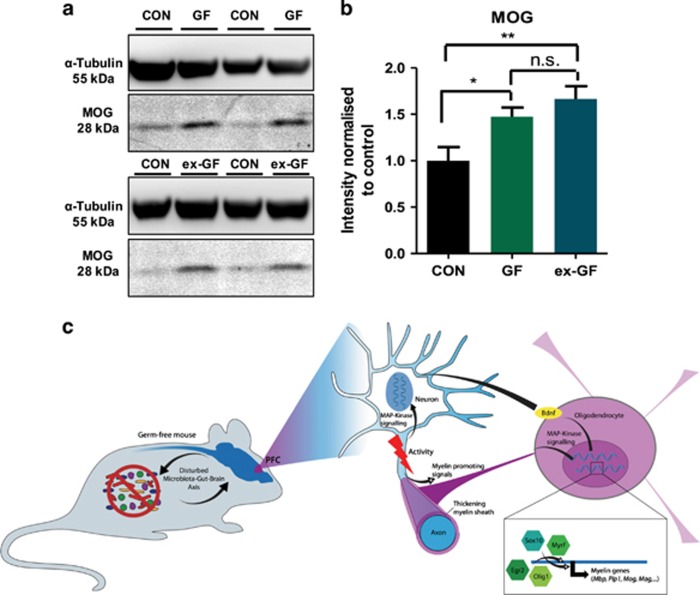
The prefrontal cortex (PFC) is a key region implicated in a range of neuropsychiatric disorders such as depression, schizophrenia and autism. In parallel, the role of the gut microbiota in contributing to these disorders is emerging. Germ-free (GF) animals, microbiota-deficient throughout life, have been instrumental in elucidating the role of the microbiota in many aspects of physiology, especially the role of the microbiota in anxiety-related behaviours, impaired social cognition and stress responsivity. Here we aim to further elucidate the mechanisms of the microbial influence by investigating changes in the homeostatic regulation of neuronal transcription of GF mice within the PFC using a genome-wide transcriptome profiling approach. Our results reveal a marked, concerted upregulation of genes linked to myelination and myelin plasticity. This coincided with upregulation of neural activity-induced pathways, potentially driving myelin plasticity. Subsequent investigation at the ultrastructural level demonstrated the presence of hypermyelinated axons within the PFC of GF mice. Notably, these changes in myelin and activity-related gene expression could be reversed by colonization with a conventional microbiota following weaning. In summary, we believe we demonstrate for the first time that the microbiome is necessary for appropriate and dynamic regulation of myelin-related genes with clear implications for cortical myelination at an ultrastructural level. The microbiota is therefore a potential therapeutic target for psychiatric disorders involving dynamic myelination in the PFC.


Figures

Figure 1
Figure 1
Differential gene expression in the prefrontal cortex of conventional (CON) and germ-free (GF) raised mice highlights myelin pathways to be significantly different. (a) Schematic of experimental design. (b) Venn diagrams representing overlaps of differentially expressed genes between experimental groups. A total of 236 genes were found to be different between groups. P-values indicate significant overlaps as determined by Fisher's exact test. (c) Functional enrichment analysis revealed significantly enriched Gene Ontology terms in GF condition associated with myelin sheath formation and regulation of action potentials amongst upregulated genes. Enrichment of downregulated genes highlights immune and defence mechanisms. (d) Gene network representing interactions and upstream regulators of genes affected in GF mice compared with CON mice. Network highlights interactions of upstream transcription factors regulating myelin component genes. (e) Fractions of upregulated genes in GF that are known to regulate myelination or are myelin component genes. Each dot represents one of 94 genes upregulated in GF mice.
Figure 2
Figure 2
Quantitative real-time PCR (qRT-PCR) validations of RNA-seq data within various brain regions of selected myelin component genes were found to be brain region specific. Germ-free status in mice results in increased myelin gene expression only in the PFC, which was normalized in exGF mice. (af) qRT-PCR of myelin gene transcripts and regulatory factors in the prefrontal cortex, frontal cortex, hippocampus, cerebellum, amygdala and striatum. Bar graphs indicate average values in 12 mice per group after β-actin normalization relative to average control levels. (g) Significant changes in oligodendrocyte-specific genes. (h) Changes in known genes involved in regulation of myelination. (i) Table representing RNA-seq and qRT-PCR fold change for individual myelin component genes used for RNA-seq validation. Fold change is in comparison with the control group. (a, g, h) Prefrontal cortex, (b) frontal cortex, (c) hippocampus, (d) cerebellum, (e) amygdala and (f) striatum. Data graphed as ±s.e.m. *P<0.05; **P<0.01; ***P<0.001.
Figure 3
Figure 3
Increased myelin sheath thickness in the prefrontal cortex (PFC) of male germ-free (GF) mice. (a) Electron micrographs of axons in the PFC of conventionally raised (CON) and GF mice. Scale bars=200 and 1000 nm. (b) Average g-ratio per animal in the PFC. (c) Scatter plot of g-ratio values in the PFC in CON (n=187 axons) and GF (n=390 axons) against axon diameter. (d) Average axonal diameter per animal. (e) Scatter plot of axon diameter against the number of lamina counted for that individual axon. (f) g-ratio for individual axonal population based on diameter range. Bar graphs shown as mean±s.e.m. NS (not significant) P>0.05; *P<0.05; **P<0.01.
Figure 4
Figure 4
Increased myelin protein in male germ-free (GF) and exGF mice. (a) Western blot analysis for MOG in the prefrontal cortex (PFC) of conventional (CON), GF and exGF. (b) Quantification of protein concentration was normalized to β-III-tubulin and expressed relative to control levels. (c) Schematic representation of findings highlighting the transcriptional network driving increased myelination. Bar graph data is shown as mean±s.e.m. *P<0.05; **P<0.01.

Similar articles

Cited by

References

    1. Morgan MA, Romanski LM, LeDoux JE. Extinction of emotional learning: contribution of medial prefrontal cortex. Neurosci Lett 1993; 163: 109–113. - PubMed
    1. Sullivan RM, Gratton A. Prefrontal cortical regulation of hypothalamic-pituitary-adrenal function in the rat and implications for psychopathology: side matters. Psychoneuroendocrinology 2002; 27: 99–114. - PubMed
    1. Davidson RJ. Anxiety and affective style: role of prefrontal cortex and amygdala. Biol Psychiatry 2002; 51: 68–80. - PubMed
    1. Goto Y, Yang CR, Otani S. Functional and dysfunctional synaptic plasticity in prefrontal cortex: roles in psychiatric disorders. Biol Psychiatry 2010; 67: 199–207. - PubMed
    1. Amaral DG, Schumann CM, Nordahl CW. Neuroanatomy of autism. Trends Neurosci 2008; 31: 137–145. - PubMed

Publication types