

Molecular Dynamics Simulations to Investigate the Influences of Amino Acid Mutations on Protein Three-Dimensional Structures of Cytochrome P450 2D6.1, 2, 10, 14A, 51, and 62
- PMID: 27046024
- PMCID: PMC4821567
- DOI: 10.1371/journal.pone.0152946
Molecular Dynamics Simulations to Investigate the Influences of Amino Acid Mutations on Protein Three-Dimensional Structures of Cytochrome P450 2D6.1, 2, 10, 14A, 51, and 62
Abstract
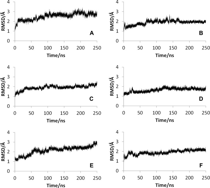
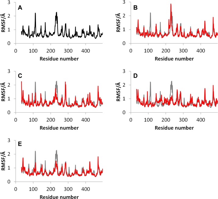
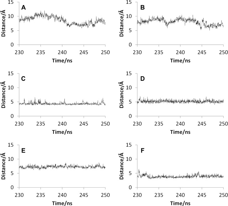
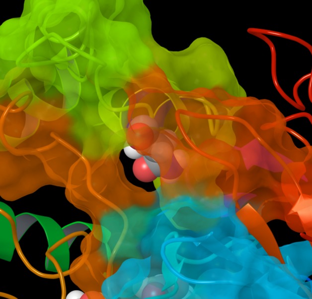
Many natural mutants of the drug metabolizing enzyme cytochrome P450 (CYP) 2D6 have been reported. Because the enzymatic activities of many mutants are different from that of the wild type, the genetic polymorphism of CYP2D6 plays an important role in drug metabolism. In this study, the molecular dynamics simulations of the wild type and mutants of CYP2D6, CYP2D6.1, 2, 10, 14A, 51, and 62 were performed, and the predictions of static and dynamic structures within them were conducted. In the mutant CYP2D6.10, 14A, and 61, dynamic properties of the F-G loop, which is one of the components of the active site access channel of CYP2D6, were different from that of the wild type. The F-G loop acted as the "hatch" of the channel, which was closed in those mutants. The structure of CYP2D6.51 was not converged by the simulation, which indicated that the three-dimensional structure of CYP2D6.51 was largely different from that of the wild type. In addition, the intramolecular interaction network of CYP2D6.10, 14A, and 61 was different from that of the wild type, and it is considered that these structural changes are the reason for the decrease or loss of enzymatic activities. On the other hand, the static and dynamic properties of CYP2D6.2, whose activity was normal, were not considerably different from those of the wild type.
Conflict of interest statement
Figures

References
-
- Nelson DR, Kamataki T, Waxman DJ, Guengerich FP, Estabrook RW, Feyereisen R, et al. The P450 superfamily: Update on new sequences, gene mapping, accession numbers, early trivial names of enzymes, and nomenclature. DNA Cell Biol. 1993;12:1–51. - PubMed
-
- Wolkers J, Witkamp RF, Nijimeijer SM, Burkow IC, de Groene EM, Lydersen C, et al. Phase I and phase II enzyme activities in Ringed seals (Phoca hispida): characterization of hepatic cytochrome P450 by activity patterns, inhibition studies, mRNA analyses, and western blotting. Aquat Toxicol. 1998;44:103–115.
-
- Meunier B, de Visser SP, Shaik S. Mechanism of oxidation reactions catalyzed by cytochrome P450 enzymes. Chem Rev. 2004;104:3947–3980. - PubMed
-
- Munro AW, Girvan HM, McLean KJ. Variations on a (t)heme–novel mechanisms, redox partners and catalytic functions in the cytochrome P450 superfamily. Nat Prod Rep. 2007;24:585–609. - PubMed
-
- Shaik S, de Visser SP. Computational approaches to cytochrome P450 function, In: de Montellano PRO, editor. Cytochrome P450: structure, mechanism, and biochemistry (3rd ed.). New York: Kluwer Academic/Plenum Publishers; 2005. pp. 45–85.
Publication types
MeSH terms
Substances
LinkOut - more resources
Full Text Sources
Other Literature Sources
