. 2016 Apr 5;12(4):e1005511.
doi: 10.1371/journal.ppat.1005511. eCollection 2016 Apr.

Transcriptome Remodeling in Trypanosoma cruzi and Human Cells during Intracellular Infection

Affiliations

Transcriptome Remodeling in Trypanosoma cruzi and Human Cells during Intracellular Infection

Yuan Li et al. PLoS Pathog. .

Abstract

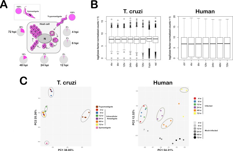
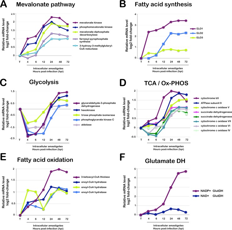
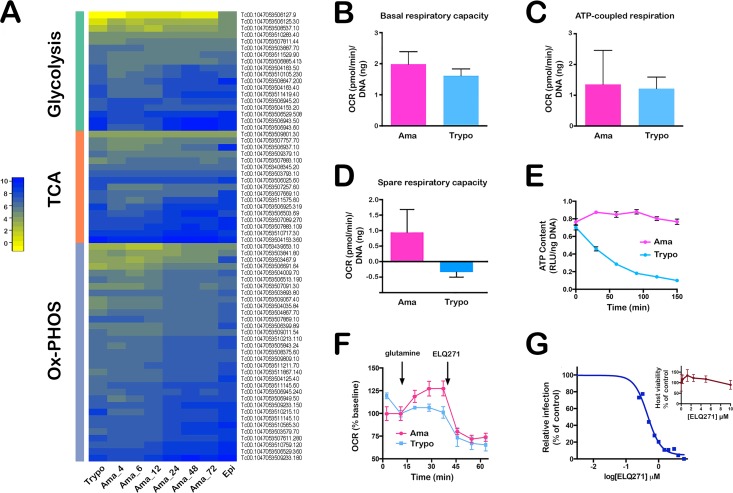
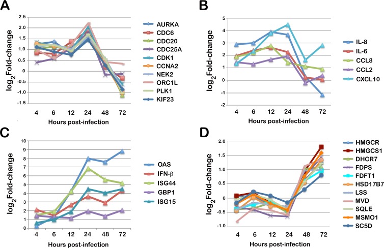
Intracellular colonization and persistent infection by the kinetoplastid protozoan parasite, Trypanosoma cruzi, underlie the pathogenesis of human Chagas disease. To obtain global insights into the T. cruzi infective process, transcriptome dynamics were simultaneously captured in the parasite and host cells in an infection time course of human fibroblasts. Extensive remodeling of the T. cruzi transcriptome was observed during the early establishment of intracellular infection, coincident with a major developmental transition in the parasite. Contrasting this early response, few additional changes in steady state mRNA levels were detected once mature T. cruzi amastigotes were formed. Our findings suggest that transcriptome remodeling is required to establish a modified template to guide developmental transitions in the parasite, whereas homeostatic functions are regulated independently of transcriptomic changes, similar to that reported in related trypanosomatids. Despite complex mechanisms for regulation of phenotypic expression in T. cruzi, transcriptomic signatures derived from distinct developmental stages mirror known or projected characteristics of T. cruzi biology. Focusing on energy metabolism, we were able to validate predictions forecast in the mRNA expression profiles. We demonstrate measurable differences in the bioenergetic properties of the different mammalian-infective stages of T. cruzi and present additional findings that underscore the importance of mitochondrial electron transport in T. cruzi amastigote growth and survival. Consequences of T. cruzi colonization for the host include dynamic expression of immune response genes and cell cycle regulators with upregulation of host cholesterol and lipid synthesis pathways, which may serve to fuel intracellular T. cruzi growth. Thus, in addition to the biological inferences gained from gene ontology and functional enrichment analysis of differentially expressed genes in parasite and host, our comprehensive, high resolution transcriptomic dataset provides a substantially more detailed interpretation of T. cruzi infection biology and offers a basis for future drug and vaccine discovery efforts.


Conflict of interest statement

The authors have declared that no competing interests exist.

Figures

Fig 1
Fig 1. Simultaneous interrogation of parasite and host transcriptomes.
(A) Intracellular T. cruzi life cycle and sample collection scheme. Extracellular T. cruzi trypomastigotes actively penetrate mammalian cells where they receive cues to differentiate into amastigote forms that replicate in the host cell cytoplasm for 3–5 days, beginning at ~22 hpi with a doubling time of ~12 hr. Amastigote division ceases on day 4 or 5 post-infection and parasites differentiate back into trypomastigotes that rupture the host cell to initiate a new cellular infection cycle. For RNA-Seq analysis, total RNA was isolated from axenic T. cruzi epimastigotes (insect vector stage), extracellular trypomastigotes and from amastigote-containing human fibroblast monolayers at 6 time points spanning 4–72 hpi. Pie charts indicate the proportion of mapped sequence reads assigned to the parasite (pink) or human (grey). (B) Distribution of global gene expression levels in a representative subset of T. cruzi and human samples. Box plots showing comparisons of the distribution of per-gene counts (log2 counts per million with an offset of 1) normalized for sequencing library size. The ends of the whiskers represent the lowest datum still within 1.5 interquartile range (IQR) of the lower quartile, and the highest datum still within 1.5 IQR of the upper quartile. Genes with extremely high or low expression levels are shown as open circles above and below the whiskers, respectively. (C) Principal component analysis plots of global transcriptome profiles. Principal component analysis (PCA) plot for RNA-Seq data with T. cruzi and human samples plotted separately. The two first principal components (PC1 and PC2) are plotted with the proportion of variance explained by each component next to the axes labels. Each sample is represented by a dot and the color label corresponding to the sample group, such as the number of hours (hr) post-infection.
Fig 2
Fig 2. Temporal expression of metabolic pathway genes in mammalian-infective stages of T. cruzi.
Relative mRNA expression of selected genes in intracellular T. cruzi amastigote stages (4–72 hpi) compared to extracellular trypomastigotes (T). Genes in the following metabolic pathways are highlighted: (A) Mevalonate pathway: mevalonate kinase (TcCLB.436521.9), mevalonate diphosphate decarboxylase (TcCLB.507993.330), squalene monooxygenase (TcCLB.509589.20), farnesyl pyrophosphate synthase (TcCLB.508323.9), 3-hydroxy-3-methylglutaryl-CoA reductase (TcCLB.509167.20). (B) Fatty Acid Synthesis: fatty acid elongase 1 (ELO1) (TcCLB.506661.30), fatty acid elongase 2 (ELO2) (TcCLB.506661.20), fatty acid elongase 3 (ELO3) (TcCLB.506661.10). (C) Glycolysis: glyceraldehyde-3-phosphate dehydrogenase (TcCLB.506943.60); hexokinase (TcCLB.508951.20), triosephosphate isomerase (TcCLB.508647.200), phosphoglycerate kinase (TcCLB.511419.40), aldolase (TcCLB.504163.40). (D) Tricarboxylic Acid / Oxidative Phosphorylation (TCA/Ox-PHOS): cytochrome b5 (TcCLB.506773.44), ATPase subunit 9 (TcCLB.503579.70), cytochrome c oxidase subunit V (TcCLB.510565.30), succinate dehydrogenase 11 (TcCLB.504035.84), succinate dehydrogenase 6 (TcCLB.507091.30), cytochrome c oxidase subunit VII (TcCLB.509233.150), cytochrome c oxidase subunit VI (TcCLB.511145.10), cytochrome c oxidase subunit IV (TcCLB.506529.360). (E) Fatty Acid Oxidation: 3-ketoacyl-CoA thiolase (TcCLB.510507.20), enoyl-CoA hydratase/isomerase (TcCLB.511529.170); enoyl-CoA hydratase, mitochondrial (TcCLB.508185.10) enoyl-CoA hydratase/isomerase (TcCLB.510997.40). (F) Glutamate Dehydrogenases (DH): NADP+-GlutDH (TcCLB.507875.20), NAD+ GlutDH (TcCLB.509445.39). All values are reported as log2 fold-change of difference between expression at in the trypomastigote stage and intracellular stages as reported in S5 Table.
Fig 3
Fig 3. Validation of predicted metabolic features of T. cruzi developmental stages.
(A) Heatmap of expression values of annotated T. cruzi genes predicted to function in intermediary metabolism with a focus on glycolysis, TCA cycle and Ox-PHOS. (B) Calculated basal respiratory capacity (C) ATP-linked respiration and (D) spare-respiratory capacity of extracellular trypomastigotes and isolated intracellular amastigotes (60 hpi) as pmol of oxygen consumed per min (oxygen consumption rate; OCR) normalized to T. cruzi DNA (ng) per well. (E) ATP content measured in isolated trypomastigotes and amastigotes in KHB buffer without a consumable carbon source at time points indicated. (F) OCR response to glutamine (10 mM) and ELQ271 (10 μM) in isolated T. cruzi trypomastigotes and amastigotes. (G) Dose-dependent inhibition of T. cruzi amastigote growth in HFF following addition of ELQ271 at 18 hpi and relative infection measured at 72 hpi. Host cell viability and growth (inset) is unaffected by the compound over the course of the assay. Graphs shown are representative of 3 independent experiments.
Fig 4
Fig 4. Dynamic host response signatures in T. cruzi-infected human fibroblasts.
Expression patterns for selected genes in the most strongly modulated pathways in T. cruzi infected HFF. Genes in the following categories are highlighted. (C) Mitotic Cell cycle: AURKA (ENSG00000087586); CDC6 (ENSG00000094804); CDC20 (ENSG00000117399); CDC25A (ENSG00000164045); CDK1 (ENSG00000170312); CCNA2 (ENSG00000145386); NEK2 (ENSG00000117650); ORC1L (ENSG00000085840); PLK1 ENSG00000137807); KIF23 (ENSG00000137807). (B) Cytokines/Chemokines: IL-8 (ENSG00000169429); IL-6 (ENSG00000136244); CCL8 (ENSG00000108700); CCL2 (ENSG00000108691); CXCL10 (ENSG00000169245). (C) Type I Interferon: OAS (ENSG00000089127); IFNB (ENSG00000171855); ISG44 (ENSG00000137959); GBP1 (ENSG00000117228); ISG15 (ENSG00000187608). (D) Mevalonate/Sterol biosynthesis: HMGCR (ENSG00000113161); HMGCS1 (ENSG00000112972); DHCR7 (ENSG00000172893); FDPS (ENSG00000160752); FDFT1 (ENSG00000079459); HSD17B7 (ENSG00000132196); LSS (ENSG00000160285); MVD (ENSG00000167508); SQLE (ENSG00000104549); MSMO1 (ENSG00000052802); SC5D (ENSG00000109929). All values are reported as log2 fold-change of the difference in expression of infected and matched uninfected controls at each time point as listed in S7 Table.

References

    1. Rassi A Jr., Rassi A, Marin-Neto JA. Chagas disease. Lancet. 2010;375(9723):1388–402. 10.1016/S0140-6736(10)60061-X - DOI - PubMed
    1. Tardieux I, Webster P, Ravesloot J, Boron W, Lunn JA, Heuser JE, et al. Lysosome recruitment and fusion are early events required for trypanosome invasion of mammalian cells. Cell. 1992;71(7):1117–30. - PubMed
    1. Tomlinson S, Vandekerckhove F, Frevert U, Nussenzweig V. The induction of Trypanosoma cruzi trypomastigote to amastigote transformation by low pH. Parasitology. 1995;110 (Pt 5):547–54. - PubMed
    1. Ley V, Robbins ES, Nussenzweig V, Andrews NW. The exit of Trypanosoma cruzi from the phagosome is inhibited by raising the pH of acidic compartments. J Exp Med. 1990;171(2):401–13. - PMC - PubMed
    1. Soll DR. The role of phenotypic switching in the basic biology and pathogenesis of Candida albicans. J Oral Microbiol. 2014;6 10.3402/jom.v6.22993 - DOI - PMC - PubMed

Publication types