. 2016 Apr 5;11(4):e0152968.
doi: 10.1371/journal.pone.0152968. eCollection 2016.

Factor XI Deficiency Alters the Cytokine Response and Activation of Contact Proteases during Polymicrobial Sepsis in Mice

Affiliations

Factor XI Deficiency Alters the Cytokine Response and Activation of Contact Proteases during Polymicrobial Sepsis in Mice

Charles E Bane Jr et al. PLoS One. .

Abstract

Sepsis, a systemic inflammatory response to infection, is often accompanied by abnormalities of blood coagulation. Prior work with a mouse model of sepsis induced by cecal ligation and puncture (CLP) suggested that the protease factor XIa contributed to disseminated intravascular coagulation (DIC) and to the cytokine response during sepsis. We investigated the importance of factor XI to cytokine and coagulation responses during the first 24 hours after CLP. Compared to wild type littermates, factor XI-deficient (FXI-/-) mice had a survival advantage after CLP, with smaller increases in plasma levels of TNF-α and IL-10 and delayed IL-1β and IL-6 responses. Plasma levels of serum amyloid P, an acute phase protein, were increased in wild type mice 24 hours post-CLP, but not in FXI-/- mice, supporting the impression of a reduced inflammatory response in the absence of factor XI. Surprisingly, there was little evidence of DIC in mice of either genotype. Plasma levels of the contact factors factor XII and prekallikrein were reduced in WT mice after CLP, consistent with induction of contact activation. However, factor XII and PK levels were not reduced in FXI-/- animals, indicating factor XI deficiency blunted contact activation. Intravenous infusion of polyphosphate into WT mice also induced changes in factor XII, but had much less effect in FXI deficient mice. In vitro analysis revealed that factor XIa activates factor XII, and that this reaction is enhanced by polyanions such polyphosphate and nucleic acids. These data suggest that factor XI deficiency confers a survival advantage in the CLP sepsis model by altering the cytokine response to infection and blunting activation of the contact (kallikrein-kinin) system. The findings support the hypothesis that factor XI functions as a bidirectional interface between contact activation and thrombin generation, allowing the two processes to influence each other.


Conflict of interest statement

Competing Interests: DG is a consultant, and receives consultant’s fees from the following companies with an interest in the role of factor XI in thrombosis: Bayer Pharma, Dyax Corp., Novartis, Isis Pharmaceuticals and Ono Pharma. None of these corporations provided support for the work presented in this manuscript. In addition. DG is on the scientific advisory board of Aronora, which also has an interest in the role of factor XI in thrombosis. Aronora did not provide financial support for the work in described in this manuscript. EIT and AG are employees of the company Aronora, and receive salary support from that entity. Other than salary support, Aronora did not provide financial support for the work in described in this manuscript. These investigators have collaborated with DG and his colleagues on projects funded by the NIH grants supporting the work on sepsis. None of the arrangements described in this sections not alters the authors' adherence to PLOS ONE policies on sharing data and materials as detailed in the guide for authors (http://www.PLOSone.org/static/editorial.action#competing).

Figures

Fig 1
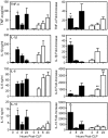
Fig 1. Plasma cytokine levels after CLP.
Shown are plasma concentrations of TNFα, IL-1β, IL-6 and IL-10 in WT (black bars) and FXI-/- (white bars) littermates at various times after high-grade CLP. Absolute cytokine levels are shown in the left-hand column and fold-increases in cytokines compared to sham treatment are shown in the right-hand column. Eight or nine mice were tested at each time point for each genotype for CLP, and three were used at each time point for sham surgery. Plasma TNFα (p = 0.006) and IL-10 (p = 0.0003) levels were significantly greater in WT mice than in FXI-/- mice 4 hr post-CLP. Fold-increases in plasma levels of TNFα, (p = 0.009), IL-1β, (p = 0.009), IL-6 (p = 0.003) and IL-10 (p = 0.0003) were significantly greater in WT mice than in FXI-/- mice 4 hr post-CLP. For IL-6, FXI-/- mice had significantly greater fold-increases in plasma levels 8 (**p = 0.005) and 24 hr (**p = 0.04) post-CLP. Error bars represent SEM.
Fig 2
Fig 2. Serum amyloid P levels post-CLP.
Plasma levels of SAP measured by ELISA in control mice (C, n = 4) not undergoing surgery, and 24 hr post-CLP (n = 8–9) or sham (Sh n = 3) surgery. Black bars are results for WT mice and white bars for FXI-/- mice. Error bars represent SEM.
Fig 3
Fig 3. Survival after CLP.
(A) Survival of male WT (…, n = 17), FXI+/- (---, n = 24), or FXI-/- (—, n = 18) mice after high-grade CLP. p = 0.03 (*) for WT vs. FXI-/-. (B) Survival for male WT (n = 17), FXI+/- (n = 28), or FXI-/- (n = 19) mice after low-grade CLP (p = 0.4 for WT vs. FXI-/-, p = 1.0 for WT vs. FXI+/-, p = 0.5 for FXI-/- vs. FXI+/-). (C-E) Survival for male mice after high-grade (—) or low-grade (…) injury. (C) FXI-/-, (D) FXI+/- and (E) WT mice. There was no difference in survival for FXI-/- mice after low- or high- grade injury (p = 0.47). Survival was significantly different between the two levels of injury for FXI+/- (p = 0.001) and WT (p = 0.0002) mice. Curves were compared by log-rank test.
Fig 4
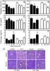
Fig 4. Effects of CLP on markers of coagulation.
(A,B) Whole blood platelet counts, (C,D) plasma thrombin-antithrombin complex (TAT) levels, and (E,F) plasma fibrinogen levels in WT (black bars) and FXI-/- (white bars) mice after CLP or sham surgery. Panels A, C and E show values at various times post-CLP. Panels B, D and F compare values 24 hour post-CLP or sham (Sh) surgery to 0 hr controls. CLP induced significant platelet reductions in WT (*p = 0.003) and FXI-/- (**p = 0.001) mice. For panels A to F, error bars represent SEM. (G) Photomicrographs (400X magnification) of paraffin embedded sections of spleen and thymus stained with hematoxylin and eosin before, and 24 hours after, CLP in WT and FXI-/- mice. Apoptotic changes (indicated by white arrows) were present in both organs for mice of both genotypes 24 hrs post-CLP.
Fig 5
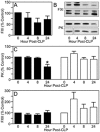
Fig 5. The effect of CLP on plasma contact proteases.
Plasma FXI, PK and FXII levels at various times after high-grade CLP were determined using densitometry of western blots and are reported as percent of baseline (0 hr) control. (A) Plasma FXI levels after CLP in WT mice (B) Examples of western blots for FXI and PK for WT mouse plasma at various times after CLP (upper panels of each pair) or sham surgery (lower panels of each pair). (C) Plasma PK levels after high-grade CLP in WT (black bars) or FXI-/- (white bars) mice. The asterisk above the 24 hr bar for PK in WT animals indicates the value is significantly different than 0 hr control (p<0.05). (D) Plasma FXII levels after high-grade CLP in WT (black bars) or FXI-/- (white bars) mice. In panels A, C, and D, each bar represents data for eight mice. For all panels, error bars represent SEM.
Fig 6
Fig 6. FXII activation by α-kallikrein or FXIa in the presence of polyanions.
(A) FXII (200 nM) was incubated with vehicle (▼), 1nM FXIa (◇), or 2 nM α-kallikrein (◆). (B-D) FXII (200 nM) incubated with (B) 5 ug/ml DNA (□,➄), (C) 5 ug/ml RNA (○,➂) or (D) 20 μg/ml Poly-P (△,▲), in the presence of 1 nM FXIa (□,○,△) or 2 nM α-kallikrein (➄,➂,▲). At the indicated times, samples were tested for FXIIa activity by chromogenic assay. Error bars are +/− one standard deviation.
Fig 7
Fig 7. Effect of HK on FXII activation in the presence of polyanions.
Plasma FXII (200 nM) was incubated with (A-C) 2 nM α-kallikrein or (D-F) 1 nM FXIa and (A,D) 20 μg/ml poly-P, (B,E) 5 μg/ml DNA or (C,F) 5 μg/ml RNA in the absence (○,□,△) or presence (➂,➄,▲) of 20 nM HK. At the indicated times, samples were tested for FXIIa activity by chromogenic assay. Error bars are +/− one standard deviation.
Fig 8
Fig 8. Poly-P-induced changes in FXII in mice.
WT C57Bl/6 mice or C57Bl/6 mice lacking FXI (XI-/-), PK (PK-/-) or FXII (XII-/-) received a bolus infusion of phosphate buffered saline (saline) or PBS containing 60 μg of poly-P into the inferior vena cava. Five minutes later blood was drawn from the inferior vena cava into sodium citrate anticoagulant. Plasma samples were analyzed by western blot for FXII under non-reducing conditions. The blot contains samples for two mice of each genotype. The position of the FXII zymogen band is indicated on the left (FXII). Free FXIIa would also run in this position. The higher molecular mass FXII specific-species likely represent FXIIa in SDS-stable complexes with plasma serine protease inhibitors (serpins).
Fig 9
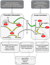
Fig 9. FXI in thrombin generation and contact activation during sepsis.
Thrombin Generation (Panel 1). Depicted are proteolytic reactions that generate thrombin at a site of vascular injury. The process is initiated by activation of factors X and IX by the factor VIIa/tissue factor (TF) complex. Vitamin-K dependent protease zymogens are shown in black type with the active protease forms indicated by a lower case “a”. The red ovals represent cofactors. During hemostasis, FXI is converted to FXIa by thrombin (white arrows). FXIa then activates FIX (green arrow). Contact activation (Panel 2). On a surface, FXII and prekallikrein (PK) undergo reciprocal activation to FXIIa and α-kallikrein. High-molecular-weight kininogen (HK) serves as a cofactor for this reaction. FXIIa can promote thrombin generation by activating FXI (white arrow). α-kallikrein cleaves HK liberating bradykinin (BK) and antimicrobial peptides (AMPs). Data presented in this manuscript raise the possibility that FXIa can also contribute to contact activation through activation of FXII (green arrow). Panels 1 and 2 list factors that could trigger or enhance thrombin generation (1) or contact activation (2). Panels 5, 6, and 7 list processes mediated by thrombin (5), FXIIa (6) or α-kallikrein (7) that could contribute to the sepsis syndrome. Panel 8 lists some of the consequences of those processes.

References

    1. Stearns-Kurosawa DJ, Osuchowski MF, Valentine C, Kurosawa S, Remick DG. The pathogenesis of sepsis. Ann Rev Path. 2011;6: 19–48. - PMC - PubMed
    1. Degen JL, Bugge TH, Goguen JD. Fibrin and fibrinolysis in infection and host defense. J Thromb Haemost. 2007. July;5 Suppl 1:24–31. - PubMed
    1. Pawlinski R, Mackman N. Tissue factor, coagulation proteases, and protease-activated receptors in endotoxemia and sepsis. Crit Care Med. 2004;32 Suppl 5: S293–297. - PubMed
    1. Pawlinski R, Mackman N. Cellular sources of tissue factor in endotoxemia and sepsis. Thromb Res. 2010. April;125 Suppl 1:S70–3. 10.1016/j.thromres.2010.01.042 - DOI - PMC - PubMed
    1. van der Poll T, Levi M. Crosstalk between inflammation and coagulation: the lessons of sepsis. Curr Vasc Pharmacol. 2012. September;10(5):632–8. - PubMed

Publication types