. 2016 Apr 5:7:11240.
doi: 10.1038/ncomms11240.

Airway bacteria drive a progressive COPD-like phenotype in mice with polymeric immunoglobulin receptor deficiency

Affiliations

Airway bacteria drive a progressive COPD-like phenotype in mice with polymeric immunoglobulin receptor deficiency

Bradley W Richmond et al. Nat Commun. .

Abstract

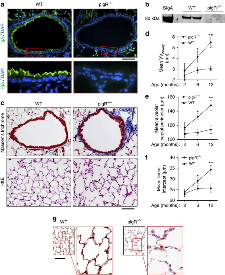
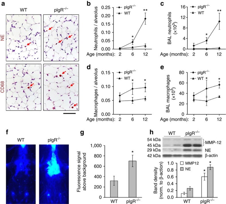
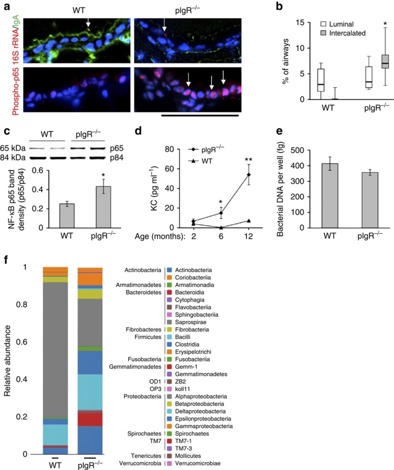
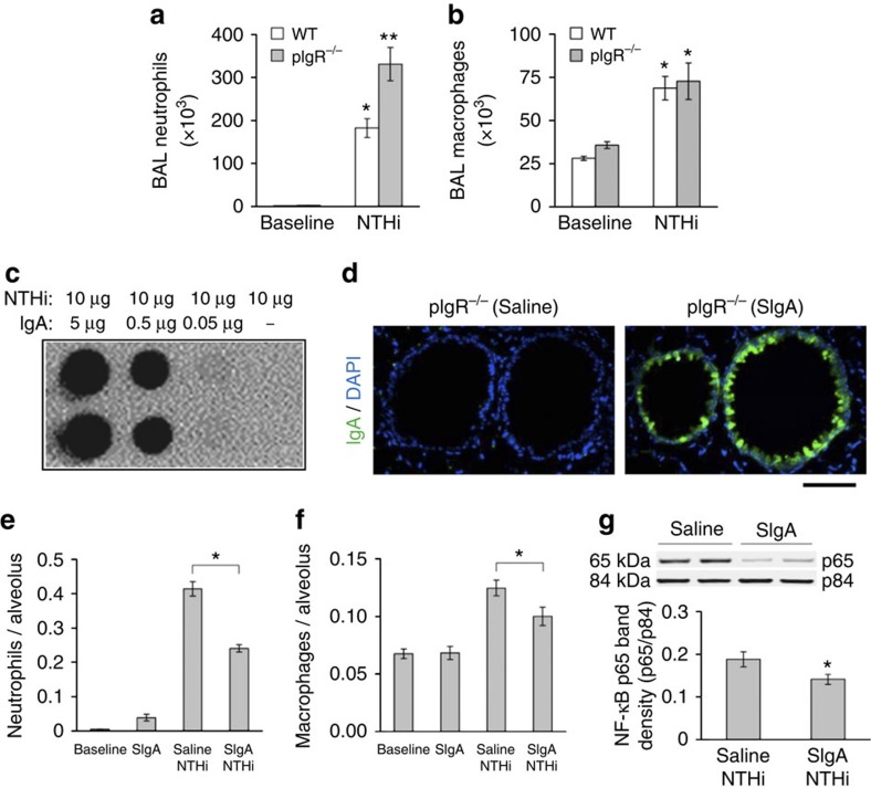
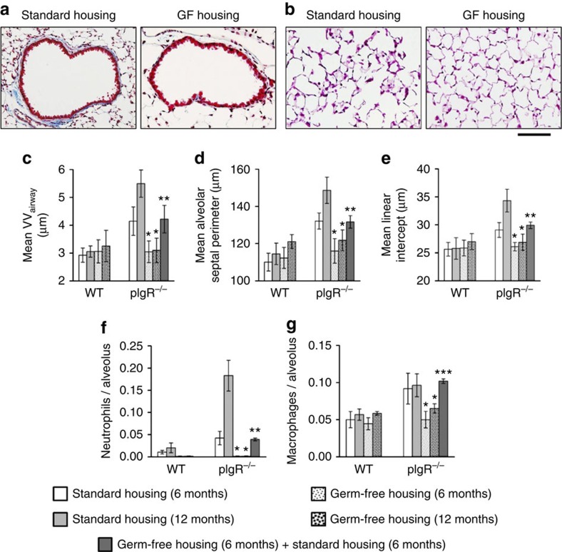
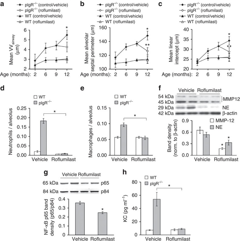
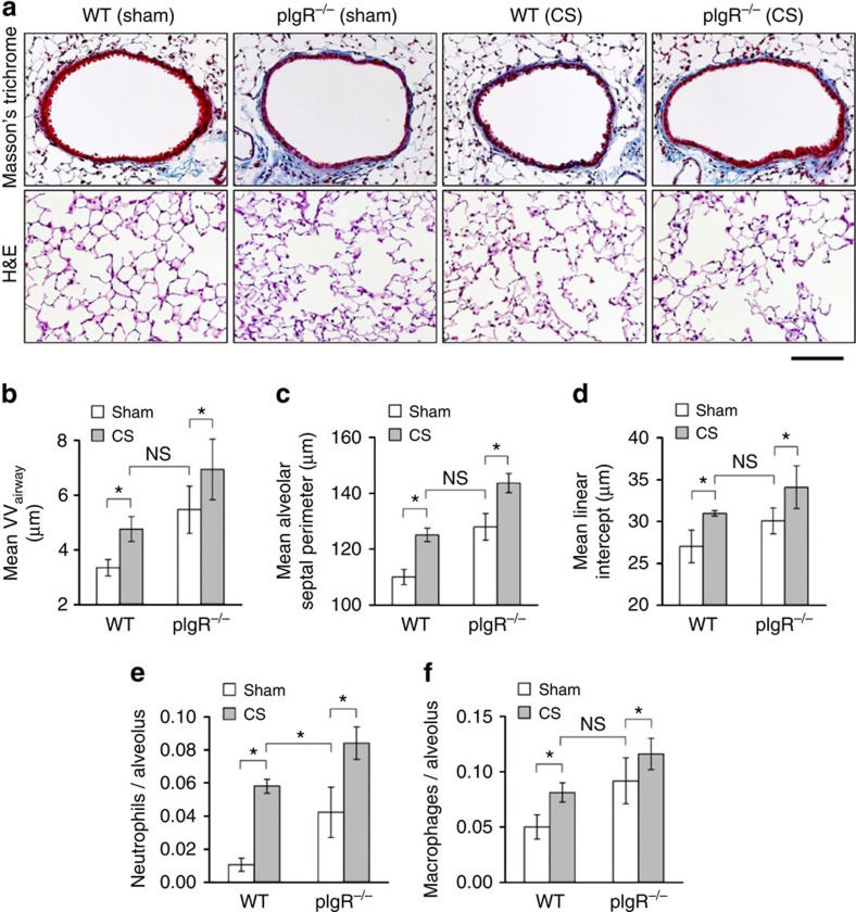
Mechanisms driving persistent airway inflammation in chronic obstructive pulmonary disease (COPD) are incompletely understood. As secretory immunoglobulin A (SIgA) deficiency in small airways has been reported in COPD patients, we hypothesized that immunobarrier dysfunction resulting from reduced SIgA contributes to chronic airway inflammation and disease progression. Here we show that polymeric immunoglobulin receptor-deficient (pIgR(-/-)) mice, which lack SIgA, spontaneously develop COPD-like pathology as they age. Progressive airway wall remodelling and emphysema in pIgR(-/-) mice are associated with an altered lung microbiome, bacterial invasion of the airway epithelium, NF-κB activation, leukocyte infiltration and increased expression of matrix metalloproteinase-12 and neutrophil elastase. Re-derivation of pIgR(-/-) mice in germ-free conditions or treatment with the anti-inflammatory phosphodiesterase-4 inhibitor roflumilast prevents COPD-like lung inflammation and remodelling. These findings show that pIgR/SIgA deficiency in the airways leads to persistent activation of innate immune responses to resident lung microbiota, driving progressive small airway remodelling and emphysema.


Figures

Figure 1
Figure 1. pIgR−/− mice develop progressive COPD-like small airway and parenchymal remodelling.
(a) Immunofluorescence staining for IgA (green) showing SIgA on the epithelial surface of a small airway from a WT mouse and no detectable SIgA on the airway surface of a pIgR−/− mouse (original magnification, × 200 and × 1,000 (insets)). Scale bar, 50 μm. (b) Western blotting for secretory component in BAL fluid from WT and pIgR−/− mice. SIgA from human colostrum was used as a positive control. (c) Representative images of small airway remodelling (Masson's trichrome, original magnification, × 200) and emphysema (haematoxylin and eosin (H&E), original magnification, × 200) in a 12-month-old pIgR−/− mouse compared with a WT control. Scale bar, 50 μm. (df) Morphometric analysis showing increased wall thickness (VVairway), mean alveolar septal perimeter length and mean linear intercept in pIgR−/− and age-matched WT littermate controls at the indicated ages. Five to ten mice per group; *P<0.01 compared with 2-month-old pIgR−/− mice and age-matched WT controls; **P<0.001 compared with all other groups (two-way analysis of variance (ANOVA)). (g) Immunostaining for elastin in 12-month-old WT and pIgR−/− mice shows reduction and fragmentation of elastin in inter-alveolar septa in a pIgR−/− mouse compared with the intact elastin network in a WT mouse (original magnification, × 100 and × 1,000 (insets)).
Figure 2
Figure 2. Lung inflammation progresses with age in pIgR−/− mice.
(a) Representative immunostains for neutrophils using antibodies to neutrophil elastase (NE) or macrophages using antibodies to CD68 in 12-month-old WT and pIgR−/− mice. Positive cells are stained brown (indicated by red arrows) (original magnification, × 200). Scale bar, 50 μm. (be) Neutrophil (NE+) and macrophage (CD68+) counts in lungs of pIgR−/− and age-matched WT littermate controls at the indicated ages, and neutrophil and macrophage counts in BAL fluid. Five to seven mice per group; *P<0.05 compared with 2-month-old pIgR−/− mice and age-matched WT mice; **P<0.01 compared with all other groups (two-way analysis of variance (ANOVA)). (f) Representative image of folate-PEG-Cy5-derived chest fluorescence 4 h after intravenous probe injection in 12-month-old WT and pIgR−/− mice. (g) Photon emission from the chest normalized to background before injection of probe. Three to four mice per group; *P<0.05 (Student's t-test). (h) Western blotting and densitometry for MMP-12 (two bands at 45 and 54 kDa) and NE (29 kDa) in lung tissue from 12-month-old WT and pIgR−/− mice. Band densities of MMP-12 and NE were normalized to β-actin. Six mice per group; *P<0.01 compared with WT mice (Student's t-test).
Figure 3
Figure 3. Bacterial invasion and NF-κB activation in airways of pIgR−/− mice.
(a) Immunofluorescent detection of bacteria (fluorescent in situ hybridization (FISH) probe for 16S rRNA, red, top panels) or NF-κB (phospho-p65 (Ser276, red, bottom panels), IgA (green, top panels) and DAPI (blue) from 12-month-old WT and pIgR−/− mice (original magnification, × 1,000). In WT mice, bacteria with bound SIgA were identified within the airway lumen (yellow on merged image, identified by arrow), whereas bacteria intercalated within the epithelium were identified in pIgR−/− mice (red, identified by arrow). Bottom panels show phopsho-p65 localized to nuclei (arrows). Scale bar, 50 μm. (b) Box and whisker plot (showing median, 25th–75th percentile and range) for intraepithelial (intercalated) and luminal bacteria in airways of 12-month-old WT and pIgR−/− mice as identified by FISH for bacterial DNA. Seven mice per group; *P<0.001. (c) Western blotting and densitometry for p65 component of NF-κB (normalized to p84) in nuclear protein extracts from lungs of 12-month-old WT and pIgR−/− mice. Six mice per group; *P<0.05 (Student's t-test). (d) KC protein levels in BAL fluid. Six mice per group; *P<0.05 compared with 2-month-old pIgR−/− mice and age-matched WT controls; **P<0.05 compared with all other groups (two-way analysis of variance (ANOVA)). (e) Total bacterial DNA was quantified from lung tissue in WT and pIgR−/− mice using quantitative PCR (qPCR) and primers specific for V1 region of prokaryotic 16S rRNA. Ten mice per group. (f) Distribution of bacterial phyla and classes in lung tissue as determined by 16S sequencing from lungs of 12-month-old WT and pIgR−/− mice (three mice studied per group but only two WT mice had sufficient bacterial DNA amplification for detailed analysis).
Figure 4
Figure 4. SIgA modulates the acute inflammatory response to NTHi in vivo.
(a,b) Neutrophil and macrophage counts in BAL fluid from 2-month-old WT and pIgR−/− mice 24 h after aerosolization of NTHi lysate (10 mg). Six to eight mice per group; *P<0.01 compared with untreated mice (baseline), **P<0.01 compared with WT mice treated with NTHi (Student's t-test). (c) Dot-blot assay demonstrating protein binding between NTHi lysates and human SIgA from colostrum. (d) Immunofluorescent detection of human SIgA (green) in the lungs of pIgR−/− mouse 1 h after i.t. delivery of SIgA or vehicle (normal saline) (original magnification, × 200). Scale bar, 50 μm. (e,f) Parenchymal neutrophil and macrophage counts 24 h after aerosol delivery of NTHi lysate to 2-month-old pIgR−/− mice pretreated with i.t. SIgA (50 μl of 0.34 mg ml−1 solution) or vehicle (normal saline). Macrophage and neutrophil numbers were quantified by immunostaining for CD68 or NE, respectively. Five to six mice per group; *P<0.05 compared with mice pretreated with saline followed by NTHi (Student's t-test). (g) Western blotting and densitometry for p65 component of NF-κB (normalized to p84) in lung nuclear protein extracts from 2-month-old pIgR−/− mice pretreated with i.t. SIgA or normal saline 1 h before NTHi nebulization and harvested 24 h later. Six mice per group; *P<0.05 (Student's t-test).
Figure 5
Figure 5. Germ-free pIgR−/− mice are protected from COPD-like lung remodelling.
(a) Representative images of small airway remodelling (Masson's trichrome, original magnification, × 200) and emphysema (b) (haematoxylin and eosin (H&E), original magnification, × 200) in a 6-month-old pIgR−/− mouse in standard housing compared with a 6-month-old pIgR−/− mouse housed in germ-free conditions. Scale bar, 50 μm. (ce) Morphometric analysis of airway wall thickness (VVairway), alveolar septal perimeter length and mean linear intercept in 6- and 12-month-old WT and pIgR−/− mice maintained in standard housing, germ-free housing or 6 months of germ-free housing followed by 6 months of standard housing as indicated. (f,g) Parenchymal neutrophil (NE+) and macrophage (CD68+) counts in lungs of 6- and 12-month-old WT and pIgR−/− mice maintained in standard housing, germ-free housing or a combination of both as indicated. Six to seven mice per group; *P<0.001 compared with all other groups, **P<0.001 compared with age-matched WT controls housed in standard conditions (two-way analysis of variance (ANOVA)).
Figure 6
Figure 6. Roflumilast blocks inflammation and COPD-like lung remodelling in pIgR−/− mice.
(ac) Morphometric analysis showing small airway wall thickness (VVairway), mean alveolar septal perimeter length and mean linear intercept at the indicated ages in pIgR−/− and WT mice treated with roflumilast or vehicle from 9 to 12 months of age. Five to ten mice per group; *P<0.01 compared with 12-month-old pIgR−/− mice treated with roflumilast, **P<0.05 compared with 6- and 9-month-old pIgR−/− mice (two-way analysis of variance (ANOVA)). (d,e) Parenchymal neutrophil and macrophage counts in 12-month-old WT and pIgR−/− mice treated for 3 months with roflumilast or vehicle. Six to seven mice per group; *P<0.01 (macrophages) or P<0.001 (neutrophils) compared with pIgR−/− mice treated with vehicle (Student's t-test). (f) Western blotting and densitometry for MMP-12 and NE in lung tissue from 12-month-old pIgR−/− mice treated with roflumilast or vehicle. Band densities of MMP-12 and NE were normalized to β-actin. Six mice per group; *P<0.05 (Student's t-test). (g) Western blotting and densitometry for p65 component of NF-κB (normalized to p84) in lung nuclear protein extracts from 12-month-old pIgR−/− mice treated with roflumilast or vehicle. Six mice per group; *P<0.01 (Student's t-test). (h) KC protein levels in BAL fluid from 12-month-old WT or pIgR−/− mice treated with roflumilast or vehicle. Six mice per group; *P<0.05 compared with pIgR−/− mice treated with vehicle (Student's t-test).
Figure 7
Figure 7. CS treatment increases airway remodelling and emphysema in pIgR−/− mice.
(a) Representative images of small airway remodelling (Masson's trichrome, original magnification, × 200X and emphysema (haematoxylin and eosin (H&E), original magnification, × 200) in WT and pIgR−/− mice treated twice daily with mainstream CS or sham control (filtered air) for 6 months (between 2 and 8 months of age). Scale bar, 50 μm. (bd) Morphometric analysis of small airway wall thickness (VVairway), mean alveolar septal perimeter length and mean linear intercept length. Fve mice per group; *P<0.01 compared with WT mice treated with sham; **P<0.01 compared with all other groups (two-way analysis of variance (ANOVA)). (e,f) Parenchymal neutrophil and macrophage counts in lung tissue from WT or pIgR−/− mice. Five mice per group; *P<0.01 compared with WT mice treated with sham; **P<0.01 compared with all other groups (two-way ANOVA).

References

    1. Vestbo J. et al. Global strategy for the diagnosis, management, and prevention of chronic obstructive pulmonary disease: GOLD executive summary. Am. J. Respir. Crit. Care Med. 187, 347–365 (2013) . - PubMed
    1. Hogg J. C., Macklem P. T. & Thurlbeck W. M. Site and nature of airway obstruction in chronic obstructive lung disease. N. Engl. J. Med. 278, 1355–1360 (1968) . - PubMed
    1. Hogg J. C. et al. The nature of small-airway obstruction in chronic obstructive pulmonary disease. N. Engl. J. Med. 350, 2645–2653 (2004) . - PubMed
    1. McDonough J. E. et al. Small-airway obstruction and emphysema in chronic obstructive pulmonary disease. N. Engl. J. Med. 365, 1567–1575 (2011) . - PMC - PubMed
    1. Baraldo S., Turato G. & Saetta M. Pathophysiology of the small airways in chronic obstructive pulmonary disease. Respiration 84, 89–97 (2012) . - PubMed

Publication types

MeSH terms