

Occurrence and Distribution of bovine tuberculosis (Mycobacterium bovis) in Slaughtered cattle in the abattoirs of Bauchi State, Nigeria
- PMID: 27047110
- PMCID: PMC4774856
- DOI: 10.14202/vetworld.2015.432-437
Occurrence and Distribution of bovine tuberculosis (Mycobacterium bovis) in Slaughtered cattle in the abattoirs of Bauchi State, Nigeria
Abstract
Aim: This study was aimed to determine the prevalence of bovine tuberculosis (bTB) in slaughtered cattle in Bauchi State, Nigeria. The cause (s) of grossly suspected bTB lesions encountered at the abattoirs during post-mortem (PM), as whether due to Mycobacterium bovis alone or together with other acid fast bacilli (AFB).
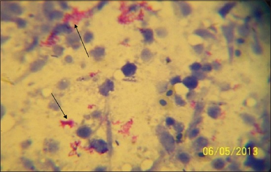
Materials and methods: A cross-sectional abattoir based study was conducted on 800 cattle slaughtered in the Northern, Central and Southern zonal abattoirs of Bauchi State, Nigeria, from June to August 2013; using PM meat inspection, Ziehl-Neelsen staining (ZN) and confirmatory polymerase chain reaction (PCR) techniques.
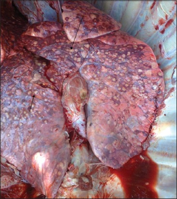
Results: The occurrence of bTB lesions from the organs of slaughtered cattle in Bauchi State, showed that the lungs had the highest number of suspected tissues 65 (54.20%), followed by the lymph nodes 28 (23.30%) while the heart, liver, spleen, intestines and mammary glands had the other 8.3%, 6.7%, 5.0%, 1.7%, and 0.8%, suspected tissues respectively. By ZN microscopic staining all 100% (2/2) of the intestines were positive for bTB, followed by the heart with 50% (5/10), then the lungs 29.23% (19/65); while the liver, lymph nodes, and spleen had 25%, 21.43% and 16.67% respectively were tested positive for bTB. It was only the mammary gland that tested negative for bTB in all the suspected tissues sampled. By PCR, the intestines had the highest positive bTB with 100% (2/2), followed by the liver with 12.5% (1/8), and then the lungs with 7.8% (5/65). The lymph nodes had 7.14% (2/28) tissues that tested positive for bTB. However, the spleen, heart and mammary gland were all tested negative with 0%; indicating that the false positive for bTB detected by ZN were confirmed by PCR. While based on the location of the abattoirs in the three senatorial zones of Bauchi State, Bauchi zonal abattoir had the highest number of suspected bTB cases 75 (62.50%), followed by Katagum zonal slaughter house with 32 (26.7%) and then Misau with 13 (10.8%). By the ZN staining technique, there were 25 (33.33%) positivity in Bauchi Zonal abattoir, while Katagum and Misau abattoirs had 9 (28.13%) and 1 (7.72%) positive respectively. By the PCR technique, 9 (12.00%), 1 (3.13%) and 0 (0.00%) positive cases were recorded for Bauchi, Katagum and Misau abattoirs respectively.
Conclusion: The present study estimated the prevalence rate of bTB in Bauchi State, using PM, ZN and PCR techniques at 15.0%, 29.16% and 8.33%, respectively. Bovine TB lesions found at PM were not all due to M. bovis alone, as other MTBC and AFB organisms may cause bTB-like lesions that were excluded by PCR specific primers. The prevalence of bTB was higher in Bauchi abattoir that supplies larger population of the state with beef. These findings also demonstrate the urgent need for public health authorities in the state to intervene in the control of the zoonotic bTB.
Keywords: Bauchi State; PCR; Ziehl-Neelsen; abattoir; bovine TB; post-Mortem.
Figures
Similar articles
-
Detection of Mycobacterium bovis in Organs of Slaughtered Cattle by DNA-Based Polymerase Chain Reaction and Ziehl-Neelsen Techniques in Bauchi State, Nigeria.J Vet Med. 2015;2015:921868. doi: 10.1155/2015/921868. Epub 2015 Feb 3. J Vet Med. 2015. PMID: 26464955 Free PMC article.
-
Public Health Implications and Risk Factors Assessment of Mycobacterium bovis Infections among Abattoir Personnel in Bauchi State, Nigeria.J Vet Med. 2015;2015:718193. doi: 10.1155/2015/718193. Epub 2015 Jan 31. J Vet Med. 2015. PMID: 26464954 Free PMC article.
-
A retrospective study of bovine tuberculosis at the municipal abattoir of Bauchi State, Northeastern Nigeria.Vet World. 2018 May;11(5):598-605. doi: 10.14202/vetworld.2018.598-605. Epub 2018 May 10. Vet World. 2018. PMID: 29915497 Free PMC article.
-
Post mortem diagnosis of Mycobacterium bovis infection in cattle.Vet Microbiol. 1994 May;40(1-2):53-63. doi: 10.1016/0378-1135(94)90046-9. Vet Microbiol. 1994. PMID: 8073629 Review.
-
A one health-focused literature review on bovine and zoonotic tuberculosis in Pakistan from the past two decades: challenges and way forward for control.One Health. 2024 May 23;18:100763. doi: 10.1016/j.onehlt.2024.100763. eCollection 2024 Jun. One Health. 2024. PMID: 38846704 Free PMC article. Review.
Cited by
-
Prevalence, Risk Factors, and Diagnostic Efficacy of Bovine Tuberculosis in Slaughtered Animals at the Chiang Mai Municipal Abattoir, Thailand.Front Vet Sci. 2022 Mar 29;9:846423. doi: 10.3389/fvets.2022.846423. eCollection 2022. Front Vet Sci. 2022. PMID: 35425824 Free PMC article.
-
Prevalence of bovine tuberculosis in slaughtered cattle and factors associated with risk of disease transmission among cattle handlers at Oko-Oba Abattoir, Lagos, Nigeria.Vet World. 2020 Aug;13(8):1725-1731. doi: 10.14202/vetworld.2020.1725-1731. Epub 2020 Aug 29. Vet World. 2020. PMID: 33061251 Free PMC article.
References
-
- WHO. World Health Organization Report, WHO Publication on Tuberculosis, Global Tuberculosis Report, 2012: Global Tuberculosis Control - Surveillance, Planning, Financing. Geneva: WHO; 2008.
-
- Tiruviluamala P, Reichman L.B. Tuberculosis. Ann. Rev. Public Health. 2002;23:403–426. - PubMed
-
- WHO. Global Tuberculosis Report: WHO Library Cataloguing-in-Publication Data. WHO Report. 2012. [Accessed on 09-10-2013]. pp. 1–100. Available from: http://www.who.int .
-
- Zignol M, Hosseini M.S, Wright A, Weezenbeek C.L, Nunn P, Watt C.J, Williams B.G, Dye C. Global incidence of multidrug-resistant tuberculosis. J. Infect. Dis. 2006;194:479–485. - PubMed
-
- Radostits O.M, Blood D.C, Hinchey K.W, Gray C.C. VeterinaryMedicine: ATextbook of Diseases of Cattle Sheep, Pigs, Goats, Horses. 10th ed. St Louis: Saunders Elsevier; 2007.
LinkOut - more resources
Full Text Sources
Other Literature Sources