

Two-dimensional and three-dimensional ultrasonography for pregnancy diagnosis and antenatal fetal development in Beetal goats
- PMID: 27047162
- PMCID: PMC4774674
- DOI: 10.14202/vetworld.2015.835-840
Two-dimensional and three-dimensional ultrasonography for pregnancy diagnosis and antenatal fetal development in Beetal goats
Abstract
Aim: The objective of this study was to compare two-dimensional (2D) and three-dimensional (3D) study of the pregnant uterus and antenatal development of the fetus.
Materials and methods: 2D and 3D ultrasound were performed from day 20 to 120 of gestation, twice in week from day 20 to 60 and once in week from day 60 to 120 of gestation on six goats. The ultrasonographic images were obtained using Toshiba, Nemio-XG (Japan) 3D ultrasound machine.
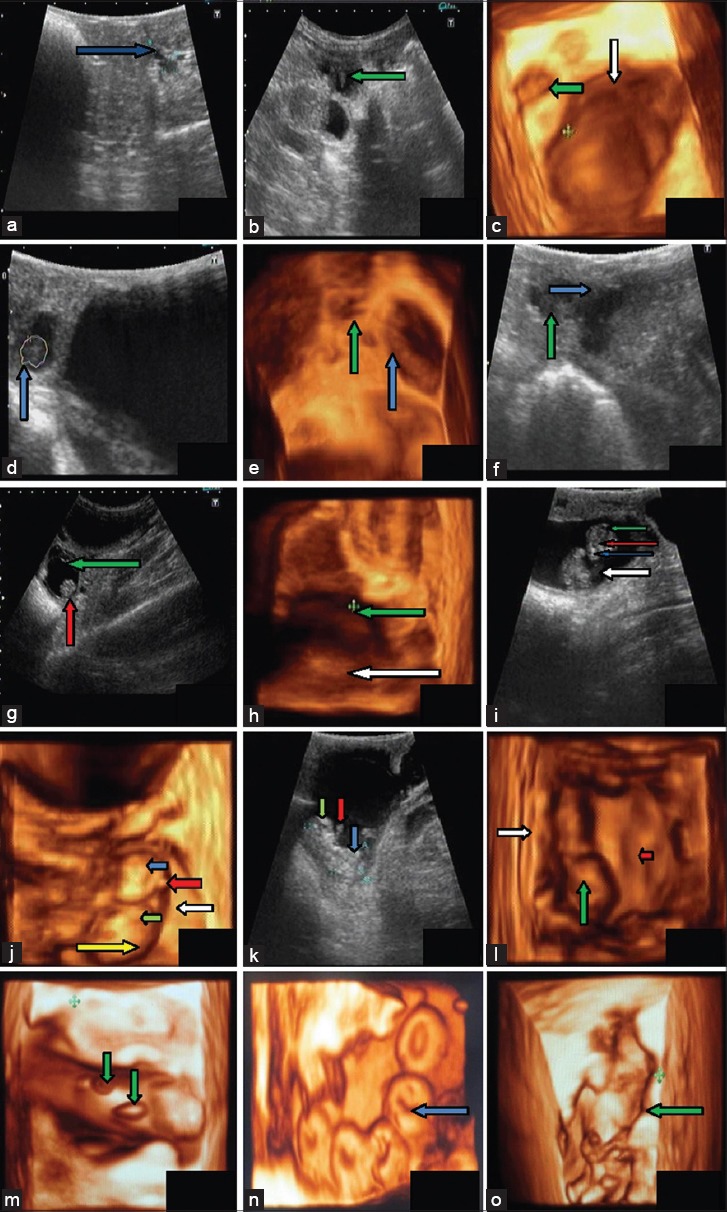
Results: On the 20(th) day of gestation, earliest diagnosis of pregnancy was done. First 3D ultrasonographic image of the conceptus, through transabdominal approach, was obtained on day 24. On 39(th) day, clear pictures of conceptus, amniotic membrane, and umbilicus were seen. On 76(th) day of gestation, internal organs of fetus viz heart, kidney, liver, urinary bladder, and stomach were seen both in 2D and 3D images. 3D imaging showed better details of uterine structures and internal organs of the fetus.
Conclusions: Comparing 3D images with 2D images, it is concluded that 2D was better in visualizing fluid while 3D images were better to view details of attachment of fetus with endometrium.
Keywords: fetal development; goats; pregnancy; three-dimensional; two-dimensional; ultrasonography.
Figures

Similar articles
-
Diagnosis of urinary bladder diseases in dogs by using two-dimensional and three-dimensional ultrasonography.Vet World. 2015 Jul;8(7):819-22. doi: 10.14202/vetworld.2015.819-822. Epub 2015 Jul 7. Vet World. 2015. PMID: 27047159 Free PMC article.
-
Diagnosis of pathological conditions of kidney by two-dimensional and three-dimensional ultrasonographic imaging in dogs.Vet World. 2016 Jul;9(7):693-8. doi: 10.14202/vetworld.2016.693-698. Epub 2016 Jul 4. Vet World. 2016. PMID: 27536028 Free PMC article.
-
A comparative study of two-dimensional and three-dimensional ultrasonography in evaluation of gastric affections in dogs.Vet World. 2015 Jun;8(6):707-12. doi: 10.14202/vetworld.2015.707-712. Epub 2015 Jun 6. Vet World. 2015. PMID: 27065634 Free PMC article.
-
The latest in ultrasound: three-dimensional imaging. Part II.Eur J Radiol. 1998 May;27 Suppl 2:S183-7. doi: 10.1016/s0720-048x(98)00077-1. Eur J Radiol. 1998. PMID: 9652520 Review.
-
Three-dimensional ultrasound with surface rendering for the study of fetal small parts.Zhonghua Yi Xue Za Zhi (Taipei). 2001 Jan;64(1):1-8. Zhonghua Yi Xue Za Zhi (Taipei). 2001. PMID: 11310366 Review.
Cited by
-
Study on development of accessory sex glands in prepubertal kids using two-dimensional ultrasonography.Vet World. 2016 Apr;9(4):346-50. doi: 10.14202/vetworld.2016.346-350. Epub 2016 Apr 6. Vet World. 2016. PMID: 27182127 Free PMC article.
References
-
- Kouamo J, Sow A, Kalandi M, Sawadogo G.J. Sensitivity, specificity, predictive value and accuracy of ultrasonography in pregnancy rate prediction in Sahelian goats after progesterone impregnated sponge synchronization. Vet. World. 2014;7(9):744–748.
-
- Suguna K, Mehrotra S, Agarwal S.K, Hoque M, Singh S.K, Shanker U, Sarath T. Early pregnancy diagnosis and embryonic and fetal development using real time B mode ultrasound in goats. Small Rumin. Res. 2008;80:80–86.
-
- Abdelghafar R.M, Bakhiet A.O, Ahmed B.H. B-mode real time ultrasonography for pregnancy diagnosis and fetal number in Saanen goats. J. Anim. Vet. Adv. 2007;6(5):702–705.
-
- Amer H.A. Ultrasonographic assessment of early pregnancy diagnosis, fetometry and sex determination in goats. Anim. Reprod. Sci. 2010;117:226–231. - PubMed
-
- Karen A.M, Fattouh E.M, Abu-Zeid S.S. Estimation of gestational age in Egyptian native goats by ultrasound fetometry. Anim. Reprod. Sci. 2009;114:167–174. - PubMed
LinkOut - more resources
Full Text Sources
Other Literature Sources