. 2015 Oct;16(5):295-303.
doi: 10.2174/1389202916666150707154719.

Non-coding RNAs in Exosomes: New Players in Cancer Biology

Affiliations

Non-coding RNAs in Exosomes: New Players in Cancer Biology

Miguel Silva et al. Curr Genomics. 2015 Oct.

Abstract

Exosomes are lipid bilayer extracellular vesicles (EVs) of 50-150nm in size, which contain nucleic acids (mRNA, ncRNAs and DNA), proteins and lipids. They are secreted by all cells and circulate in all body fluids. Exosomes are key mediators of several processes in cancer that mediate tumor progression and metastasis. These nano-vesicles, when secreted from cancer cells, are enriched in non-coding RNAs (e.g. microRNAs) complexed with the RNA-Induced Silencing Complex (RISC), that mediate an efficient and rapid silencing of mRNAs at the recipient cell, reprogramming their transcriptome. MicroRNAs in circulation encapsulated in exosomes are protected from degradation by a lipid bilayer and might serve as potential non-invasive diagnostic and screening tools to detect early stage cancer, to facilitate treatment options and possible help in curative surgical therapy decisions. Additionally, engineered exosomes can be used as therapy vehicles for targeted delivery of RNAi molecules, escaping the immune system detection.

Keywords: Exosomes; Extracellular vesicles; Non-coding RNAs.; microRNA biogenesis; microRNAs.


Figures
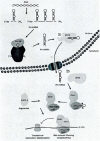
Fig. (1)
Fig. (1)
MiRNAs biogenesis pathway.
Fig. (2)
Fig. (2)
Schematic representation of exosomes biogenesis. Following endocytosis into early endosomes (A) ILVs are formed through an inward budding of the membrane (B). MVBs can then fuse with the plasma membrane (C), resulting in the release of the ILVs as exosomes into the extracellular space (D).
Fig. (3)
Fig. (3)
Overall composition of exosomes.
Fig. (4)
Fig. (4)
Proposed mechanisms of intercellular communication between exosomes and target cells: A) Direct interaction with receptors in the target cell and activation of intracellular signaling; B) Interaction with the target cell through ligation of previous cleaved fragments released from exosomes; C) Fusion of the exosomes with the membrane of target cell; D) Exosomes are phagocyted by the target cell.

References

    1. Higgs P.G., Lehman N. The RNA World: molecular cooperation at the origins of life. Nat. Rev. Genet. 2015;16(1):7–17. doi: 10.1038/nrg3841. - DOI - PubMed
    1. Mattick J.S., Makunin I.V. Non-coding RNA. Hum. Mol. Genet. 2006;15(Spec No 1):R17–R29. doi: 10.1093/hmg/ddl046. - DOI - PubMed
    1. Esteller M. Non-coding RNAs in human disease. Nat. Rev. Genet. 2011;12(12):861–874. doi: 10.1038/nrg3074. - DOI - PubMed
    1. Mercer T.R., Dinger M.E., Mattick J.S. Long non-coding RNAs: insights into functions. Nat. Rev. Genet. 2009;10(3):155–159. doi: 10.1038/nrg2521. - DOI - PubMed
    1. Taft R.J., Pang K.C., Mercer T.R., Dinger M., Mattick J.S. Non-coding RNAs: regulators of disease. J. Pathol. 2010;220(2):126–139. doi: 10.1002/path.2638. - DOI - PubMed

LinkOut - more resources