

Hippocampal Pathway Plasticity Is Associated with the Ability to Form Novel Memories in Older Adults
- PMID: 27047376
- PMCID: PMC4801877
- DOI: 10.3389/fnagi.2016.00061
Hippocampal Pathway Plasticity Is Associated with the Ability to Form Novel Memories in Older Adults
Abstract
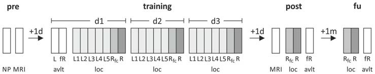
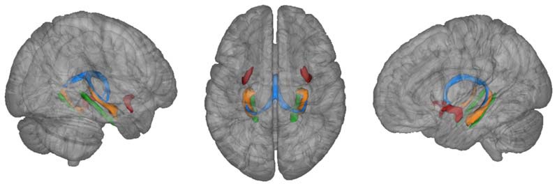
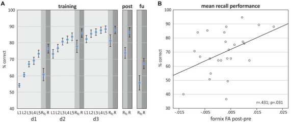
White matter deterioration in the aging human brain contributes to cognitive decline. The fornix as main efferent hippocampal pathway is one of the tracts most strongly associated with age-related memory impairment. Its deterioration may predict conversion to Alzheimer's dementia and its precursors. However, the associations between the ability to form novel memories, fornix microstructure and plasticity in response to training have never been tested. In the present study, 25 healthy older adults (15 women; mean age (SD): 69 (6) years) underwent an object-location training on three consecutive days. Behavioral outcome measures comprised recall performance on the training days, and on 1-day and 1-month follow up assessments. MRI at 3 Tesla was assessed before and after training. Fornix microstructure was determined by fractional anisotropy and mean diffusivity (MD) values from diffusion tensor imaging (DTI). In addition, hippocampal volumes were extracted from high-resolution images; individual hippocampal masks were further aligned to DTI images to determine hippocampal microstructure. Using linear mixed model analysis, we found that the change in fornix FA from pre- to post-training assessment was significantly associated with training success. Neither baseline fornix microstructure nor hippocampal microstructure or volume changes were significantly associated with performance. Further, models including control task performance (auditory verbal learning) and control white matter tract microstructure (uncinate fasciculus and parahippocampal cingulum) did not yield significant associations. Our results confirm that hippocampal pathways respond to short-term cognitive training, and extend previous findings by demonstrating that the magnitude of training-induced structural changes is associated with behavioral success in older adults. This suggests that the amount of fornix plasticity may not only be behaviorally relevant, but also a potential sensitive biomarker for the success of training interventions aimed at improving memory formation in older adults, a hypothesis to be evaluated in future studies.
Keywords: cognitive training; diffusion tensor imaging; fornix; learning; white matter microstructure.
Figures
References
LinkOut - more resources
Full Text Sources
Other Literature Sources
Medical