

Dynamic Progression of White Matter Hyperintensities in Alzheimer's Disease and Normal Aging: Results from the Sunnybrook Dementia Study
- PMID: 27047377
- PMCID: PMC4805606
- DOI: 10.3389/fnagi.2016.00062
Dynamic Progression of White Matter Hyperintensities in Alzheimer's Disease and Normal Aging: Results from the Sunnybrook Dementia Study
Abstract
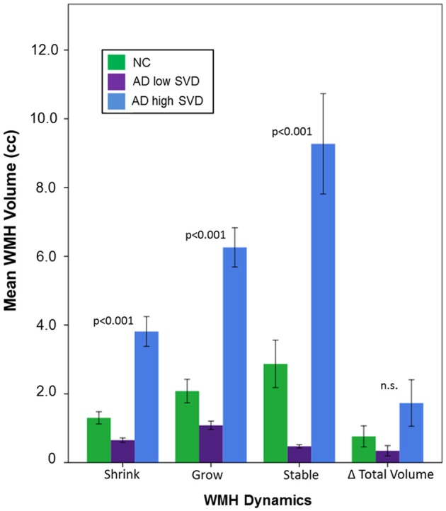
Although white matter hyperintensities (WMH), markers of cerebral small vessel disease (SVD), are believed to generally increase over time, some studies have shown sharp decreases after therapeutic intervention, suggesting that WMH progression may be more dynamic than previously thought. Our primary goal was to examine dynamic progression of WMH in a real-world sample of Alzheimer's disease (AD) patients and normal elderly (NC), with varying degrees of SVD. WMH volumes from serial magnetic resonance imaging (MRI; mean = 1.8 years) were measured from NC (n = 44) and AD patients (n = 113) with high and low SVD burden. Dynamic progression for each individual was measured using spatial overlap images to assess shrinkage, growth, and stable WMH volumes. Significant group differences were found for shrinkage (p < 0.001), growth (p < 0.001) and stable (p < 0.001) WMH, where the AD high SVD group showed the largest changes relative to low SVD and NC. Our results suggest spatial progression measured at the individual patient level may be more sensitive to the dynamic nature of WMH.
Keywords: Alzheimer’s disease; aging; dementia; longitudinal progression; small vessel disease; white matter hyperintensities.
Figures


References
-
- Apostolova L. G., Green A. E., Babakchanian S., Hwang K. S., Chou Y. Y., Toga A. W., et al. . (2012). Hippocampal atrophy and ventricular enlargement in normal aging, mild cognitive impairment (MCI) and Alzheimer Disease. Alzheimer Dis. Assoc. Disord. 26, 17–27. 10.1097/WAD.0b013e3182163b62 - DOI - PMC - PubMed
-
- Barnes J., Carmichael O. T., Leung K. K., Schwarz C., Ridgway G. R., Bartlett J. W., et al. . (2013). Vascular and Alzheimer’s disease markers independently predict brain atrophy rate in Alzheimer’s Disease Neuroimaging Initiative controls. Neurobiol. Aging 34, 1996–2002. 10.1016/j.neurobiolaging.2013.02.003 - DOI - PMC - PubMed
LinkOut - more resources
Full Text Sources
Other Literature Sources