. 2016 Mar 22:7:77.
doi: 10.3389/fimmu.2016.00077. eCollection 2016.

B Cell Development in the Bone Marrow Is Regulated by Homeostatic Feedback Exerted by Mature B Cells

Affiliations

B Cell Development in the Bone Marrow Is Regulated by Homeostatic Feedback Exerted by Mature B Cells

Gitit Shahaf et al. Front Immunol. .

Abstract

Cellular homeostasis in the B cell compartment is strictly imposed to balance cell production and cell loss. However, it is not clear whether B cell development in the bone marrow is an autonomous process or subjected to regulation by the peripheral B cell compartment. To specifically address this question, we used mice transgenic for human CD20, where effective depletion of B lineage cells is obtained upon administration of mouse anti-human CD20 antibodies, in the absence of any effect on other cell lineages and/or tissues. We followed the kinetics of B cell return to equilibrium by BrdU labeling and flow cytometry and analyzed the resulting data by mathematical modeling. Labeling was much faster in depleted mice. Compared to control mice, B cell-depleted mice exhibited a higher proliferation rate in the pro-/pre-B compartment, and higher cell death and lower differentiation in the immature B cell compartment. We validated the first result by analysis of the expression of Ki67, the nuclear protein expressed in proliferating cells, and the second using Annexin V staining. Collectively, our results suggest that B lymphopoiesis is subjected to homeostatic feedback mechanisms imposed by mature B cells in the peripheral compartment.

Keywords: B lymphocytes; BrdU; computer simulation; homeostatic feedback; mathematical modeling.


Figures
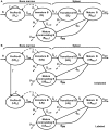
Figure 1
Figure 1
Model of B cell population dynamics in the bone marrow and spleen. (A) The model of maturing B-cell population dynamics in the bone marrow and spleen is shown. All population processes – differentiation, proliferation, and death – are described by arrows. The rate of each process is given near the corresponding arrow. S is the source of B lineage precursors (cells/6 h), parameters δ denote differentiation rates, μ mortality rates, γ proliferation rates, and ϕ flow rates. (B) The model for labeling dynamics of developing B cells in the bone marrow assumes that cells in the unlabeled pro-/pre-B cell compartment move to the corresponding labeled compartment upon dividing. The model also assumes that each labeled cell remains labeled for the duration of the experiment.
Figure 2
Figure 2
Selective depletion of BM B cell subsets in an hCD20Tg mouse. (A) BM cells from hCD20Tg mouse were stained for B220, IgM, AA4.1, and hCD20. For analysis, viable lymphocyte were defined by forward and side light scatter, and gates were set to analyze B220+/AA4.1+ early developing B cells (pro-/pre-B and immature B cells) and to exclude circulated B220+/AA4.1 and non-B lineage cells. Gated cells were analyzed for expression of IgM and hCD20. (B) hCD20Tg mice were injected with anti-hCD20 antibodies. Three days after injection, BM cells were stained for B220 and IgM and analyzed for the following B cell subsets: pre-/pro- (B220+/IgM), immature (B220+/IgM+), and circulated mature (B220hi/IgM+) B cells, relative to control hCD20Tg mice that were not treated. The results shown are representative of three to four mice in each group.
Figure 3
Figure 3
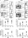
Control and hCD20-depleted mice (34 days after depletion) were injected with BrdU and analyzed for BrdU labeling as detailed in Section “Materials and Methods.” (A) BM and spleen cells from the control and the depleted mice were stained for IgM, AA4.1 and BrdU labeling. For analysis, viable lymphocytes were defined by forward and side light scatter, and the relative BrdU labeling in BM immature (IgM+/AA4.1+) and mature-circulated (IgM+/AA4.1), and in spleen transitional (IgM+/AA4.1+) and mature (IgM+/AA4.1) B cells was determined using gates set as shown (representative example). (B) BrdU labeling kinetics in B cell-depleted (black, open circles for each single mouse and band for the mean value) and control (gray, filled triangles for each single mouse and band for the mean value) mice. For total cell numbers, see Figure 4.
Figure 4
Figure 4
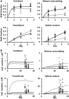
(A) BrdU labeling kinetics obtained by a simulation of the B cell development model using the parameter value set that gave the best fit to the data, shown (as lines) along with the data (symbols), for control (filled gray triangles and gray lines), and depleted mice (open black circles and black lines), in the immature, mature recirculating, transitional, and splenic mature B cell subsets. The x-axis shows the time from the start of labeling. (B) The ranges of total cell numbers in each B cell subset in the experimental measurements of control and depleted mice and the corresponding simulations [symbols and lines are as in (A)]. The x-axis shows the time from the start of B cell depletion. The parameter values used in these simulations are given in Table 1.
Figure 5
Figure 5
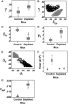
Box Plots of the parameter value distributions for each rate parameter that was found to significantly differ between the simulations of control and depleted mice. (A) The proliferation rate of the Boe subpopulation. (B) The proliferation rate of the Boe subpopulation plotted against its differentiation rate. (C) The rate of exit of immature B cell from the BM. (D) Immature B cell death rate. (E) The rate of exit of immature B cell from the BM plotted against their death rate. (F) Box Plots of the sources of mature recirculating B cell subpopulations, that is, the total cell number entering in each time step of 6 h. The white boxes are the flows from the splenic mature B cell subpopulation (ϕs × BMspl) and the gray boxes are the flows from the BM immature B cell subpopulation (δi_re × Bi). (G) The exit rate from the splenic mature B cell subpopulation.
Figure 6
Figure 6
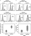
BM cells from control and hCD20-depleted mice (34 days after depletion) were stained for B220 IgM AA4.1 and Ki67 or Annexin V. For analysis, viable lymphocytes were defined by forward and side light scatter and the relative Ki67 or Annexin V staining in pro-/pre-B (B220+/AA4.1+/IgM) or immature (B220+/AA4.1+/IgM+) B cell subsets was determined using gates set as shown in Figure 2A. (A) Histograms showing Ki67 expression in the gated pro-/pre- and immature B cell subsets in the control and depleted mice (representative example). (B) Ki67+ cell numbers in the pro-/pre-B cell compartment in control and B cell-depleted mice. (C) Histograms showing Annexin V expression in the pro-/pre-B and immature cell subsets in control and depleted mice (representative of four mice in each group). (D) Percentages of Annexin V+ cells in the pro-/pre-B and immature B cell compartments in control and B cell-depleted mice.

Similar articles

Cited by

References

    1. Rajewsky K. Clonal selection and learning in the antibody system. Nature (1996) 381:751–8.10.1038/381751a0 - DOI - PubMed
    1. Edry E, Melamed D. Receptor editing in positive and negative selection of B lymphopoiesis. J Immunol (2004) 173:4265–71.10.4049/jimmunol.173.7.4265 - DOI - PubMed
    1. Melchers F, Rolink A, Grawunder U, Winkler TH, Karasuyama H, Ghia P, et al. Positive and negative selection events during B lymphopoiesis. Curr Opin Immunol (1995) 7:214–27.10.1016/0952-7915(95)80006-9 - DOI - PubMed
    1. Hardy RR, Carmack CE, Shinton SA, Kemp JD, Hayakawa K. Resolution and characterization of pro-B and pre-pro-B cell stages in normal mouse bone marrow. J Exp Med (1991) 173:1213–25.10.1084/jem.173.5.1213 - DOI - PMC - PubMed
    1. Rolink AG, Schaniel C, Andersson J, Melchers F. Selection events operating at various stages in B cell development. Curr Opin Immunol (2001) 13:202–7.10.1016/S0952-7915(00)00205-3 - DOI - PubMed

LinkOut - more resources