. 2016:2016:8283062.
doi: 10.1155/2016/8283062. Epub 2016 Mar 7.

Viscoat Assisted Inverted Internal Limiting Membrane Flap Technique for Large Macular Holes Associated with High Myopia

Affiliations

Viscoat Assisted Inverted Internal Limiting Membrane Flap Technique for Large Macular Holes Associated with High Myopia

Zongming Song et al. J Ophthalmol. 2016.

Abstract

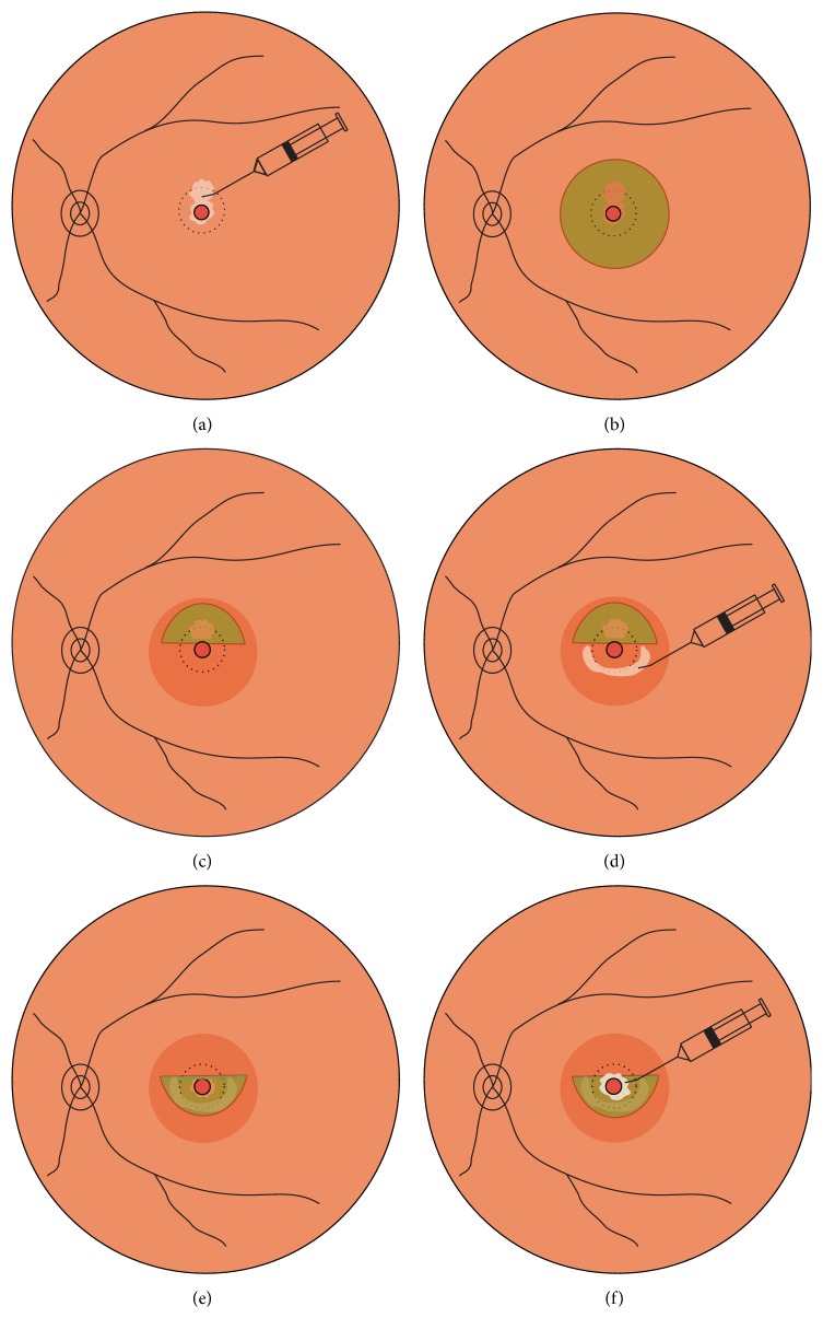
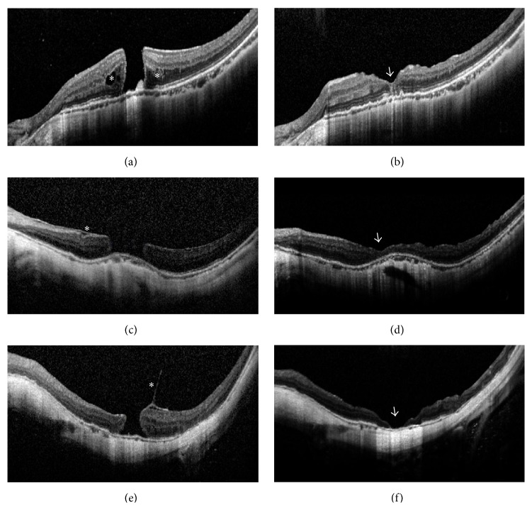
Purpose. To investigate the surgical outcomes of Viscoat® assisted inverted internal limiting membrane (ILM) flap technique for large macular holes (MHs) associated with high myopia. Design. Prospective, interventional case series. Methods. Fifteen eyes of 15 patients with high myopia underwent vitrectomy and Viscoat assisted inverted ILM flap technique to treat MH without RD. Patients were followed up over 6 months. The main outcome measures were MH closure evaluated by optical coherence tomography (OCT) and best-corrected visual acuities (BCVAs). Result. MH closure was observed in all eyes (100%) following the initial surgery. Type 1 closure was observed in 13 eyes (86.7%); type 2 closure was observed in the remaining 2 eyes (13.3%). Compared to the preoperative baseline, the mean BCVA (logarithm of the minimum angle of resolution) improved significantly at 3 months and 6 months after surgery (P = 0.025, 0.019, resp.). The final BCVA improved in 10 eyes (66.7%), remained unchanged in 3 eyes (20.0%), and worsened in 2 eyes (13.3%). Conclusion. Vitrectomy combined with Viscoat assisted inverted ILM flap technique is an effective treatment for large MHs in highly myopic eyes. It may increase the success rate of the initial surgery and enhance the anatomical and functional outcomes.


Figures

Figure 1
Figure 1
Schematic drawings showing the Viscoat assisted single-layered inverted internal limiting membrane (ILM) flap technique. (a) After performing a 23-gauge 3-port pars plana vitrectomy, Viscoat is injected into the macular hole (MH) and its mirror symmetrical area superior to MH. (b) Indocyanine green (ICG, 0.125% solution) is then applied around the MH within the arcade to stain the ILM. Excessive ICG as well as Viscoat is removed by suction. (c) The ILM is peeled off in a circular fashion for approximately 2.5 disc diameters around the MH. The inferior ILM is completely peeled off, while the superior ILM is not removed completely but left attached to the edge of the MH, forming an ILM flap of about 1 disk diameter. (d) Viscoat is injected and smeared in an arch around the MH in the lower half part of the macular as an adhesive. (e) The ILM flap is flipped by inverting it using the intraocular forceps to cover the whole MH and gently massaged to make it flattened. (f) Supplementary Viscoat is applied on top of the inverted ILM flap as ballast before air-fluid exchange is performed.
Figure 2
Figure 2
Optical coherence tomography (OCT) images of large myopic macular holes (MHs) in selected cases before and after surgery. (a) Case Number 5 before surgery. An MH of 563 μm and retinoschisis around the MH (asterisks) was present. Best-corrected visual acuities (BCVAs) = 0.02. (b) Case Number 5 at 6 months after surgery. Type 1 closure in U-shape and restoration of the inner and outer segment junction line was achieved (arrowhead). BCVA = 0.3. (c) Case Number 12 before surgery. A large-sized MH of 812 μm and epiretinal membrane (asterisk) was present. BCVA = FC/50 cm. (FC = finger counting) (d) Case Number 12 at 6 months after surgery. Type 1 closure in shallow V-shape was present (arrowhead). BCVA = 0.3. (e) Case Number 14 before surgery. A longstanding MH of 491 μm and vitreomacular traction (asterisk) was present. BCVA = HM/BE. (HM = hand motion; BE = before eye) (f) Case Number 14 at 6 months after surgery. Type 2 closure with bare retinal pigment epithelium in the center (arrowhead) was present. BCVA = HM/BE.

References

    1. Xu L., Li J., Cui T., et al. Refractive error in urban and rural adult Chinese in Beijing. Ophthalmology. 2005;112(10):1676–1683. doi: 10.1016/j.ophtha.2005.05.015. - DOI - PubMed
    1. Liu H. H., Xu L., Wang Y. X., Wang S., You Q. S., Jonas J. B. Prevalence and progression of myopic retinopathy in Chinese adults: the Beijing eye study. Ophthalmology. 2010;117(9):1763–1768. doi: 10.1016/j.ophtha.2010.01.020. - DOI - PubMed
    1. Stirpe M., Michels R. G. Retinal detachment in highly myopic eyes due to macular holes and epiretinal traction. Retina. 1990;10(2):113–114. doi: 10.1097/00006982-199004000-00004. - DOI - PubMed
    1. Kelly N. E., Wendel R. T. Vitreous surgery for idiopathic macular holes: results of a pilot study. Archives of Ophthalmology. 1991;109(5):654–659. doi: 10.1001/archopht.1991.01080050068031. - DOI - PubMed
    1. Kadonosono K., Yazama F., Itoh N., et al. Treatment of retinal detachment resulting from myopic macular hole with internal limiting membrane removal. American Journal of Ophthalmology. 2001;131(2):203–207. doi: 10.1016/S0002-9394(00)00728-5. - DOI - PubMed