

PLANTAR THROMBOPHLEBITIS: MAGNETIC RESONANCE IMAGING FINDINGS
- PMID: 27047898
- PMCID: PMC4799486
- DOI: 10.1016/S2255-4971(15)30036-7
PLANTAR THROMBOPHLEBITIS: MAGNETIC RESONANCE IMAGING FINDINGS
Abstract
Objective: Demonstrate the magnetic resonance imaging (MRI) findings in plantar thrombophlebitis.
Methods: Retrospective review of twenty patients with pain in the plantar region of the foot, in which the MRI findings indicated plantar thrombophlebitis.
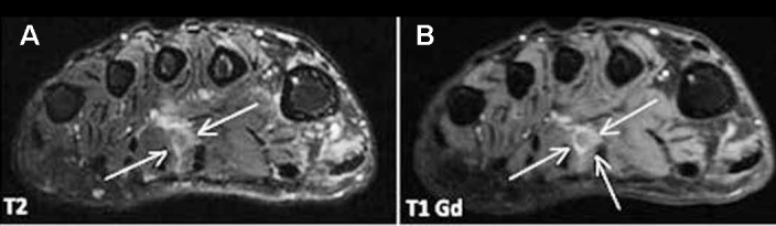
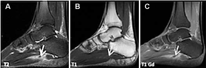
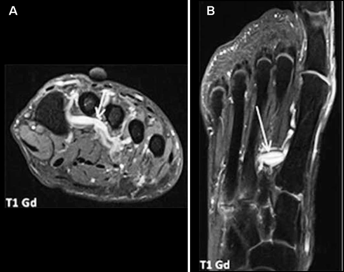
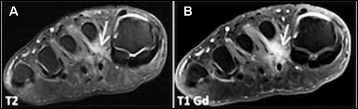
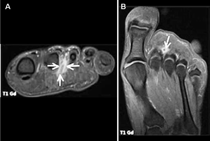
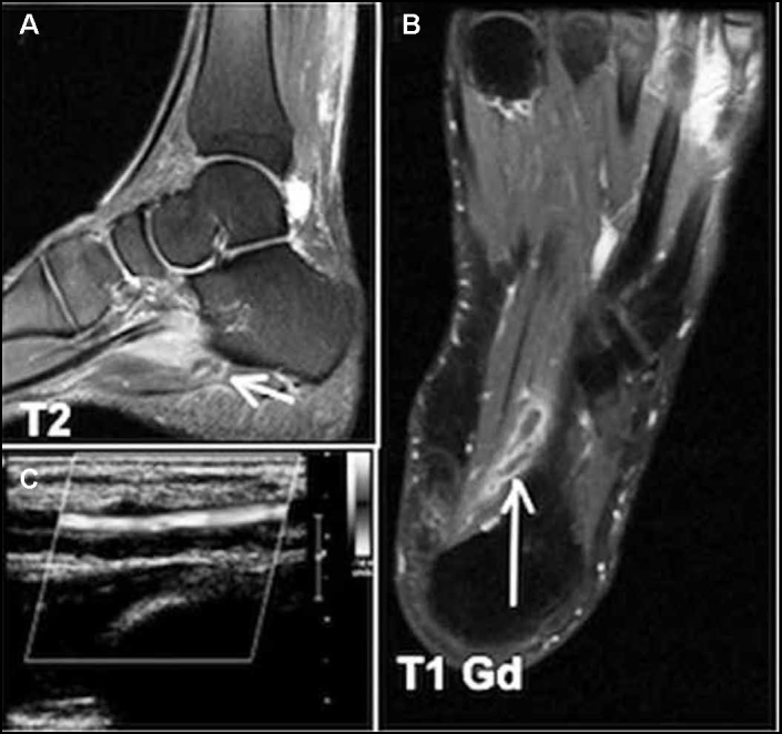
Results: A total of fourteen men and six women, mean age 46.7 years were evaluated. Eight of these patients also underwent Doppler ultrasonography, which confirmed the thrombophlebitis. The magnetic resonance images were evaluated in consensus by two radiologists with experience in musculoskeletal radiology (more than 10 years each), showing perivascular edema in all twenty patients (100%) and muscle edema in nineteen of the twenty patients (95%). All twenty patients had intraluminal intermediate signal intensity on T2-weighted (100%) and venous ectasia was present in seventeen of the twenty cases (85%). Collateral veins were visualized in one of the twenty patients (5%). All fourteen cases (100%), in which intravenous contrast was administered, showed perivenular tissues enhancement and intraluminal filling defect. Venous ectasia, loss of compressibility and no flow on Doppler ultrasound were also observed in all eight cases examined by the method.
Conclusion: MRI is a sensitive in the evaluation of plant thrombophlebitis in patients with plantar foot pain.
Keywords: Foot; Magnetic Resonance Imaging; Thrombophlebitis.
Figures
Similar articles
-
Magnetic Resonance Imaging Features of Plantar Vein Thrombosis.Diagnostics (Basel). 2024 Jun 8;14(12):1215. doi: 10.3390/diagnostics14121215. Diagnostics (Basel). 2024. PMID: 38928631 Free PMC article.
-
Imaging Features of Plantar Vein Thrombosis: An Easily Overlooked Condition in the Differential Diagnosis of Foot Pain.Diagnostics (Basel). 2024 Jan 5;14(2):126. doi: 10.3390/diagnostics14020126. Diagnostics (Basel). 2024. PMID: 38248003 Free PMC article. Review.
-
Foot pain after a plantar fasciotomy: an MR analysis to determine potential causes.J Comput Assist Tomogr. 1999 Sep-Oct;23(5):707-12. doi: 10.1097/00004728-199909000-00013. J Comput Assist Tomogr. 1999. PMID: 10524852
-
Idiopathic and diabetic skeletal muscle necrosis: evaluation by magnetic resonance imaging.Skeletal Radiol. 2005 Apr;34(4):203-9. doi: 10.1007/s00256-004-0881-8. Epub 2005 Feb 8. Skeletal Radiol. 2005. PMID: 15700181
-
Diagnostic imaging in deep vein thrombosis of the limbs.Rays. 1996 Jul-Sep;21(3):328-39. Rays. 1996. PMID: 9063053 Review. English, Italian.
Cited by
-
Venous thrombosis: a mimic of musculoskeletal injury on MR imaging.Skeletal Radiol. 2023 Jul;52(7):1263-1276. doi: 10.1007/s00256-022-04258-4. Epub 2022 Dec 19. Skeletal Radiol. 2023. PMID: 36534142 Review.
-
Magnetic Resonance Imaging Features of Plantar Vein Thrombosis.Diagnostics (Basel). 2024 Jun 8;14(12):1215. doi: 10.3390/diagnostics14121215. Diagnostics (Basel). 2024. PMID: 38928631 Free PMC article.
-
Erythema nodosum-like lesion on heel in a patient assumed Behçet's disease.BMJ Case Rep. 2019 Nov 19;12(11):e232056. doi: 10.1136/bcr-2019-232056. BMJ Case Rep. 2019. PMID: 31748365 Free PMC article. No abstract available.
-
Plantar vein thrombosis: a rare differential diagnosis in patients with plantar pain.Radiol Bras. 2015 Nov-Dec;48(6):399-400. doi: 10.1590/0100-3984.2015.0075. Radiol Bras. 2015. PMID: 26811559 Free PMC article. No abstract available.
-
Imaging Features of Plantar Vein Thrombosis: An Easily Overlooked Condition in the Differential Diagnosis of Foot Pain.Diagnostics (Basel). 2024 Jan 5;14(2):126. doi: 10.3390/diagnostics14020126. Diagnostics (Basel). 2024. PMID: 38248003 Free PMC article. Review.
References
-
- Siegal DS, Wu JS, Brennan DD, Challies T, Hochman MG. Plantar vein thrombosis: a rare cause of plantar foot pain. Skeletal Radiol. 2008;37(3):267–269. - PubMed
-
- Bernathova M, Bein E, Bendix N, Bodner G. Sonographic diagnosis of plantar vein thrombosis: report of 3 cases. J Ultrasound Med. 2005;24(1):101–103. - PubMed
-
- Gray H. Anatomy of the human body. Philadelphia: Lea & Febiger, 1918; Bartleby.com, 2000. Disponível: www.bartleby.com/107/
-
- White JV, Katz ML, Cisek P, Kreithen J. Venous outflow of the leg: anatomy and physiologic mechanism of the plantar venous plexus. J Vasc Surg. 1996;24(5):819–824. - PubMed
-
- Cavezzi A. Isolated thrombosis of plantar veins. Case report. Minerva Cardioangiol. 1999;47(9):309–313. - PubMed
LinkOut - more resources
Full Text Sources