. 2016:2016:2517514.
doi: 10.1155/2016/2517514. Epub 2016 Mar 7.

Human Umbilical Cord Mesenchymal Stem Cells Therapy in Cyclophosphamide-Induced Premature Ovarian Failure Rat Model

Affiliations

Human Umbilical Cord Mesenchymal Stem Cells Therapy in Cyclophosphamide-Induced Premature Ovarian Failure Rat Model

Dan Song et al. Biomed Res Int. 2016.

Abstract

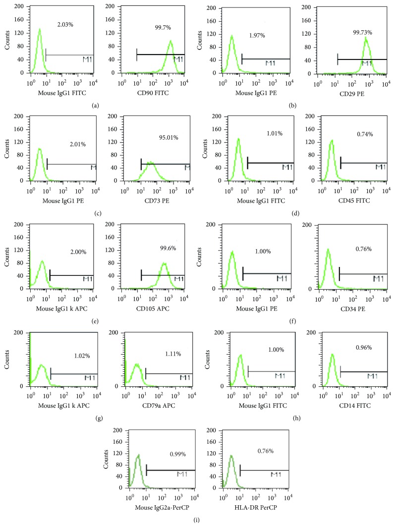
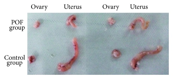
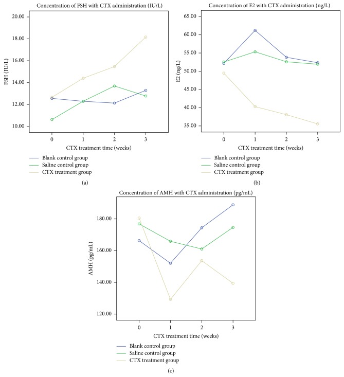
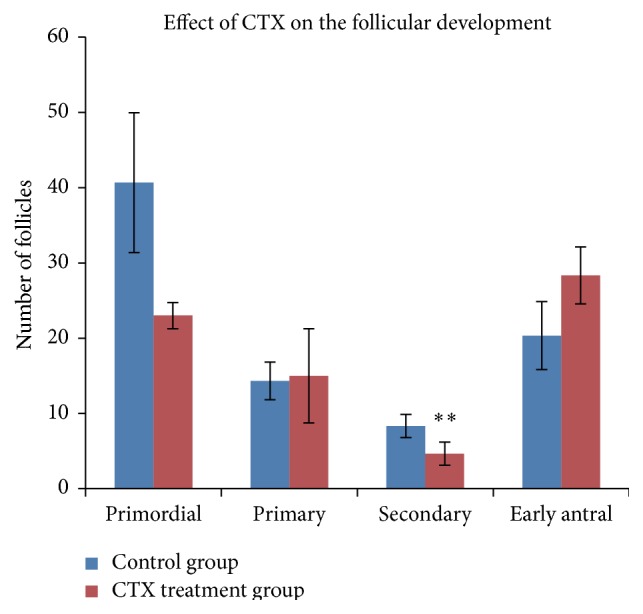
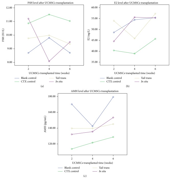
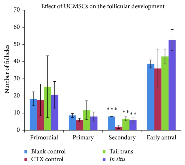
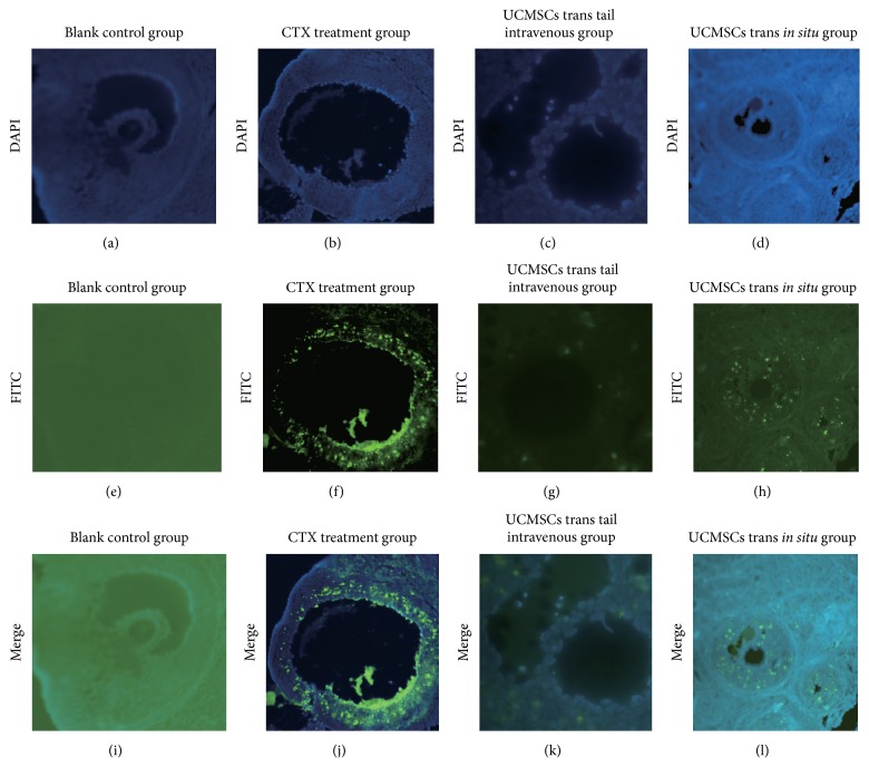
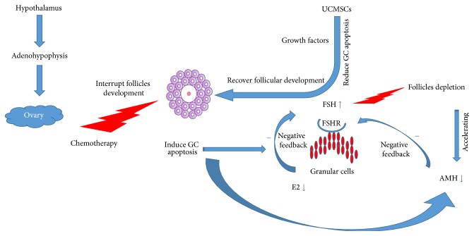
Premature ovarian failure (POF) is one of the most common causes of infertility in women. In our present study, we established cyclophosphamide- (CTX-) induced POF rat model and elucidated its effect on ovarian function. We detected the serum estrogen, follicle stimulating hormone, and anti-Müllerian hormone of mice models by ELISA and evaluated their folliculogenesis by histopathology examination. Our study revealed that CTX administration could severely disturb hormone secretion and influence folliculogenesis in rat. This study also detected ovarian cells apoptosis by deoxy-UTP-digoxigenin nick end labeling (TUNEL) and demonstrated marked ovarian cells apoptosis in rat models following CTX administration. In order to explore the potential of human umbilical cord mesenchymal stem cells (UCMSCs) in POF treatment, the above indexes were used to evaluate ovarian function. We found that human UCMSCs transplantation recovered disturbed hormone secretion and folliculogenesis in POF rat, in addition to reduced ovarian cell apoptosis. We also tracked transplanted UCMSCs in ovaries by fluorescence in situ hybridization (FISH). The results manifested that the transplanted human UCMSCs could reside in ovarian tissues and could survive for a comparatively long time without obvious proliferation. Our present study provides new insights into the great clinical potential of human UCMSCs in POF treatment.


Figures

Figure 1
Figure 1
Flow cytometry analysis of human UCMSCs. Flow cytometry analysis of human UCMSCs showed their expression for CD90/CD29/CD73/CD45/CD105/CD34/CD79a/CD14/HLA-DR.
Figure 2
Figure 2
Photographs of ovaries removed from CTX treated groups and control group.
Figure 3
Figure 3
The trend for average level of FSH, AMH, and E2 in each group (blank control group, saline treatment group, and CTX treatment group) during three weeks after CTX treatment.
Figure 4
Figure 4
Effect of CTX on the follicles development. Ovarian H&E staining pathological sections were from control group and CTX treatment group. Follicles were counted and classified. Data were means ± SD of counts of different stages follicles (n = 3) (primordial, primary, and early antral follicles of POF group versus control group: p > 0.05, secondary follicles of POF group versus control group: p = 0.004). ∗∗ p < 0.01.
Figure 5
Figure 5
The trend for average level of FSH, AMH, and E2 in each group during six weeks after UCMSCs transplantation.
Figure 6
Figure 6
Effect of UCMSCs transplantation on follicular development. Ovarian H&E staining sections were from blank control group, CTX control group, and UCMSCs transplantation groups (by tail intravenously or in situ). Follicles were numbered and classified. Data were means ± SD of counts of different stages follicles in three experiments (secondary follicles of UCMSCs treatment groups versus POF control group: p < 0.01; primordial, primary, and early antral follicles of UCMSCs treatment groups versus POF control group: p > 0.05, n = 3). ∗∗ p < 0.01; ∗∗∗ p < 0.001.
Figure 7
Figure 7
Cell apoptosis assay: UCMSCs transplantation reduced cell apoptosis of ovaries in CTX-induced POF rats. (a, b, c, d) Blue fluorescence indicates cell nucleus stained by DAPI (blank control group, CTX treatment group, tail intravenous trans group, and in situ UCMSCs trans group). (e, f, g, h) Green fluorescence (FITC) stained cells illustrating the degree and site of apoptosis of cells in the four groups. (i, j, k, l) merged the two stainings.
Figure 8
Figure 8
FISH analysis of UCMSCs, tracking of the transplanted human UCMSCs in rats ovarian tissue. (a, b, c, d) Distribution of human UCMSCs in rats ovaries after transplantation for six weeks (blank control group, CTX control group, tail intravenous trans group, and in situ trans group). (e, f, g, h) Distribution of human UCMSCs in rats ovaries after transplantation for eight weeks. The location of the UCMSCs was determined using orange (SRY/Y chromosome) and green (CEP/X chromosome) signals. Double-labeled staining (red and green fluorescence) cells were defined as human UCMSCs.
Figure 9
Figure 9
The network of steroidal hormones regulation system during pathological process of POF and UCMSCs therapeutic mechanism. With the hypothalamus regulation, adenohypophysis could synthesize and produce gonadotropins (FSH), which could stimulate follicles growth. GCs of the growth follicles could produce E2, having a feedback negative effect on FSH production, thus reducing follicles excessive growth and avoiding their depletion. AMH could also inhibit FSH production. The negative feedback effect of E2 and AMH on the follicles growth was interrupted by CTX administration. Lack of inhibition of E2 and AMH causes uncontrolled elevating FSH level, which promotes follicles pool exhausted very soon, leading to POF. The transplanted human UCMSCs may secrete kinds of growth factors, which could rescue the overapoptosis of GCs and ultimately improve follicles development and retargeted hormonal balance.

References

    1. Coulam C. B. Premature gonadal failure. Fertility and Sterility. 1982;38(6):645–655. - PubMed
    1. Davis C. J., Davison R. M., Payne N. N., Rodeck C. H., Conway G. S. Female sex preponderance for idiopathic familial premature ovarian failure suggests an X chromosome defect: opinion. Human Reproduction. 2000;15(11):2418–2422. doi: 10.1093/humrep/15.11.2418. - DOI - PubMed
    1. Goswami R., Goswami D., Kabra M., Gupta N., Dubey S., Dadhwal V. Prevalence of the triple X syndrome in phenotypically normal women with premature ovarian failure and its association with autoimmune thyroid disorders. Fertility and Sterility. 2003;80(4):1052–1054. doi: 10.1016/S0015-0282(03)01121-X. - DOI - PubMed
    1. Simpson J. L., Rajkovic A. Ovarian differentiation and gonadal failure. American Journal of Medical Genetics. 1999;89(4):186–200. - PubMed
    1. Brougham M. F. H., Wallace W. H. B. Subfertility in children and young people treated for solid and haematological malignancies. British Journal of Haematology. 2005;131(2):143–155. doi: 10.1111/j.1365-2141.2005.05740.x. - DOI - PubMed

Publication types

Substances

LinkOut - more resources